
-
Paper Information
- Next Paper
- Paper Submission
-
Journal Information
- About This Journal
- Editorial Board
- Current Issue
- Archive
- Author Guidelines
- Contact Us
American Journal of Medicine and Medical Sciences
p-ISSN: 2165-901X e-ISSN: 2165-9036
2025; 15(10): 3470-3473
doi:10.5923/j.ajmms.20251510.40
Received: Sep. 3, 2025; Accepted: Oct. 1, 2025; Published: Oct. 25, 2025

Biochemical and Immunological Parameters of Blood Serum of Patients with Tuberculosis in the Bukhara Region
 Abstract
 Reference
 Full-Text PDF
 Full-text HTML
Jalolitdin Sulton Nazarov
Department of Microbiology, Virology and Immunology, Bukhara State Medical Institute named after Abu Ali ibn Sino, Gijduvan St. 23, Bukhara, Uzbekistan
Correspondence to: Jalolitdin Sulton Nazarov, Department of Microbiology, Virology and Immunology, Bukhara State Medical Institute named after Abu Ali ibn Sino, Gijduvan St. 23, Bukhara, Uzbekistan.
| Email: |  |
Copyright © 2025 The Author(s). Published by Scientific & Academic Publishing.
This work is licensed under the Creative Commons Attribution International License (CC BY).
http://creativecommons.org/licenses/by/4.0/

Objective: To characterize biochemical and immunological parameters in the blood serum of patients with a verified diagnosis of tuberculosis (TB) in the Bukhara region and to explore a multimarker model with emphasis on multidrug-resistant (MDR) and extensively drug-resistant (XDR) TB. Methods: We examined serum samples from 147 TB patients before initiation and after completion of anti-tuberculosis therapy at the Bukhara Regional Center of Phthisiology and Pulmonology; 20 practically healthy adults served as controls. Biochemical profiling (ALT, AST, glucose, creatinine, urea, total protein) and immunological assays (IgA, IgM, IgG, IgE; cytokines including IFNγ, IL‑6, IL‑1β, TNF‑α) were performed using standard analyzers and ELISA. Results: Following therapy, ALT/AST elevations were common, consistent with treatment-related hepatocellular stress. Comorbidities were frequent—hypertension (33%), coronary heart disease (29%), angina (23%), and diabetes mellitus (27%). In the MDR/XDR subset (n=15), IFNγ and IL‑6 were detected in all patients; IL‑1β and TNF‑α in 7 and 6 patients, respectively; anti-inflammatory cytokines were largely normal (IL‑10 elevated in 2, IL‑4 in none). Elevated immunoglobulins were observed predominantly for IgA (14/15), IgM (12/15), and IgE (12/15), with more modest IgG elevations (7/15). Conclusions: A composite of pro-inflammatory cytokines (IFNγ, IL‑6, IL‑1β) together with immunoglobulins (IgA, IgM, IgE) shows promise as a diagnostic and prognostic signature in MDR/XDR-TB. These markers may inform patient stratification and immunocorrective approaches alongside standard chemotherapy.
Keywords: Tuberculosis, Biochemical markers, Cytokine profile, MDR-TB, XDR-TB, Immunoglobulins
Cite this paper: Jalolitdin Sulton Nazarov, Biochemical and Immunological Parameters of Blood Serum of Patients with Tuberculosis in the Bukhara Region, American Journal of Medicine and Medical Sciences, Vol. 15 No. 10, 2025, pp. 3470-3473. doi: 10.5923/j.ajmms.20251510.40.
Article Outline
1. Introduction
- Tuberculosis (TB) remains among the most consequential infectious diseases, with billions living with latent infection and a substantial annual burden of active disease. Beyond microbiological confirmation, complementary biochemical and immunological markers can refine diagnosis, disease monitoring, and prognosis. Humoral responses—particularly serum immunoglobulins—show class- and antigen-specific patterns during active versus latent TB, and may modulate cellular immunity [1-3,7-9,11-13].Cytokines orchestrate host-pathogen interactions in TB; dysregulated pro-inflammatory signaling (e.g., IFNγ, IL‑6, TNF‑α) can drive tissue damage and poor outcomes, whereas balanced responses are essential for control [5,6,17]. Serum biochemistry (e.g., acute-phase proteins, hepatocellular enzymes, renal indices) complements immune profiling by capturing systemic effects of disease and therapy [14-16].Here we report a single-center study from the Bukhara region evaluating serum biochemical parameters and a targeted panel of immunoglobulins and cytokines in TB patients before and after chemotherapy, with focused analyses in MDR/XDR subgroups. We propose a practical multimarker set for clinical use.
2. Materials and Methods
2.1. Study Design and Participants
- We conducted a cross-sectional and longitudinal analyses of patients with bacteriologically confirmed TB treated at the Bukhara Regional Center of Phthisiology and Pulmonology (BrcPaP). A total of 147 patients provided venous blood samples at two time points: prior to initiation of anti-tuberculosis therapy and after completion of the treatment course. A control group of 20 practically healthy adults was also assessed.Inclusion criteria: adult patients with verified TB diagnosis receiving care at BrcPaP and able to provide serum samples at the specified time points. Exclusion criteria: [to be specified, e.g., inability to provide consent, acute conditions precluding phlebotomy, etc.].Study period: 2022 – 2024 years.
2.2. Data Collection Instruments
- Biochemical analyses were performed on a Biossays 240 plus automatic biochemical analyzer; erythrocyte sedimentation rate (ESR) was assessed on a Sysmex CA‑600 series analyzer (Siemens). Leukocyte counts used an SL‑1 manual counter (Kyiv, 1964) and a Zeiss Axiostar plus binocular optical microscope.Immunological assays included enzyme-linked immunosorbent assays (ELISA) for IgA, IgM, IgG, IgE and selected cytokines using: ELISA Reader (DR‑200B), ELISA Microplate Washer (DRW‑320, Diatek), and ELISA Thermo Shaker (MB‑100‑2A, Allsheng); devices manufactured in 2023.
2.3. Procedures
- Whole blood was collected between 07:00–08:00 while fasting, via antecubital venipuncture (5 mL) using disposable syringes. Samples were transported to the laboratory within 1.5 hours. After centrifugation at room temperature (Cence TDZ4K, 1500 rpm, 10–15 minutes), serum was aliquoted into sterile 2 mL PP cryovials using a filtered single-channel pipette (1000 μL tips). Serum aliquots were stored at −30°C (Liebherr) until analysis.Clinical biochemistry and ELISAs were performed according to reagent and instrument manufacturer instructions.
2.4. Ethical Considerations
- The study protocol was approved by the Institutional Review Board of the Bukhara State Medical Institute (BSMI), Bukhara, Uzbekistan; approval code [1847/45 issued on [18 March 2023]. All procedures complied with the Declaration of Helsinki (2013). Written informed consent was obtained from all participants; data were anonymized prior to analysis.
2.5. Data Analysis
- Analyses were conducted using [software, e.g., SPSS vXX or R v4.X]. Continuous variables are reported as mean ± standard error (SE) unless otherwise specified. Within-patient pre/post differences were assessed using paired tests (paired t-test for approximately normal data; Wilcoxon signed-rank otherwise). Categorical comparisons used χ² or Fisher’s exact tests. Statistical significance was set at p<0.05 (two-tailed).
3. Results
3.1. Biochemical Parameters
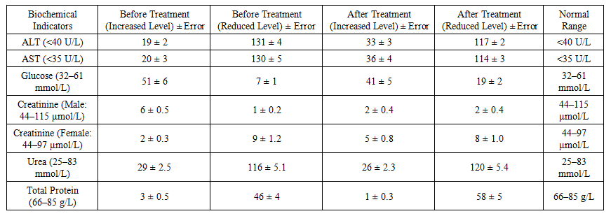 | Table 1. Biochemical parameters of 147 BrcPaP patients before and after treatment (as reported) |
3.2. Comorbidities
- Cardiovascular comorbidity was prevalent: hypertension in 49 patients (33%; 29 women [19.7%], 20 men [13.6%]); coronary heart disease in 43 (29.2%; 24 women [16.3%], 19 men [12.9%]); angina pectoris in 34 (23.1%).Diabetes mellitus ranked second among comorbidities (40 patients; 27.2%; similar distribution across sexes). Hepatitis B/C was confirmed in 17 patients (11.5%).
3.3. Cytokine Profile in MDR/XDR Subset (n=15)
- In patients with MDR‑TB and XDR‑TB (n=15), pro-inflammatory cytokines demonstrated the highest diagnostic yield: IFNγ and IL‑6 were detected in all patients, IL‑1β in 7, and TNF‑α in 6.
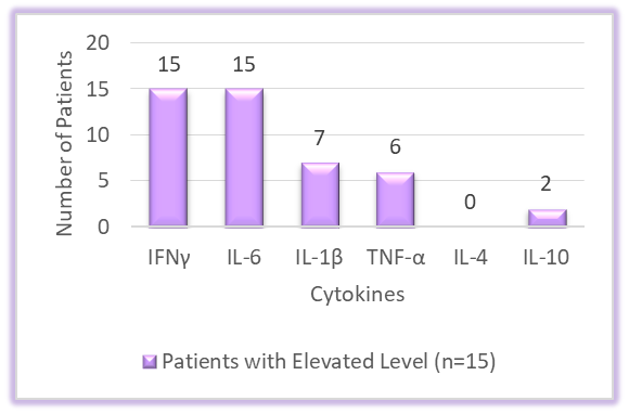 | Figure 1. Cytokine profile (counts of patients with elevated levels) in MDR/XDR-TB (n=15) |
3.4. Immunoglobulins in MDR/XDR Subset (n=15)
- Elevated IgA was observed in 14/15 patients, followed by IgM and IgE (each 12/15). IgE showed considerable intra-individual variability across time points, potentially reflecting responses to chemotherapy. IgG elevations were less frequent (7/15), with normalization in some patients after treatment.
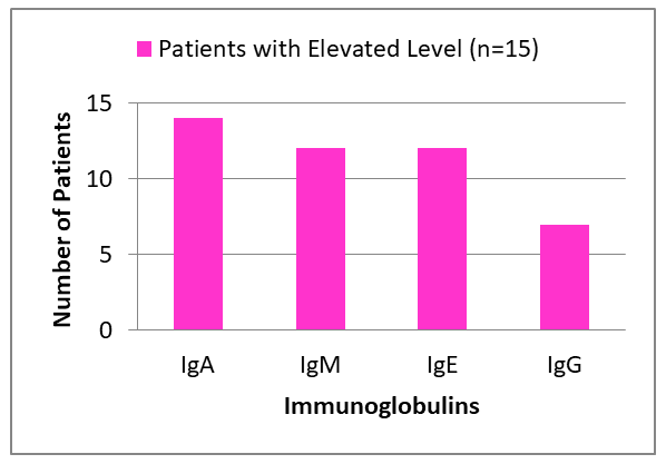 | Figure 2. Immunoglobulin titer (counts of patients with elevated levels) in MDR/XDR-TB (n=15) |
4. Discussion
- This study demonstrates that routine biochemical parameters and targeted immunological markers provide complementary insights into TB pathobiology and treatment response. Post-therapy elevations in ALT/AST are consistent with known hepatic stress from anti-TB regimens and warrant monitoring. The high prevalence of cardiovascular disease and diabetes mellitus underscores a syndemic burden that can complicate management and outcomes.The MDR/XDR subset revealed a robust pro-inflammatory signature dominated by IFNγ and IL‑6, in line with established roles in TB immunity and pathology [5,6,17]. Serological markers—especially IgA, IgM, and IgE—showed discriminative potential, echoing prior reports that combinations of immunoglobulin classes against selected mycobacterial antigens can improve diagnostic performance [1-3,11,12].Integrating these signals into a composite clinical algorithm could facilitate early risk stratification, therapeutic monitoring, and potentially guide immunocorrective strategies alongside chemotherapy.
5. Study Limitations
- • Single-center design; limited generalizability beyond the Bukhara region.• Incomplete standardization of timing for follow-up sampling; potential confounding from comorbidities and concomitant medications.• Small MDR/XDR sample (n=15) limits power for subgroup analyses and dynamic (pre/post) modeling.• Some laboratory reference intervals and unit notations require verification against the local laboratory’s standards prior to resubmission (e.g., glucose and urea ranges).
6. Conclusions
- Serum biochemical indices together with a focused panel of pro-inflammatory cytokines (IFNγ, IL‑6, IL‑1β) and immunoglobulins (IgA, IgM, IgE) may form a practical multimarker signature for TB—particularly MDR/XDR—supporting diagnosis, monitoring, and prognostication alongside standard therapy.
ACKNOWLEDGEMENTS
- We thank the Bukhara Regional Center of Phthisiology and Pulmonology and the Bukhara branch of the Institute of Human Immunology and Genomics, Academy of Sciences of the Republic of Uzbekistan, for laboratory support and collaboration.
Authors’ Contribution
- JSN conceived the study, collected and processed samples, performed analyses, interpreted data, and drafted the manuscript.
Conflicts of Interest
- The author declares no conflicts of interest.
Funding
- No specific funding was received for this work. [If applicable, insert funding details here].