-
Paper Information
- Next Paper
- Paper Submission
-
Journal Information
- About This Journal
- Editorial Board
- Current Issue
- Archive
- Author Guidelines
- Contact Us
American Journal of Medicine and Medical Sciences
p-ISSN: 2165-901X e-ISSN: 2165-9036
2025; 15(10): 3449-3452
doi:10.5923/j.ajmms.20251510.35
Received: Sep. 1, 2025; Accepted: Oct. 3, 2025; Published: Oct. 16, 2025

Experimental Morphological Changes in the Spleen Under the Influence of Heavy Metal Salts and Its Relationship with Hypodynamic
 Abstract
 Reference
 Full-Text PDF
 Full-text HTML
Khasanova Dilnoza Akhrorovna1, Bozorov Ilkhomjon Kholmurodovich2, Razhabova Gulcekhra Hamroyevna3
1Associate Professor of the Department of Anatomy, Clinical Anatomy (OSTA) of the Bukhara State Medical Institute named after Abu Ali ibn Sino, Uzbekistan
2Bukhara State Medical Institute named after Abu Ali ibn Sino Uzbekistan
3Associate Professor of Internal Medicine in Family Medicine Bukhara State Medical Institute named after Abu Ali ibn Sino, Uzbekistan
Correspondence to: Khasanova Dilnoza Akhrorovna, Associate Professor of the Department of Anatomy, Clinical Anatomy (OSTA) of the Bukhara State Medical Institute named after Abu Ali ibn Sino, Uzbekistan.
| Email: |  |
Copyright © 2025 The Author(s). Published by Scientific & Academic Publishing.
This work is licensed under the Creative Commons Attribution International License (CC BY).
http://creativecommons.org/licenses/by/4.0/

It was found that in the hypodynamia of white-bred rats, aluminum salts rapidly spread through the blood to the body and quickly combine with tissue proteins, causing acute local changes, causing their accumulation in the walls of the central artery and sinusoids in the spleen tissue, having a strong effect on blood circulation in the early stages and developing hyperplasia. Hypodynamia, a condition that aggravates and aggravates the course of intoxication, leads to a decrease in the reactive center and marginal zone of the white pulp, cessation of the proliferation of T and B lymphocytes, accumulation of blood in the red pulp, expansion of the sinuses, sclerosis of their walls, ruptured focal hemorrhages, an increase in damaged erythrocytes, the appearance and proliferation of numerous siderophages in the red pulp, an increase in hemosiderin in the intercellular structures of the spleen, rapid destruction of the components of the spleen by aluminum salts were detected.
Keywords: Hypodynamia, White pulp, Red pulp, Spleen, Morphology, Morphometry
Cite this paper: Khasanova Dilnoza Akhrorovna, Bozorov Ilkhomjon Kholmurodovich, Razhabova Gulcekhra Hamroyevna, Experimental Morphological Changes in the Spleen Under the Influence of Heavy Metal Salts and Its Relationship with Hypodynamic, American Journal of Medicine and Medical Sciences, Vol. 15 No. 10, 2025, pp. 3449-3452. doi: 10.5923/j.ajmms.20251510.35.
1. Introduction
- The spleen is highly sensitive to the effects of factors of various origins and is one of the first in the body to respond to adaptive changes in morphological organization. These facts determine the possibility of using the spleen as an experimental object for assessing the immunomodulatory effect of external factors.According to the International Metabolic Disorders Report, it can be concluded that the combined effect of heavy metal compounds, in particular, pollutants of major metals, causes significant disorders in the body, differing in the degree of damage from the effects caused by each element separately. In many studies, an increase in the process of lipoperoxidation plays an important role in the pathogenesis of chronic heavy metal poisoning. Lipid peroxidation products cause secondary damage, primarily to the formation of cell membranes, reduce the function of enzymes, regenerative processes, and from a certain point serve as the main pathogenetic factor in the development of intoxication syndrome. The effect of dioxins and a number of heavy metals reduces the phagocytic activity of macrophages, the proliferation and maturation of thymocytes. Many heavy metals (lead, mercury, cadmium, cobalt, thallium, titanium, tungsten), dioxins, polychlorinated and polycyclic hydrocarbons have a depressant effect on local and systemic immunity. [1]The problem of pollution of the environment and the human body with heavy metals has led to the need to study in detail their removal, distribution and accumulation in the body. Determining the laws that determine the state and behavior of heavy metals in the environment is one of the important scientific tasks. Metals are present in the human body in insignificant quantities and play an important role, are part of biologically active substances that regulate the normal functioning of organisms, but their combined effect on the organs and systems of the human body and the consequences of these effects have not been fully studied. Taking this into account, the need to continue morphological and experimental studies on this problem has not lost its relevance.The purpose of the study is to study the effect of heavy metal salts on the morphology of the spleen of rats during physical inactivity.
2. Materials and Methods
- The study used 160 male outbred white rats at the age of 3, 6, 9, 12 months, kept in ordinary vivarium conditions. All laboratory animals were divided into 2 groups: group 1 - animals in standard vivarium conditions and with an intact diet; group 2 - laboratory animals on a background of physical inactivity with heavy metal salts in a standard diet.The preparation of histological preparations from the spleen consisted of four stages and was carried out using traditional methods. A mechanical rotary microtome (China) was used to prepare the preparations, and the prepared sections were stained with hematoxylin and eosin. For this, the sections were immersed in a hematoxylin solution for 3-5 min, then washed with distilled water. When the cell nuclei were stained purple when observed under a microscope, they were stained in an eosin solution for 1.5 min, washed in distilled water, and alcohols of increasing strength (from 70° to 100°) were used for dehydration. To remove the alcohol from the prepared histological preparation and to fix it, it was sequentially placed in a 1/3 solution of O-xylene and placed in Canada balsam. [2]
3. Results and Discussions
- In our study, we studied the morphological and cumulative properties of heavy metal salts and aluminum compounds in the spleen of 3-month-old white outbred rats in a state of hypodynamia under experimental conditions. It was found that the spleen capsule and its trabeculae thickened, and the delicate connective tissue expanded. Spleen tissue participates in the immune-protective function to a high degree, and subsequently, as a result of increased toxicity and increased hypoxic processes, atrophic changes occur in the spleen tissue. In the white pulp area of the spleen tissue; we can see that the reactive center-germinative zone has decreased, as well as the mantle and marginal ridges. Due to the increase in toxic substances in the blood, the PALS area (T-lymphocytes decreased) also decreased, the arterial blood vessel thickened, and deformed. Neutrophilic infiltrates appeared in the red pulp, splenic bands shrank, sinusoids narrowed, and erythrocyte hemolysis began (Figure 1). The above pathological processes were relatively milder in 3-month-old white rats. They led to a deficiency in the immune defense system and rapid development of atrophy of the spleen tissue.
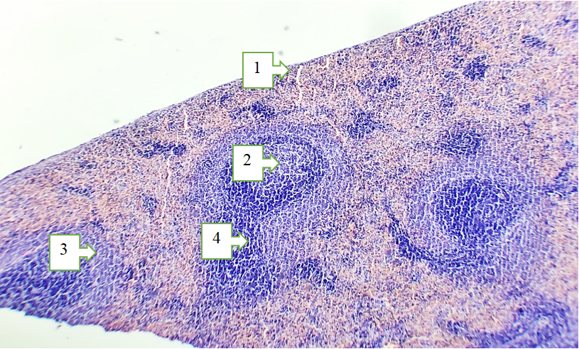 | Figure 1. Morphological appearance of the spleen tissue of 3-month-old white outbred rats in a state of hypodynamia. Staining G-E. Ob 10x10 ok. 1. The spleen capsule is thinned. 2. White pulp area: Reactive center-germinative zone is reduced. 3. Mantle and marginal zones are reduced. 4. PALS zone is reduced |
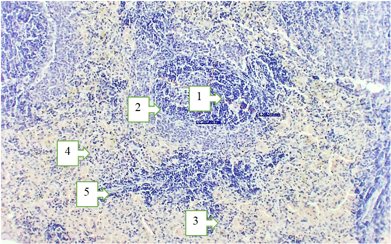 | Figure 2. Morphological appearance of the spleen tissue of 6-month-old white outbred rats in a state of hypodynamia. Staining G-E. Ob 10x10 ok. 1. White pulp area: Reactive center-germinative zone is reduced (in morphometric dimensions). 2. Mantle and marginal zones are reduced. 3. Neutrophilic infiltration in the red pulp. 4. Severe hemolysis of erythrocytes. 5. Splenic arachnoid thickening (in pink) |
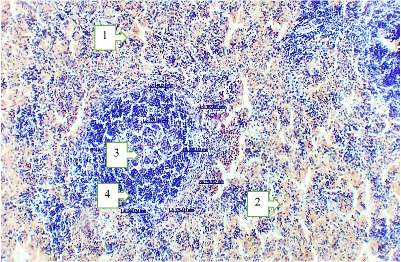 | Figure 3. Morphological appearance of the spleen tissue of 9-month-old white outbred rats in a state of hypodynamia. Staining G-E. Ob 10x10 ok. 1. Neutrophilic infiltration in the red pulp. 2. Severe hemolysis of erythrocytes. 3. White pulp area: Reactive center-germinative zone is reduced (in morphometric dimensions). 4. Mantle and marginal zones are reduced (in morphometric dimensions) |
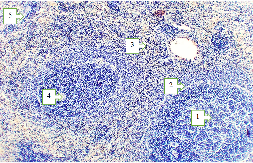 | Figure 4. Morphological appearance of the spleen tissue of 12-month-old white outbred rats in a state of hypodynamia. Staining G-E. Ob 10x10 ok. 1. White pulp area: Reactive center-germinative zone is very small, hypoplasia. 2. Mantle and marginal zones are very small. 3. Neutrophilic infiltration in the red pulp. 4. PALS zone is small, the vessel wall is sclerotic. 5. Splenic trabeculae are thickened |
4. Conclusions
- In our study, we studied the morphological and cumulative properties of heavy metal salts and aluminum compounds in the spleen of 3-6-9-12-month-old white outbred rats in a state of hypodynamia under experimental conditions. The spleen capsule and its trabeculae were markedly thickened and expanded (when stained with Van Gieson's dye, they were stained in a dark pink color). We can see that the spleen tissue is highly involved in the immune-protective function and as a result, atrophic changes in the spleen tissue occur in the white pulp area: the reactive center-germinative zone is reduced, as well as the mantle and marginal zones are reduced. As a result of the increase in toxic substances in the blood, the PALS area (decreased T-lymphocytes) also decreased, neutrophil infiltration in the red pulp increased, splenic bands decreased, sinusoids narrowed, and erythrocyte hemolysis increased. The above pathological processes increased with age in 3-6-9-12-month-old white non-breed rats. They led to the rapid development of immunodeficiency, hypoplasia, and atrophy of the spleen tissue.