American Journal of Medicine and Medical Sciences

p-ISSN: 2165-901X    e-ISSN: 2165-9036

2025;  15(9): 3269-3278

doi:10.5923/j.ajmms.20251509.94

Received: Aug. 22, 2025; Accepted: Sep. 21, 2025; Published: Sep. 30, 2025


Molecular-Genetic Predictors of Atrial Fibrillation in Patients with Arterial Hypertension in the Uzbek Population

Abdullaeva G. Zh.1, Mashkurova Z. T.1, Zakirova D. V.2, Radzhabova G. M.1, Yusupova Kh. F.1, Ibragimov N. N.1, Abdullaev A. A.2

1Republican Specialized Scientific and Practical Medical Center of Cardiology, Tashkent, Uzbekistan

2Center for Advanced Technologies, Tashkent, Uzbekistan

Copyright © 2025 The Author(s). Published by Scientific & Academic Publishing.

This work is licensed under the Creative Commons Attribution International License (CC BY).
http://creativecommons.org/licenses/by/4.0/

Abstract

To investigate the association between polymorphisms of the PITX2 (rs6817105), CAV1 (rs11773845, rs3807990), and KCNN3 (rs13376333) genes and the development of atrial fibrillation (AF) in patients with arterial hypertension (AH) in the Uzbek population. The study included 154 patients with AH who were initially diagnosed with paroxysmal AF (n = 21; 13.64%), persistent AF (n = 44; 28.57%), and permanent AF (n = 89; 57.79%). The mean age of patients with AH and AF at baseline was 64.1 ± 10.35 years. The control group (n = 91) consisted of patients with AH without AF, with a mean age of 56.03 ± 12.18 years. Clinical and instrumental examinations were performed (ECG, echocardiography, Holter monitoring), as well as molecular genetic analysis of extracted DNA using real-time PCR. Associations between polymorphisms and AF were evaluated using logistic regression and different inheritance models. Statistical analysis was performed with Statistica 10.0 and SNPassoc (R). Statistically significant associations were identified between several polymorphisms and the risk of atrial fibrillation in patients with arterial hypertension in the Uzbek population. The rs6817105 polymorphism of the PITX2 gene demonstrated a strong association with AF: carriage of the minor C allele increased the risk of AF 2.76-fold (OR = 2.76; p = 3.27×10⁻⁷) under the log-additive inheritance model. For rs11773845 of the CAV1 gene, an association was found at both the allele and genotype levels: presence of the A allele (p = 6.22×10⁻¹¹) and the heterozygous AC genotype (OR = 2.43; p = 0.001) significantly increased the likelihood of AF. Interestingly, analysis of rs3807990 of the same CAV1 gene revealed the opposite effect: carriage of the C allele was associated with a reduced risk of AF (OR = 0.71; p = 0.049), suggesting a potential protective role. Finally, the rs13376333 polymorphism of the KCNN3 gene also showed a significant association with AF: the C allele was more frequent in patients with rhythm disturbances, increasing AF risk (OR = 1.63; p = 0.020) under the log-additive model. These findings confirm the role of the studied genetic markers in the pathogenesis of atrial fibrillation in patients with arterial hypertension and highlight the importance of genetic testing in this group of patients. The present data demonstrate, for the first time, associations of several single nucleotide polymorphisms with atrial fibrillation in patients with arterial hypertension in the Uzbek population. These results emphasize the importance of integrating molecular genetic markers into clinical practice for personalized risk assessment and prevention of cardiac arrhythmias.

Keywords: Arterial hypertension, Atrial fibrillation, PITX2 rs6817105 polymorphism, CAV1 rs11773845 polymorphism, CAV1 rs3807990 polymorphism, KCNN3 rs13376333 polymorphism

Cite this paper: Abdullaeva G. Zh., Mashkurova Z. T., Zakirova D. V., Radzhabova G. M., Yusupova Kh. F., Ibragimov N. N., Abdullaev A. A., Molecular-Genetic Predictors of Atrial Fibrillation in Patients with Arterial Hypertension in the Uzbek Population, American Journal of Medicine and Medical Sciences, Vol. 15 No. 9, 2025, pp. 3269-3278. doi: 10.5923/j.ajmms.20251509.94.

1. Introduction

Atrial fibrillation (AF) is one of the most common arrhythmias, associated with high morbidity and mortality. According to the European Society of Cardiology, the prevalence of AF in the adult population ranges from 1% to 4%, with the rate increasing with age, reaching 10–15% among individuals over 80 years old [1]. AF significantly increases the risk of stroke, heart failure, and cognitive impairment, making the study of factors influencing its development critically important. In addition to traditional risk factors such as age, arterial hypertension (AH), obesity, and diabetes mellitus, genetic factors play a substantial role in the pathogenesis of AF. Current research suggests that up to 40% of AF cases may be attributable to hereditary predisposition, and one of the key directions in cardiogenetics is the study of single nucleotide polymorphisms (SNPs) that affect the structural and electrophysiological properties of the atria [2]. Particular interest lies in the study of AF in patients with AH, since hypertension is one of the main predisposing factors for the development of this arrhythmia. However, not all patients with AH develop AF, indicating a significant role of genetic predisposition. Studies conducted among various ethnic groups have demonstrated that the prevalence of AF-associated polymorphisms varies depending on racial and ethnic background. The Uzbek population was formed under the influence of Central Asian, Iranian, Turkic, and Mongoloid ethnic groups, which may have led to an altered frequency of specific SNPs affecting cardiovascular diseases [3]. However, data on AF in the Uzbek population are virtually absent, despite its unique features shaped by genetic factors. Analysis of the literature from European, Chinese, and other populations has identified several polymorphic variants of genes associated with AF risk. In particular, the rs13376333 polymorphism of the KCNN3 gene, which encodes small-conductance calcium-activated potassium channels (SK3), has been identified as a risk factor for AF. A study in the Han Chinese population showed that the T allele of rs13376333 was significantly more frequent in patients with AF compared to the control group (7.6% vs. 3.6%; p < 0.001), confirming its importance as a potential marker of arrhythmia susceptibility [4]. Furthermore, a meta-analysis including 24,339 participants confirmed that this SNP increases the risk of overall AF (OR = 1.33) and lone AF (OR = 1.58) [5]. Another important genetic biomarker is the rs6817105 polymorphism of the PITX2 gene, located on chromosome 4q25, which has been identified as one of the most significant predictors of AF. This SNP influences the development of the cardiac conduction system, left atrial morphology, and sinus node function. According to Tomomori et al. [6], the minor allele of rs6817105 increases AF risk more than twofold (OR = 2.12; p = 4.9×10⁻²⁶). Importantly, the frequency of this polymorphism varies among different ethnic groups, and its investigation in the Uzbek population will help determine its clinical significance in this cohort. Another promising marker is the rs3807990 variant of the CAV1 gene, which encodes caveolin-1, a protein involved in the regulation of vascular tone, lipid metabolism, and inflammatory processes. A study conducted in 2019 showed that this SNP is associated with hypercholesterolemia and endothelial dysfunction, potentially contributing to the development of AH and AF [7]. In turn, the rs11773845 polymorphism in the same gene was linked to metabolic syndrome and elevated triglyceride levels in the Latin American population, highlighting its possible relevance to cardiovascular disease development in other ethnic groups as well [8]. The genetic predisposition to AF has also been confirmed by Sasano et al. [2], who identified that SNPs rs6817105, rs3807989, rs10824026, and rs2106261 can be used for AF and stroke risk stratification. Their risk prediction model demonstrated an AUC = 0.631 for AF and an AUC = 0.950 for stroke, indicating high predictive value of these polymorphisms. Given the high prevalence of AH in Uzbekistan and the lack of molecular genetic research on AF-associated markers in this population, there is a strong need for local studies aimed at identifying genetic predictors of AF in hypertensive patients in the Uzbek population.

2. Materials and Methods

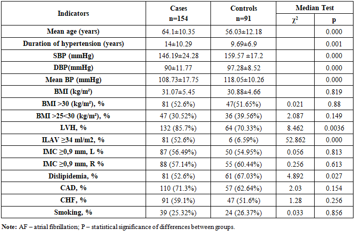
The study included 154 patients with arterial hypertension (AH), in whom atrial fibrillation (AF) was initially diagnosed as paroxysmal in 21 cases (13.64%), persistent in 44 cases (28.57%), and permanent in 89 cases (57.79%). The mean age of AH patients with AF at baseline was 64.1 ± 10.35 years (Table 1). The control group (n = 91) consisted of AH patients without AF, with a mean age of 56.03 ± 12.18 years. As shown in Table 1, patients with AH and AF differed from the control group in terms of age, duration of hypertension, and office blood pressure measurements. Notably, the age of AH patients with AF was significantly higher compared with the control group: 64.1 ± 10.35 years versus 56.03 ± 12.18 years (p = 0.000). The duration of hypertension was also longer in the AF group than in the control group, amounting to 14.0 ± 10.29 years versus 9.69 ± 6.9 years, respectively (p = 0.001). The number of patients with left ventricular hypertrophy (LVH) was significantly higher in the AH + AF group than in AH patients without AF: 132 (85.7%) versus 64 (70.33%) (χ² = 8.462, p = 0.0036). It is important to note that the proportion of patients with left atrial volume index (LAVI) ≥ 34 ml/m² was substantially greater among AH patients with AF compared with those without AF: 52.6% versus 6.59%, respectively (χ² = 52.862, p = 0.000). It should be noted that the two groups did not differ in the number of patients with CAD and CHF.
Table 1. Clinical characteristics of patients with arterial hypertension with and without atrial fibrillation
     
AF was classified as paroxysmal, persistent, and permanent according to the ACC/AHA/ESC guidelines on AF [9]. The diagnosis of AF was based on ECG findings and/or Holter ECG monitoring according to standard diagnostic criteria [10]. To verify atrial fibrillation, Holter ECG monitoring was performed using the Cardiospy monitor (LABTECH LTD, Hungary). The study protocol was reviewed and approved by the Ethics Committee of the Republican Specialized Scientific and Practical Medical Center of Cardiology. All participants provided written informed consent. Echocardiographic (EchoCG) examination was performed using the Clearview-350 “Affiniti 30” ultrasound system (PHILIPS, Netherlands) in accordance with the recommendations of the American Society of Echocardiography in M- and B-modes [11]. In M-mode, measurements were obtained via a parasternal long-axis approach according to the Penn Convention method [12]. The following intracardiac hemodynamic parameters were assessed: left ventricular end-diastolic and end-systolic dimensions (EDD, ESD), wall thickness of the left ventricle: interventricular septum (IVS) and posterior wall (LVPW) in diastole. LV mass (LVM) was calculated using the Devereux B.R. formula [12]:
LVM = 1.04 × [(LVEDD + IVS + LVPW)³ – (LVEDD)³] – 13.6 (g).
Left ventricular mass index (LVMI) was calculated as the ratio of LVM to body surface area. Left ventricular hypertrophy (LVH) was diagnosed at LVMI >115 g/m² for men and >95 g/m² for women [13]. The size of the left atrium (LA) and its indexed volume (LAVI) were also evaluated. Exclusion criteria at baseline included: patients with unstable angina, stable angina class III–IV, CHF class III–IV according to NYHA, history of cardiac surgery, rheumatic valvular defects, artificial pacemaker, QT >480 ms, documented sick sinus syndrome, previous sustained ventricular arrhythmias, WPW syndrome, Brugada syndrome, arterial hypotension (systolic BP <95 mmHg), bradycardia (HR <60 bpm), severe hepatic or renal dysfunction, severe pulmonary insufficiency, AV block grade II–III, LA thrombus, confirmed thyroid dysfunction, and decompensated diabetes mellitus. For genotyping of blood samples for rs6817105 polymorphism of the PITX2 gene, rs11773845 polymorphism of the CAV1 gene, rs3807990 polymorphism of the CAV1 gene, and rs13376333 polymorphism of the KCNN3 gene, genomic DNA was extracted from whole blood using the “ArtDNA MiniSpin” kit (LLC “ArtBioTech,” Belarus) according to the manufacturer’s protocol. DNA quantity and quality were assessed by gel electrophoresis and a NanoDrop spectrophotometer (Thermo Scientific, USA). PCR was performed using a QuantStudio 5 amplifier (Applied Biosystems). Reactions were carried out in 10 µl volumes using the TaqMan® Genotyping Assays kit (Thermo Fisher Scientific, USA) according to the manufacturer’s protocol. The reaction mixture included 10 ng of genomic DNA, 5 µl of TaqMan Genotyping Master Mix, and 0.5 µl of TaqMan Genotyping Assays. Genotyping results were analyzed using the “Design & Analysis 2.6.0 2021” software (Thermo Fisher Scientific) and exported to Microsoft Excel-2019 for primary processing. Statistical analysis of the obtained data was performed using the software package Statistica 10.0. Descriptive statistics were presented as mean (M) and standard deviation (SD). Comparative analysis of genotype and allele frequencies of gene polymorphisms was performed using logistic regression analysis with the R programming language and the SNPassoc package. General, dominant, recessive, and additive inheritance models were applied.

3. Results

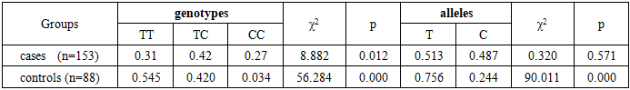
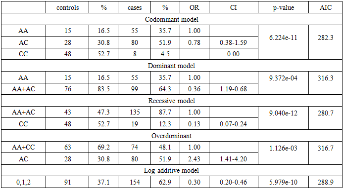
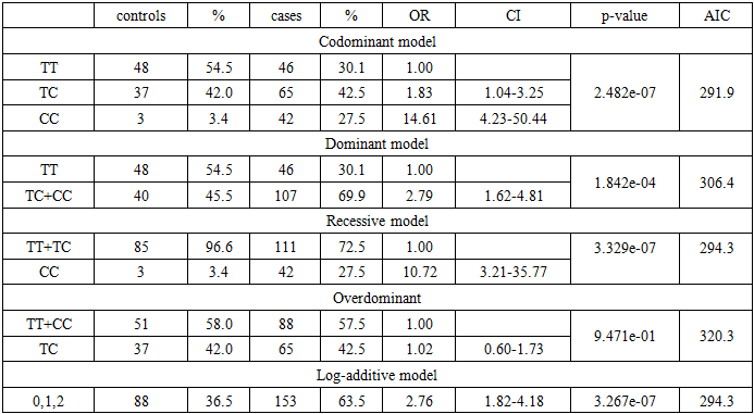
A total of 154 hypertensive patients with atrial fibrillation (cases) and 91 hypertensive patients without atrial fibrillation (controls), all from the Uzbek population, were genotyped. Below are the results of molecular-genetic analysis and the determination of correlations between genetic markers and the case and control groups using the statistical method of logistic regression. An analysis of the distribution of allele and genotype frequencies was conducted for the following gene polymorphisms involved in the development of atrial fibrillation in hypertensive patients in the Uzbek population: rs6817105 polymorphism of the PITX2 gene, rs11773845 polymorphism of the CAV1 gene, rs3807990 polymorphism of the CAV1 gene, and rs13376333 polymorphism of the KCNN3 gene.
rs6817105 polymorphism of the PITX2 gene and atrial fibrillation
To study the association of the rs6817105 polymorphism of the PITX2 gene with atrial fibrillation in hypertensive patients, 153 hypertensive patients with AF (cases) and 88 hypertensive patients without AF (controls), all from the Uzbek population, were genotyped. When analyzing the distribution of genotype and allele frequencies of the rs6817105 polymorphism of the PITX2 gene among hypertensive patients with AF, the following genotype distribution was observed: TT genotype – identified in 46 (30.1%) patients, TC genotype – in 65 (42.5%), CC genotype – in 42 (27.5%), χ2=8.882, p=0.012 (Table 2). The allelic distribution was nearly equal: T allele – 51.3%, C allele – 48.7%, χ2=0.320, p=0.571. The opposite pattern was observed among hypertensive patients without AF. In particular, the allelic distribution showed a significant predominance of the T allele compared with the C allele: 75.6% versus 24.4%, respectively, χ2=90.011, p=0.000. The genotype ratio TT:TC:CC was as follows: 54.5% : 42.0% : 3.4%, χ2=56.284, p=0.000. Thus, the obtained results indicate a significantly greater accumulation of the T allele and TT genotype of the rs6817105 polymorphism of the PITX2 gene among hypertensive patients without AF compared with hypertensive patients with AF, in whom, on the contrary, the C allele prevailed.
Table 2. Frequencies of genotypes and alleles of the rs6817105 polymorphism of the PITX2 gene in hypertensive patients with atrial fibrillation and in controls
     
The obtained data in both groups were consistent with Hardy–Weinberg equilibrium (HWE):
Hardy–Weinberg test for controls (χ² = 0.90, p = 0.34):
• Genotype TT: 0.545 (HWE 0.571)
• Genotype TC: 0.420 (HWE 0.369)
• Genotype CC: 0.034 (HWE 0.060)
Hardy–Weinberg test for cases (χ² = 1.74, p = 0.19):
• Genotype TT: 0.301 (HWE 0.263)
• Genotype TC: 0.425 (HWE 0.500)
• Genotype CC: 0.275 (HWE 0.237)
The results of the genetic analysis using inheritance models in the case and control groups (the case group consisted of hypertensive patients with atrial fibrillation, the control group – hypertensive patients without atrial fibrillation) demonstrated a correlation between the rs6817105 polymorphism of the PITX2 gene and atrial fibrillation under the log-additive inheritance model (OR = 2.76, p = 3.267e-07, AIC = 294.3), as shown in Table 3. Other models (codominant p = 2.482e-07, dominant p = 1.842e-04, recessive p = 3.329e-07, overdominant p = 9.471e-01) also revealed statistically significant correlations; however, the most optimal and reliable genetic models in this statistical analysis were the codominant and log-additive models. Analysis of the inheritance models indicated a significant association under the log-additive model for the C allele (OR = 2.76, 95% CI: 1.82–4.18, p = 3.267e-07) and under the overdominant model for the CC genotype (OR = 1.02, 95% CI: 0.60–1.73, p = 9.471e-01) with atrial fibrillation in hypertensive patients of the Uzbek population.
Table 3. Results of correlation–regression analysis of the association between atrial fibrillation and the rs6817105 polymorphism of the PITX2 gene
     
rs11773845 polymorphism of the CAV1 gene and atrial fibrillation
To investigate the association of the rs11773845 polymorphism of the CAV1 gene with atrial fibrillation (AF) in hypertensive patients, 154 hypertensive patients with AF (cases) and 91 hypertensive patients without AF (controls), all from the Uzbek population, were genotyped. When analyzing the distribution of genotype and allele frequencies of the rs11773845 polymorphism of the CAV1 gene among hypertensive patients with AF, the following genotype distribution was identified:
AA genotype – 55 patients (35.7%)
AC genotype – 80 patients (52.0%)
CC genotype – 19 patients (12.3%)
χ² = 54.955, p = 0.000 (Table 4).
Table 4. Frequencies of genotypes and alleles of the rs11773845 polymorphism of the CAV1 gene in hypertensive patients with atrial fibrillation and in controls
     
Allelic distribution revealed a predominance of the A allele:
A allele – 61.7%
C allele – 38.3%
χ² = 33.662, p = 0.000.
In contrast, the opposite pattern was observed among hypertensive patients without AF. Specifically, allele distribution showed a significant predominance of the C allele compared with the A allele:
C allele – 68.1%
A allele – 31.9%
χ² = 47.868, p = 0.000.
The genotype distribution in the control group was as follows:
AA – 16.5%
AC – 30.8%
CC – 52.7%
χ² = 27.33, p = 0.000.
Thus, the results indicate a significantly higher accumulation of the A allele and the AC genotype of the rs11773845 polymorphism of the CAV1 gene among hypertensive patients with AF compared with those without AF, in whom, on the contrary, the C allele predominated.
The obtained data in both groups were consistent with Hardy–Weinberg equilibrium (HWE):
Hardy–Weinberg test for controls (χ² = 4.02, p = 0.05):
Genotype AA: 0.165 (HWE 0.102)
Genotype AC: 0.308 (HWE 0.434)
Genotype CC: 0.527 (HWE 0.464)
Hardy–Weinberg test for cases (χ² = 0.84, p = 0.36):
Genotype AA: 0.357 (HWE 0.381)
Genotype AC: 0.519 (HWE 0.473)
Genotype CC: 0.123 (HWE 0.147)
The results of the genetic analysis using inheritance models in the case and control groups (the case group consisted of hypertensive patients with atrial fibrillation, the control group – hypertensive patients without atrial fibrillation) demonstrated a correlation between the rs11773845 polymorphic marker of the CAV1 gene and atrial fibrillation under the codominant inheritance model (OR = 1.00, p = 6.224e-11), as shown in Table 5. Other models (dominant p = 9.372e-04, recessive p = 9.040e-12, overdominant p = 1.126e-03, and log-additive p = 5.979e-10) also showed statistically significant correlations; however, the codominant model proved to be the most optimal and reliable genetic model in this statistical analysis. Analysis of inheritance models demonstrated a strong association:
- under the codominant model for the A allele (OR = 1.00, p = 6.224e-11), and
- under the overdominant model for the AC genotype (OR = 2.43, 95% CI: 1.41–4.20, p = 1.126e-03) with atrial fibrillation in hypertensive patients of the Uzbek population.
Table 5. Results of correlation–regression analysis of the association between atrial fibrillation and the rs11773845 polymorphism of the CAV1 gene
     
rs3807990 polymorphism of the CAV1 gene and atrial fibrillation
To investigate the association of the rs3807990 polymorphism of the CAV1 gene with atrial fibrillation (AF) in hypertensive patients, 154 hypertensive patients with AF (cases) and 96 hypertensive patients without AF (controls), all from the Uzbek population, were genotyped. When analyzing the distribution of genotype and allele frequencies of the rs3807990 polymorphism of the CAV1 gene among hypertensive patients with AF, the following genotype distribution was identified:
CC genotype – 68 patients (44.2%)
CT genotype – 59 patients (38.3%)
TT genotype – 27 patients (17.5%)
χ² = 27.136, p = 0.000 (Table 6).
Table 6. Frequencies of genotypes and alleles of the rs3807990 polymorphism of the CAV1 gene in hypertensive patients with atrial fibrillation and in controls
     
Allelic distribution revealed a predominance of the C allele:
C allele – 63.3%
T allele – 36.7%
χ² = 43.662, p = 0.000.
Among hypertensive patients without AF, no significant differences in allele distribution were observed:
C allele – 53.6%
T allele – 46.4%
χ² = 2.042, p = 0.153.
The genotype distribution in the control group was as follows:
CC – 33.3%
CT – 40.6%
TT – 26.0%
χ² = 4.594, p = 0.100.
Thus, the obtained results indicate a significantly higher accumulation of the C allele of the rs3807990 polymorphism of the CAV1 gene among hypertensive patients with AF compared with hypertensive patients without AF.
The obtained data in both groups were consistent with Hardy–Weinberg equilibrium (HWE):
Hardy–Weinberg test for controls (χ² = 1.54, p = 0.21):
Genotype CC: 0.333 (HWE 0.288)
Genotype CT: 0.406 (HWE 0.497)
Genotype TT: 0.260 (HWE 0.215)
Hardy–Weinberg test for cases (χ² = 2.31, p = 0.13):
Genotype CC: 0.442 (HWE 0.401)
Genotype CT: 0.383 (HWE 0.465)
Genotype TT: 0.175 (HWE 0.135)
The results of the genetic analysis using inheritance models in the case and control groups (the case group consisted of hypertensive patients with atrial fibrillation, the control group – hypertensive patients without atrial fibrillation) demonstrated a correlation between the C allele of the rs3807990 polymorphic marker of the CAV1 gene and atrial fibrillation in hypertensive patients of the Uzbek population under the log-additive inheritance model (OR = 0.71, 95% CI: 0.51–1.00, p = 0.049), as shown in Table 7.
Table 7. Results of correlation–regression analysis of the association between atrial fibrillation and the rs3807990 polymorphism of the CAV1 gene
     
rs13376333 polymorphism of the KCNN3 gene and atrial fibrillation
To investigate the association of the rs13376333 polymorphism of the KCNN3 gene with atrial fibrillation (AF) in hypertensive patients, 154 hypertensive patients with AF (cases) and 96 hypertensive patients without AF (controls), all from the Uzbek population, were genotyped. When analyzing the distribution of genotype and allele frequencies of the rs13376333 polymorphism of the KCNN3 gene among hypertensive patients with AF, the following genotype distribution was identified:
CC genotype – 82 patients (53.2%)
CT genotype – 57 patients (37.0%)
TT genotype – 15 patients (9.7%)
χ² = 66.994, p = 0.000 (Table 8).
Table 8. Frequencies of genotypes and alleles of the rs13376333 polymorphism of the KCNN3 gene in hypertensive patients with atrial fibrillation and in controls
     
Allelic distribution revealed a predominance of the C allele:
C allele – 71.8%
T allele – 28.2%χ² = 116.597, p = 0.000.
A similar pattern was observed among hypertensive patients without AF. In particular, allelic distribution showed a significant predominance of the C allele compared with the T allele:
C allele – 81.3%
T allele – 18.8%
χ² = 150.0, p = 0.000.
The genotype distribution in the control group was as follows:
CC – 67.7%
CT – 27.1%
TT – 5.2%
χ² = 86.906, p = 0.000.
Thus, the obtained results indicate a significantly greater accumulation of the C allele and CC genotype of the rs13376333 polymorphism of the KCNN3 gene both among hypertensive patients with AF and among those without AF.
The obtained data in the control group did not conform to Hardy–Weinberg equilibrium (HWE):
Hardy–Weinberg test for controls (χ² = 0.69, p = 0.41):
Genotype CC: 0.677 (HWE 0.660)
Genotype CT: 0.271 (HWE 0.305)
Genotype TT: 0.052 (HWE 0.035)
Hardy–Weinberg test for cases (χ² = 0.60, p = 0.44):
Genotype CC: 0.532 (HWE 0.515)
Genotype CT: 0.370 (HWE 0.405)
Genotype TT: 0.097 (HWE 0.080)
The results of the genetic analysis using inheritance models in the case and control groups (the case group consisted of hypertensive patients with AF, the control group – hypertensive patients without AF) demonstrated a correlation between the rs13376333 polymorphism of the KCNN3 gene and atrial fibrillation under the log-additive inheritance model (OR = 1.63, p = 0.020, AIC = 331.6), as shown in Table 9. A statistically significant correlation was also observed under the dominant inheritance model (p = 0.023). However, the most optimal and reliable genetic model in this statistical analysis was the log-additive model. Thus, analysis of inheritance models revealed a significant association under the log-additive model for the C allele (OR = 1.63, 95% CI: 1.07–2.50, p = 0.020) and under the dominant model for the CC genotype (OR = 1.00, p = 0.023) with atrial fibrillation in hypertensive patients of the Uzbek population.
Table 9. Results of correlation–regression analysis of the association between atrial fibrillation and the rs13376333 polymorphism of the KCNN3 gene
     
Thus, an association of the C allele and CC genotype of the rs13376333 polymorphism of the KCNN3 gene with the risk of atrial fibrillation (AF) in hypertensive patients of the Uzbek population has been demonstrated.

4. Discussion

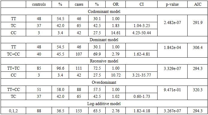
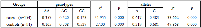
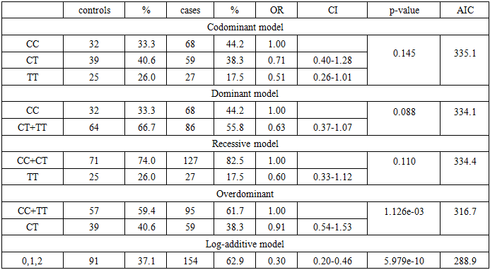
It is well known that hypertension (HTN) contributes to the development of cardiac rhythm disturbances, in particular ventricular arrhythmias, but most frequently atrial fibrillation [14], which represents a manifestation of hypertensive cardiomyopathy [15]. Even high-normal blood pressure is associated with an increased risk of AF [16], and HTN is the most common comorbid diagnosis in patients with AF.
Altered geometry of the left ventricle (LV) is often associated with diastolic dysfunction [17, 18]. Enlargement of the left atrium (LA) is also frequently observed in patients with HTN, which is associated with adverse cardiovascular outcomes [19, 20], an increased incidence of AF [21], and diastolic dysfunction [22, 23]. In the present study, the age-dependent nature of AF development in patients with HTN was confirmed, as well as the undeniable influence of the duration of HTN, which is associated with the severity of cardiovascular remodeling. Specifically, in the group of hypertensive patients with AF compared to controls without AF, a significantly higher number of cases of left ventricular hypertrophy (LVH) and increased left atrial volume index (LAVI) were observed.
To elucidate the genetic mechanisms of electrophysiological disturbances of the myocardium in HTN, various genes encoding key physiological systems were investigated. The literature describes mutations in the cardiac sodium channel gene (SCN5A) and in KCNQ genes, which may cause rhythm disturbances, including AF. Over the past decade, several genome-wide association studies (GWAS) have identified the ATFB5 gene with the rs2200733 single-nucleotide polymorphism at locus 4q25 as the most common chromosomal variant present in patients with AF [24]. In our previous study, we analyzed the potential relationship between rs2200733 (ATFB5) and the development of AF in individuals of Uzbek nationality [25]. The distribution of genotypes and alleles of rs2200733 among all enrolled patients coincided with results from previous studies conducted in various European countries [26, 27]. Our findings were consistent with the results of a meta-analysis [28] and indicated a significant association between the rare mutant variant (TT genotype) and a fourfold increased risk of AF. In the present study, we examined associations with polymorphisms most pathognomonic for AF, as described in the literature: rs6817105 of the PITX2 gene, rs11773845 of the CAV1 gene, rs3807990 of the CAV1 gene, and rs13376333 of the KCNN3 gene. We identified associations of:
- C allele (OR = 2.76, 95% CI: 1.82–4.18, p = 3.267e-07) and CC genotype (OR = 1.02, 95% CI: 0.60–1.73, p = 0.947) of rs6817105 (PITX2);
- AC genotype (OR = 2.43, 95% CI: 1.41–4.20, p = 1.126e-03) and A allele (OR = 1.00, p = 6.224e-11) of rs11773845 (CAV1);
- C allele of rs3807990 (CAV1) (OR = 0.71, 95% CI: 0.51–1.00, p = 0.049);
- C allele (OR = 1.63, 95% CI: 1.07–2.50, p = 0.020) and CC genotype (OR = 1.00, p = 0.023) of rs13376333 (KCNN3).
with the risk of atrial fibrillation in hypertensive patients of the Uzbek population. The present study, aimed at identifying molecular–genetic markers of AF in hypertensive patients, has high scientific and practical significance. The data obtained allow us to refine the prevalence of SNPs associated with AF in this population, as well as their contribution to the development of arrhythmias in hypertensive patients. This may contribute to the development of custom genetic panels for AF risk assessment, optimization of treatment strategies, and implementation of a personalized approach to the therapy of hypertensive patients with a predisposition to AF. The introduction of modern genetic technologies into clinical practice will make it possible to more accurately assess the risk of arrhythmias in patients and to apply preventive measures to reduce morbidity and mortality associated with this rhythm disorder.

5. Conclusions

This study is the first to demonstrate associations of the C allele and CC genotype of rs6817105 (PITX2), the AC genotype and A allele of rs11773845 (CAV1), the C allele of rs3807990 (CAV1), and the C allele and CC genotype of rs13376333 (KCNN3) with the risk of atrial fibrillation in hypertensive patients of the Uzbek population.

References

[1]  Van Gelder IC, Rienstra M, Bunting KV, Casado-Arroyo R, Caso V, et al; ESC Scientific Document Group. 2024 ESC Guidelines for the management of atrial fibrillation developed in collaboration with the European Association for Cardio-Thoracic Surgery (EACTS). Eur Heart J. 2024 Sep 29; 45(36): 3314-3414. doi: 10.1093/eurheartj/ehae176. Erratum in: Eur Heart J. 2025 Jul 07:ehaf306. doi: 10.1093/eurheartj/ehaf306. PMID: 39210723.
[2]  Sasano T, Ihara K, Tanaka T, Furukawa T. Risk stratification of atrial fibrillation and stroke using single nucleotide polymorphism and circulating biomarkers. PLoS One. 2023 Oct 12; 18(10): e0292118. doi: 10.1371/journal.pone.0292118. PMID: 37824462; PMCID: PMC10569505.
[3]  Kim JA, Chelu MG, Li N. Genetics of atrial fibrillation. Curr Opin Cardiol. 2021 May 1; 36(3): 281-287. doi: 10.1097/HCO.0000000000000840. PMID: 33818546; PMCID: PMC8211390.
[4]  Luo Z., Yan C., Zhang W. et al. Association between SNP rs13376333 and rs1131820 in the KCNN3 gene and atrial fibrillation in the Chinese Han population. Clinical Chemistry and Laboratory Medicine. 2014 Dec;  52(12):  1867–1873. doi: 10.1515/cclm-2014-0491.
[5]  Yao JL, Zhou YF, Yang XJ, Qian XD, Jiang WP. KCNN3 SNP rs13376333 on Chromosome 1q21 Confers Increased Risk of Atrial Fibrillation. International Heart Journal. 2015; 56(5): 511–515. doi:10.1536/ihj.15133. PMID: 26370375.
[6]  Tomomori S, Nakano Y, Ochi H, et al. Chromosome 4q25 Variant rs6817105 Brings Sinus Node Dysfunction and Left Atrial Enlargement. Scientific Reports. 2018 Oct1; 8(1): 13557. DOI:10.1038/s41598-018-32453-8.PMID: 30275441.
[7]  Ilikay S, Coşkunpınar E, KurnazGömleksiz Ö, Buğra Z, et al. Effects of common variations of NOS3 and CAV1 genes on hypercholesterolemic profile in coronary heart disease. Istanbul J Pharm. 2019; 49(2): 53–60. DOI: 10.26650/IstanbulJPharm.2019.18010.
[8]  Gustavo MoraGarcía, Doris GómezCamargo, Ángelo Alario, Claudio GómezAlegría и др. A Common Variation in the Caveolin 1 Gene Is Associated with High Serum Triglycerides and Metabolic Syndrome in an Admixed Latin American Population. Metabolic Syndrome and Related Disorders. 2018; 16(5): 234240. doi:10.1089/met.2018.0004.
[9]  2016 ESC Guidelines for The Management of Atrial Fibrillation Developed in Collaboration With Eacts. Eur Heart J (2016) 37 (38): 2893-2962. DOI: .
[10]  Camm AJ, Kirchhof P, Lip GY, et al. Guidelines for the management of atrial fibrillation: the Task Force for the Management of Atrial Fibrillation of the European Society of Cardiology (ESC). Europace. 2010; 12: 1360-1420.
[11]  Sahn D.J., Demaria A., Kisslo J. et al. Recommendation regarding quantitation in M-mode echocardiography Results of a servey of echocardiographic measerments // Circulation. - 1987. - Vol. 58. - P. 1072-1082.
[12]  Devereux R.B., Reichek N. Echocardiographyic determination of left ventricular mass in man // Circulation. - 1977. -№55. - P. 613-618.
[13]  The Task Force for management of arterial hypertension of the European Society of Cardiolody (ESC) and the European Society of Hypertension (ESH). European Heart Journal 2018 – doi/10.1093/eurheartj/ehy339.
[14]  Zafrir B., Lund L., Laroche C., Ruschitzka F., Crespo-Leiro M., Coats A., et al; On behalf of the ESC-HFA HF Long-Term Registry Investigators. Prognostic implications of atrial fibrillation in heart failure with reduced, mid-range, and preserved ejection fraction: a report from 14 964 patients in the European Society of Cardiology Heart Failure Long-Term Registry. European Heart Journal (2018) 39, 4277–4284.
[15]  Stanley Nattel MD MasahideHaradaMD, PhD. Atrial Remodeling and Atrial Fibrillation: Recent Advances and Translational Perspectives (2014) 22, 2365-2345 .
[16]  Ott A., Breteler M.M., de Bruyne M.C. et al. Atrial fibrillation and dementia in a population-based study. The Rotterdam Study. Stroke. 1997; 28: 316–321.
[17]  Marwick TH, Gillebert TC, Aurigemma G, Chirinos J, Derumeaux G, Galderisi M, et al. Recommendations on the use of echocardiography in adult hypertension: a report from the European Association of Cardiovascular Imaging (EACVI) and the American Society of Echocardiography (ASE). Eur Heart J Cardiovasc Imaging 2015; 16: 577–605.
[18]  de Simone G, Kitzman DW, Chinali M, Oberman A, Hopkins PN, Rao DC, et al. Left ventricular concentric geometry is associated with impaired relaxation in hypertension: the HyperGEN study. Eur Heart J 2005; 26: 1039–1045.
[19]  Domanski M, Mitchell G, Pfeffer M, Neaton JD, Norman J, Svendsen K, et al, MRFIT Research Group. Pulse pressure and cardiovascular disease-related mortality: follow-up study of the Multiple Risk Factor Intervention Trial (MRFIT). JAMA 2002; 287: 2677–2683.
[20]  Yaghi S, Moon YP, Mora-McLaughlin C, Willey JZ, Cheung K, Di Tullio MR, et al. Left atrial enlargement and stroke recurrence: the Northern Manhattan Stroke Study. Stroke 2015; 46: 1488–1493.
[21]  Losi MA, Izzo R, De Marco M, Canciello G, Rapacciuolo A, Trimarco V, et al. Cardiovascular ultrasound exploration contributes to predict incident atrial fibrillation in arterial hypertension: the Campania Salute Network. Int J Cardiol 2015;199:290–295.
[22]  Douglas PS. The left atrium: a biomarker of chronic diastolic dysfunction and cardiovascular disease risk. J Am Coll Cardiol 2003; 42: 1206–1207.
[23]  Kuznetsova T, Haddad F, Tikhonoff V, Kloch-Badelek M, Ryabikov A, Knez J, et al, European Project On Genes in Hypertension Investigators. Impact and pitfalls of scaling of left ventricular and atrial structure in population-based studies. J Hypertens 2016; 34: 1186–1194.
[24]  Sinner MF, Ellinor PT, Meitinger T, Benjamin EJ, Kääb S. Genome-wide association studies of atrial fibrillation: past, present, and future. Cardiovasc Res. 2011; 89: 701-709.
[25]  Abdullaeva G.J., Abdullaev A.A., Kevorkov A.G., Abduvalieva G.A., Zakirov N.U., Kurbanov R.D. INTERRELATION BETWEEN rs2200733 POLYMORPHISM OF ATFB5 GENE AND ATRIAL FIBRILLATION IN UZBEK PATIENTS. Turk Kardiyol Dern Ars 2021; 49(5): 404-409 doi: 10.5543/tkda.2021.08434.
[26]  Ferran A., Alegret J.M., Subirana I. et al. Association Between rs2200733 and rs7193343 Genetic Variants and Atrial Fibrillation in Spanish Population, and Meta-analysis of Previous Studies. Rev. Esp. Cardiol. 2014; 67(10): 822-829.
[27]  Kalinderi K, Fragakis N. et al. Association Between rs2200733 Polymorphism on Chromosome 4q25 and Atrial Fibrillation in a Greek Population. Hellenic J Cardiol 2015; 56: 224-229.
[28]  Rattanawong P., Chenbhanich J. et al. A Chromosome 4q25 variant is Associated with Atrial Fibrillation Reccurence After Catheter Ablation: A systematic Review and Meta-analysis. J Atr Fibrillation. 2018 Apr; 10(6): 1666.