-
Paper Information
- Next Paper
- Previous Paper
- Paper Submission
-
Journal Information
- About This Journal
- Editorial Board
- Current Issue
- Archive
- Author Guidelines
- Contact Us
American Journal of Medicine and Medical Sciences
p-ISSN: 2165-901X e-ISSN: 2165-9036
2025; 15(9): 3214-3218
doi:10.5923/j.ajmms.20251509.82
Received: Sep. 9, 2025; Accepted: Sep. 28, 2025; Published: Sep. 29, 2025

Features of Changes in Visual Functions in Children with Beta-Thalassemia
 Abstract
 Reference
 Full-Text PDF
 Full-text HTML
Odilova Guldjamol Rustamovna1, Yanchenko Sergei Vladimirovich2, Saidova Nigora Farkhodovna3
1Doctor of Medical Sciences, Associate Professor, Head of the Department of Ophthalmology, Bukhara State Medical Institute, A. Navoi str., 1, Bukhara, Uzbekistan
2MD, Professor Bukhara State Medical Institute named after Abu Ali ibn Sino, A. Navoi str., 1, Bukhara, Uzbekistan
3Postgraduate, Assistant Bukhara State Medical Institute named after Abu Ali ibn Sino Department of Ophthalmology, A. Navoi str., 1, Bukhara, Uzbekistan
Correspondence to: Odilova Guldjamol Rustamovna, Doctor of Medical Sciences, Associate Professor, Head of the Department of Ophthalmology, Bukhara State Medical Institute, A. Navoi str., 1, Bukhara, Uzbekistan.
| Email: |  |
Copyright © 2025 The Author(s). Published by Scientific & Academic Publishing.
This work is licensed under the Creative Commons Attribution International License (CC BY).
http://creativecommons.org/licenses/by/4.0/

Background: Beta-thalassemia is a hereditary blood disorder characterized by reduced or absent synthesis of β-globin chains, leading to chronic anemia and tissue hypoxia. These systemic effects may impact ocular structures, especially the retina and optic nerve, during critical developmental periods in children. Although visual complaints are often absent, subclinical alterations may already exist. Methods: This cross-sectional study involved 30 children (60 eyes) with genetically confirmed beta-thalassemia and 20 age- and sex-matched healthy controls (40 eyes). All participants underwent comprehensive ophthalmic evaluation, including computer-based perimetry and optical coherence tomography angiography (OCT-A). Key parameters analyzed included Mean Deviation (MD), Pattern Standard Deviation (PSD), Retinal Nerve Fiber Layer (RNFL) thickness, and peripapillary perfusion. Results: MD was decreased in all thalassemia patients: 50% exhibited mild visual field loss (up to –6 dB) and 50% showed moderate loss (–6 to –12 dB). PSD was elevated in 87% of eyes, indicating localized field defects. OCT findings showed RNFL thinning in 60% of cases, especially in the superior and nasal sectors. OCT-A revealed reduced peripapillary perfusion (75–80%) compared to controls (80–85%), though macular vascular density remained preserved. Conclusions: Children with beta-thalassemia exhibit early functional and microvascular retinal changes despite preserved central vision. Perimetry and OCT-A offer valuable non-invasive tools for early detection and monitoring of subclinical neuroretinal damage in this population.
Keywords: Beta thalassemia, Children, Computer perimetry, OCT angiography, Fundus
Cite this paper: Odilova Guldjamol Rustamovna, Yanchenko Sergei Vladimirovich, Saidova Nigora Farkhodovna, Features of Changes in Visual Functions in Children with Beta-Thalassemia, American Journal of Medicine and Medical Sciences, Vol. 15 No. 9, 2025, pp. 3214-3218. doi: 10.5923/j.ajmms.20251509.82.
Article Outline
1. Introduction
- Beta-thalassemia is a hereditary blood disorder in which the synthesis of β-chains of hemoglobin is disrupted, leading to chronic hypochromic anemia and multiple organ complications, including the visual analyzer [1]. In children, this condition is accompanied by prolonged tissue hypoxia, which can negatively affect the development of the visual system, especially during critical periods of visual analyzer formation [2].According to Abdel-Malak RR et al. [3], children with β-thalassemia have a decrease in the thickness of the retinal nerve fiber layer (RNFL), which confirms the risk of subclinical optic neuropathy. Studies by Kazanci EG et al. [2] have shown that microcirculatory changes in the peripapillary region can be recorded using OCT angiography (OCT-A), even in the absence of pronounced clinical symptoms. Also, Taneja M. [9] emphasizes the importance of using perimetry and OCT as non-invasive methods for early diagnosis of hypoxic changes in the retina in anemic conditions.Thus, a comprehensive assessment of visual functions using computer perimetry and OCT angiography in children with β-thalassemia has high clinical significance, as it allows identifying the initial manifestations of neuroretinal disorders before the appearance of pronounced symptoms.The aim of the study was to identify early changes in visual functions in children with beta-thalassemia based on data from computed perimetry and optical coherence tomography angiography (OCT-A).
2. Materials and Methods
- Study participants:• Main group: 30 children with a confirmed diagnosis of beta-thalassemia (60 eyes), aged 6 to 18 years. The diagnosis was established based on clinical and hematological criteria and molecular diagnostic data.• Control group: 20 clinically healthy children (40 eyes), matched by age and gender.Participation in the study was approved by the local ethics committee. Written consent was obtained from the parents (see section 8).Research methods:1. Computer perimetry (Tomey AP device):Humphrey 30-2 or Central 22 protocol. Indicators:• MD (Mean Deviation):average photosensitivity;• PSD (Pattern Standard Deviation):severity of local deviations of the visual field.As indicated by Gupta MP et al. [5], these parameters reliably reflect functional changes in the visual pathway in children with systemic diseases.2. OCT and OCT angiography (Optopol device):• OCT:o Retinal nerve fiber layer (RNFL) thickness;o The structure of the macula, the presence of edema and thinning;o Ganglion cell status (GCC), volume losses (VFL, VGL).• OCT-A:o Optic nerve disc (OND) perfusion;o Vascular density of the macular and peripapillary zones;o Foveal avascular zone (FAZ) area.According to Osman E. et al. [12], OCT-A allows to detect even minimal vascular changes in children with systemic hypoxia.3. Statistical processing:The analysis was carried out in the SPSS v.26 program. The mean values were compared using the Student's t-test; differences were considered significant at p < 0.05.
3. Results
3.1. Perimetry Results
- According to automated perimetry data, the following changes were revealed in children with beta-thalassemia:Example 1Patient: boy, 7 years old, right eyeTest: Central 22, Fast Threshold• AP_MD: –7.91 dB — significant decrease in light sensitivity• AP_PSD: 2.96 dB - local segmental deviations• GHT: "No defects", but some abnormalities were found in the periphery
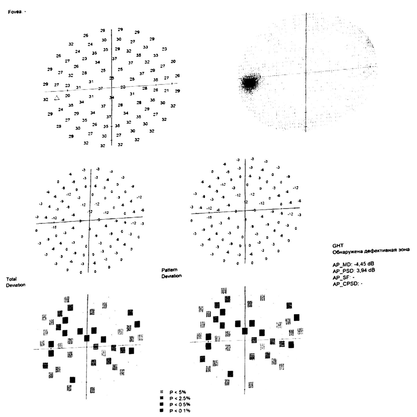 | Figure 1. Perimetry of the right eye of a 7-year-old boy (Figure 1) |
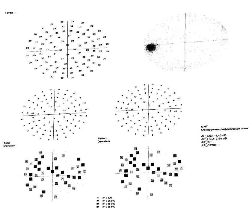 | Figure 2. Perimetry of the left eye of a 13-year-old girl (Figure 2) |
|
3.2. OCT and OCT Angiography Results
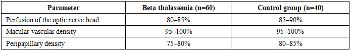
- • Structure of the macula preserved in most patients.• RNFL: In 60% of eyes, moderate thinning was observed in the superior and nasal sectors.• OCT-A: peripapillary perfusion was reduced (up to 75–80%) compared to the control (80–85%).
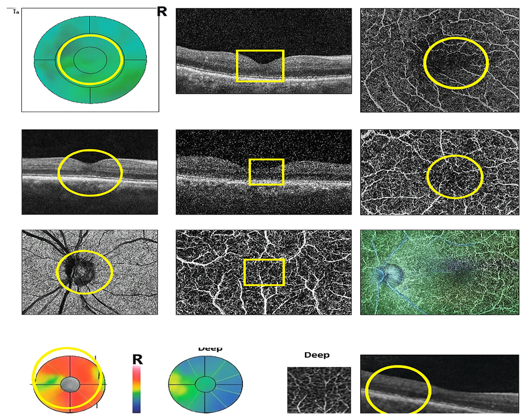 | Figure 3. OCT and OCTA image of a patient with thalassemia: macular thinning, decreased capillary network density in the FAZ and periphery (Figure 3) |
|
4. Discussion
- The results of the present study demonstrate the presence of subclinical changes in visual functions in children with beta-thalassemia, even in the absence of pronounced morphological changes according to OCT and OCTA data.Perimetric changes:• A decrease in MD was observed in all patients, which may reflect the general hypoxic load on the optic nerve [9].• An elevated PSD in 87% of cases indicates the presence of focal visual field defects, which confirms possible early ganglion cell damage [14].Similar data are described in the work of Abdel-Malak RR et al. [3], where a decrease in RNFL was also recorded in children with β-thalassemia, without obvious ophthalmological complaints.Morphological changes according to OCT and OCT-A data:• In some patients, RNFL thinning was detected, especially in the superonasal quadrants, which correlated with local disturbances in perimetry (Fig. 1, 2).• Despite the preserved vascular density in the macular zone, hypoperfusion was noted in the peripapillary region, a typical finding in chronic anemia in children [7,15].Research by Sharma R. and Mehta S. [15] showed that in children with anemia, OCTA can record vascular disorders even before clinical manifestations of vision loss.Thus, the combined use of functional (perimetry) and morphological (OCT/OCT-A) methods allows us to identify early signs of neuroretinal disorders in β-thalassemia, even in the asymptomatic phase.
5. Conclusions
- Based on the data obtained, the following conclusions can be drawn:1. Children with beta-thalassemia have decreased visual field sensitivity, which is reflected in decreased MD and increased PSD during computer perimetry.2. Subclinical thinning of the RNFL layer was detected in 60% of patients, predominantly in the superonasal quadrant.3. OCT angiography data did not demonstrate significant differences in macular perfusion between the main and control groups, however, hypoperfusion in the peripapillary zone was recorded in a number of patients.4. Comprehensive ophthalmological diagnostics using perimetry and OCT-A is highly informative for the early detection of neuroretinal changes in hemoglobinopathies.It is recommended to conduct regular ophthalmological examinations in children with hemopathies for the purpose of dynamic monitoring and prevention of irreversible changes in vision.