American Journal of Medicine and Medical Sciences

p-ISSN: 2165-901X    e-ISSN: 2165-9036

2025;  15(9): 3178-3183

doi:10.5923/j.ajmms.20251509.75

Received: Sep. 9, 2025; Accepted: Sep. 25, 2025; Published: Sep. 29, 2025


Asymmetric Dimethylarginine and Metabolic Dysregulation: Novel Insights into Links with Insulin Resistance and Atherogenic Dyslipidemia

Munisakhon M. Makhkamova1, Nargiza M. Nurillayeva2

1PhD Researcher at the Department of “Internal Diseases in Family Medicine and Fundamentals of Preventive Medicine №1” at Tashkent State Medical University, Tashkent, Uzbekistan

2DSc, Professor, Head of the Department of “Internal Diseases in Family Medicine and Fundamentals of Preventive Medicine №1” at Tashkent State Medical University, Tashkent, Uzbekistan

Copyright © 2025 The Author(s). Published by Scientific & Academic Publishing.

This work is licensed under the Creative Commons Attribution International License (CC BY).
http://creativecommons.org/licenses/by/4.0/

Abstract

Objective. To examine associations of asymmetric dimethylarginine (ADMA) with metabolic biomarkers reflecting insulin resistance and dyslipidemia in patients with cardiometabolic disorders. Methods. The study included 149 patients. In all participants, ADMA concentrations were measured by ELISA; indices of carbohydrate metabolism (glucose, insulin, HOMA-IR), anthropometrics (BMI, waist circumference, WHR), and lipid profile (total cholesterol, triglycerides \[TG], HDL-C, LDL-C, VLDL-C, atherogenic index) were assessed. Correlation analysis was performed using Spearman’s rank coefficient. Results. The median ADMA level was 241.7 μmol/L (Q1–Q3: 152.1–591.4). Statistically significant positive correlations were observed between ADMA and insulin (ρ=0.18; p=0.026), HOMA-IR (ρ=0.19; p=0.019), body mass index (ρ=0.23; p=0.005), and waist circumference (ρ=0.16; p=0.047). Associations with glucose, hip circumference, and WHR were not statistically significant. No significant correlations were identified within the lipid profile, except for a trend toward an inverse association with HDL-C (ρ=−0.15; p=0.071). Conclusions. These findings support the role of ADMA as a biomarker reflecting the interplay between endothelial dysfunction and components of the metabolic syndrome. The strongest associations were observed with indices of insulin resistance and adiposity.

Keywords: Asymmetric dimethylarginine, Insulin resistance, HOMA-IR, Dyslipidemia, Endothelial dysfunction, Metabolic syndrome, Biomarkers

Cite this paper: Munisakhon M. Makhkamova, Nargiza M. Nurillayeva, Asymmetric Dimethylarginine and Metabolic Dysregulation: Novel Insights into Links with Insulin Resistance and Atherogenic Dyslipidemia, American Journal of Medicine and Medical Sciences, Vol. 15 No. 9, 2025, pp. 3178-3183. doi: 10.5923/j.ajmms.20251509.75.

1. Introduction

Cardiometabolic disorders converge on a web of metabolic and vascular abnormalities in which insulin resistance, atherogenic dyslipidemia, and endothelial dysfunction reinforce one another to accelerate atherosclerotic risk [1] [16]. (Picture 1) Asymmetric dimethylarginine (ADMA), an endogenous inhibitor of nitric oxide synthase, has been advanced as both a biomarker and mediator within this nexus because it impairs nitric-oxide–dependent endothelial signaling [2]. Foundational clinical and experimental studies have linked higher circulating ADMA to impaired endothelium-dependent vasodilation and to pro-atherogenic vascular phenotypes across diverse populations [3].
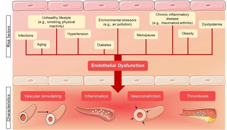
Picture 1. Risk factors for endothelial cell dysfunction and its pathological consequences. Created with Smart Servier Medical Art
This conceptual frame-endothelial dysfunction as a pivot between metabolic stress and vascular disease-continues to guide inquiry into whether ADMA marks, mediates, or merely mirrors insulin resistance and dyslipidemic risk [2] [3]. The enzymology that governs ADMA homeostasis centers on dimethylarginine dimethylaminohydrolase 1 (DDAH1), which hydrolyzes ADMA and thereby preserves nitric oxide bioavailability [4].
Recent multicenter work and contemporary reviews have clarified that the DDAH2 isoform does not metabolize ADMA in vivo, resolving a two-decade controversy and refocusing mechanistic attention on DDAH1-dependent pathways [5]. These developments underscore that interventions targeting the ADMA–DDAH1–NO axis remain biologically plausible levers to influence cardiometabolic risk profiles [6].
They also caution against attributing changes in circulating dimethylarginines to DDAH2 activity, which appears to act via ADMA-independent mechanisms in other tissues [7].
Methodological choices in ADMA quantification complicate cross-study comparisons and can distort effect estimates if not addressed analytically [8]. Head-to-head comparisons show that commercial immunoassays tend to overestimate ADMA relative to liquid-chromatography–mass-spectrometry (LC-MS/MS), with biases large enough to shift clinical interpretation [9].
Population data derived with chromatographic methods suggest typical reference intervals for plasma ADMA cluster near the sub-micromolar range, which helps anchor outlier detection in clinical cohorts [4, 6]. A meta-analytic synthesis has further quantified reference ranges in ostensibly healthy individuals and emphasized the magnitude of disagreement between ELISA and LC-MS/HPLC platforms. Consequently, studies must disclose assay modality and address potential measurement error to avoid spurious correlations with metabolic phenotypes [10] [14]. Attention to pre-analytical variables-sample handling, storage, and timing-remains equally important given their outsized contribution to total laboratory error [11].
Insulin resistance (IR) is a core pathophysiological substrate of cardiometabolic disease and a strong predictor of atherosclerotic cardiovascular events [12]. The homeostasis model assessment (HOMA-IR) offers a practical surrogate of IR derived from fasting glucose and insulin and has been widely adopted across epidemiologic and clinical studies [13]. Scholarly guidance, however, urges careful interpretation of HOMA metrics with awareness of assay differences and model assumptions [14] [23]. At the interface of metabolic and vascular biology, impaired nitric oxide signaling-a downstream consequence of elevated ADMA-can hinder skeletal-muscle glucose uptake and microvascular perfusion, plausibly linking endothelial dysfunction to systemic insulin resistance [15]. In clinical cohorts spanning diabetes, prehypertension, obesity, and kidney disease, higher ADMA has correlated with HOMA-IR and related glycemic measures [16], albeit with heterogeneity by population, assay, and covariate adjustment [17].
Atherogenic dyslipidemia-a triad of hypertriglyceridemia, low HDL-cholesterol, and a predominance of small dense LDL particles-clusters with IR and amplifies residual cardiovascular risk even under statin therapy [18]. Mechanistically, insulin resistance drives hepatic VLDL overproduction and triglyceride enrichment of lipoproteins, seeding the sdLDL-rich phenotype that is more atherogenic than larger LDL subfractions [19]. Multiple lines of evidence, including prospective data, associate sdLDL-C with incident coronary events beyond traditional lipids, motivating its use in refined risk stratification where available [20]. Because particle number influences atherogenic potential, apolipoprotein B-representing the concentration of atherogenic lipoprotein particles [8] [21]-has emerged as a superior marker for risk assessment relative to LDL-C across a range of clinical contexts. Consensus statements and expert guidance increasingly endorse apoB targets to complement or, in selected scenarios, supersede cholesterol-based metrics.
Guideline evolution has tracked this evidence base, with the 2025 ESC/EAS Focused Update [22] integrating new recommendations on risk stratification and lipid-lowering intensity on top of the 2019 framework. Against this background, the biological and clinical logic for interrogating ADMA alongside lipid phenotypes is strong [2] [23]. Experimental and early clinical data suggest that ADMA-driven nitric-oxide deficiency may promote atherogenic lipoprotein remodeling indirectly via endothelial dysfunction, tissue inflammation, and altered metabolic flexibility [24]. Yet observational studies linking ADMA with triglycerides, HDL-C, non-HDL-C, or apoB have yielded mixed results, raising questions about confounding, power, and analytical heterogeneity [25]. Clarifying these relationships requires cohorts with careful phenotyping, robust adjustment for lifestyle and pharmacologic factors, and explicit attention to analytic modality for both ADMA and lipid measurements [26].
Prospective and cross-sectional analyses have linked ADMA with chronic kidney disease progression and with reduced glomerular filtration, although the strength of association can vary depending on whether metabolism or clearance predominates in a given population [6] [13]. These methodological and pathophysiological considerations motivate a focused clinical analysis in patients with cardiometabolic disturbances to assess whether ADMA tracks with insulin resistance indices [4] [26] and with atherogenic lipid features beyond traditional risk factors [27]. Robust estimation strategies should include prespecified covariates that influence both ADMA [28] and metabolic phenotypes, such as age, sex, adiposity, renal function, smoking status, physical activity, and contemporary cardiometabolic therapies [5] [28]. Given assay-related dispersion in absolute ADMA values, reporting should emphasize internal ranks or quantiles and, where relevant, provide sensitivity analyses by analytic platform [21]. Parallel attention to lipid particle burden-through apoB or non-HDL-C-and to qualitative traits-through sdLDL metrics where available-can illuminate whether endothelial dysfunction markers and atherogenic lipoprotein signatures intersect in clinically meaningful ways [5] [17]. Finally, alignment with evolving guideline frameworks ensures interpretability and translational relevance for risk communication and therapy selection in real-world cardiometabolic care [15].
In summary, ADMA sits at a biologically plausible crossroads linking nitric-oxide–dependent endothelial function with metabolic dysregulation [28], yet inference has been limited by assay variability, confounding by renal function, and inconsistent lipid phenotyping [3] [15]. A carefully phenotyped clinical cohort assessed with standardized methods affords an opportunity to clarify whether ADMA associates with HOMA-IR and with atherogenic dyslipidemic markers-triglycerides, HDL-C, non-HDL-C, apoB, and sdLDL-after rigorous adjustment [4] [20]. Such evidence would refine the positioning of ADMA within cardiometabolic risk assessment and could help determine whether this endothelial dysfunction marker provides additive value alongside contemporary lipid metrics and guideline-directed risk stratification.

2. Methods

This prospective observational study enrolled patients with ischemic heart disease (IHD) presenting with stable angina of functional class I–II. Follow-up was conducted from January 1, 2024 to June 1, 2025 at the Multidisciplinary Clinic of the Tashkent State Medical University (TSMU) and the Republican Specialized Scientific and Practical Medical Centre of Cardiology (RSSPMC Cardiology). The study cohort comprised 149 patients with IHD, functional class I–II. A control group included 20 apparently healthy individuals without clinical or instrumental signs of cardiovascular disease.
At the initial evaluation, medical history and physical examination were performed, with systematic assessment of IHD symptoms (angina episodes, exercise tolerance). Liver status was evaluated by abdominal ultrasonography or FibroScan. Laboratory testing included routine hematology and biochemistry with alanine aminotransferase (ALT), aspartate aminotransferase (AST), a complete lipid profile, and indices of glucose metabolism. Cardiac function was assessed by a standard 12-lead electrocardiogram (ECG) and transthoracic echocardiography.
Inclusion criteria. Confirmed diagnosis of IHD with stable effort angina, functional class I–II. Age 35 to 75 years; Provision of written informed consent to participate.
Exclusion criteria. Unstable angina, acute coronary syndrome, or prior myocardial infarction; Chronic heart failure of New York Heart Association (NYHA) class III–IV or left ventricular ejection fraction < 40%; Severe comorbid conditions potentially affecting ADMA levels or endothelial function, including chronic kidney disease with estimated glomerular filtration rate (eGFR)<45 mL/min/1.73 m²; liver cirrhosis or severe chronic liver diseases, active malignancy, or pronounced systemic inflammatory/autoimmune disorders; Decompensated type 1 diabetes mellitus; poorly controlled type 2 diabetes mellitus with HbA1c > 9% at enrollment (such patients required treatment optimization prior to evaluation); Use of agents known to influence ADMA metabolism within 4 weeks prior to study procedures (e.g., high-dose L-arginine, phosphodiesterase-5 inhibitors, etc.).
In all participants, general clinical information was obtained and anthropometry was performed: body weight (kg) and height (cm) were measured to calculate body mass index (BMI, kg/m²), waist circumference (cm) and hip circumference (cm) were recorded to derive the waist-to-hip ratio (WHR). Criteria for abdominal obesity were applied as follows: waist circumference ≥94 cm in men and ≥80 cm in women. The atherogenic index (coefficient of atherogenicity) was calculated as (total cholesterol − HDL-cholesterol) / HDL-cholesterol. Fasting venous blood was collected from all subjects for biochemical analyses. Serum ADMA concentration was measured by enzyme-linked immunosorbent assay (ELISA) using a commercial kit (Beijing Ltd., China) and expressed in μmol/L. Plasma glucose was determined by the hexokinase method (mmol/L). Immunoreactive insulin was measured by ELISA (μIU/mL). Insulin resistance was assessed using the HOMA-IR index, calculated as: glucose (mmol/L) × insulin (μIU/mL) / 22.5. The serum lipid profile (mmol/L) included triglycerides (TG), total cholesterol (TC), high-density lipoprotein cholesterol (HDL-C), and calculated low-density lipoprotein cholesterol (LDL-C). Lipid measurements were performed using enzymatic colorimetric methods on an automated analyzer. All measurements were conducted in a single certified laboratory with adherence to internal and external quality control procedures.
Statistical analyses were performed using MS Excel. Correlations were assessed using Spearman’s rank correlation coefficient (ρ). Statistical significance for correlations was set at p<0.05. Where indicated, multiple regression analysis (linear regression) was employed to identify independent predictors of ADMA levels, entering variables that were significant in univariable analyses.

3. Results

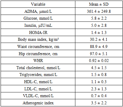
A total of 149 individuals were examined, of whom 55.7% were women and 44.3% were men. The median age was 46 years [38–54]. The median BMI was 30.2 kg/m² [27.8–32.7], corresponding to excess body weight on average in the cohort. The median waist circumference was 88.5 cm [85.8–92.1]; in 57% of participants it exceeded sex-specific thresholds, indicating abdominal obesity. The waist-to-hip ratio (WHR) was 0.914 [0.902–0.929], exceeding the conventional normal values (0.85 for women, 0.90 for men) in most participants.
The median serum ADMA concentration was 361.4 ± 249.8 nmol/L. Median fasting plasma glucose was 5.1 mmol/L [4.4–6.4]; in 13% of patients, glycemia exceeded 7 mmol/L, suggestive of impaired glucose tolerance or diabetes. Median immunoreactive insulin was 3.8 μIU/mL [3.1–5.9], and HOMA-IR was 0.95 [0.64–1.57]. Elevated HOMA-IR > 2.5 (insulin-resistance threshold) was observed in 11% of participants. The lipid profile showed overall moderate abnormalities: total cholesterol 4.0 mmol/L [3.5–4.8], triglycerides 1.50 mmol/L [1.10–1.90], HDL-C 1.00 mmol/L [0.95–1.22], and calculated LDL-C 2.22 mmol/L [1.55–3.03]. In 42% of participants, HDL-C was below target (< 1.0 mmol/L in men and < 1.2 mmol/L in women). The atherogenic coefficient ranged from −0.39 to 16.5 (median 3.20 [2.06–4.49]). Values above the recommended limit of 3.0 were recorded in more than half of the cohort (56%), indicating a high prevalence of atherogenic dyslipidemia in the sample.
In the correlation analysis, statistically significant associations were identified between ADMA levels and several indices of metabolic status. Serum ADMA concentration showed a significant positive correlation with fasting insulin (Spearman’s ρ=0.18; p=0.026) and with HOMA-IR (ρ=0.19; p=0.019), indicating that higher ADMA levels were associated with greater insulin resistance. Table 1 presents a scatterplot illustrating the relationship between ADMA and HOMA-IR, demonstrating an upward trend in HOMA-IR with increasing ADMA concentration. Positive correlations were also observed between ADMA and measures of general and central adiposity-BMI (ρ=0.23; p=0.005) and waist circumference (ρ=0.16; p=0.047). Thus, individuals with greater excess weight tended to exhibit higher ADMA levels. (Table 1)
Table 1. Descriptive characteristics of the study cohort (n = 149)
     
Table 1 summarizes baseline anthropometric, glycemic, and lipid parameters together with circulating ADMA for the full cohort. Data are presented as mean ± standard deviation to reflect central tendency under a parametric summary, alongside the median and interquartile range (Q1–Q3) to capture distributional skewness. Several variables-including ADMA, fasting insulin, and HOMA-IR-exhibit right-skew (mean exceeding the median), which is typical for metabolic biomarkers influenced by adiposity and insulin resistance. BMI centers in the obese range, while waist circumference and WHR indicate a predominance of central adiposity. The lipid profile shows modest hypertriglyceridemia with relatively low HDL-C and a median atherogenic index above 3.0, consistent with an atherogenic dyslipidemia phenotype in a substantial proportion of participants. These summaries provide the empirical backdrop for subsequent correlation and regression analyses examining the relationships between ADMA, insulin resistance (HOMA-IR), and atherogenic lipid traits.
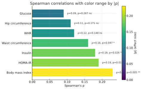
Figure 1. Correlations of ADMA with metabolic and anthropometric variables
ADMA demonstrated statistically significant positive correlations with fasting insulin (ρ=0.18, p=0.026) and HOMA-IR (ρ=0.19, p=0.019), indicating that higher ADMA levels are associated with greater insulin resistance. The associations with adiposity indices were similar in direction: BMI (ρ=0.23, p=0.005) and waist circumference (ρ=0.16, p=0.047) were both positively related to ADMA, suggesting that general and central obesity track with elevated ADMA. Correlations with glucose, hip circumference, and WHR did not reach statistical significance.
The associations between ADMA and lipid parameters were less pronounced. No statistically significant correlations were observed with triglycerides (ρ = −0.05; p = 0.52), LDL-cholesterol (ρ = −0.02; p = 0.77), or the atherogenic index (ρ = 0.10; p = 0.22).
Nevertheless, there was a trend toward an inverse correlation between ADMA and HDL-cholesterol (ρ = −0.15; p = 0.071): higher ADMA values were associated with slightly lower HDL-C, although this association did not reach statistical significance. The correlation between ADMA and the waist-to-hip ratio (WHR) was also non-significant (ρ = 0.12; p = 0.15). (Table 2)
Table 2. Correlations of ADMA with lipid profile variables
     

4. Discussion

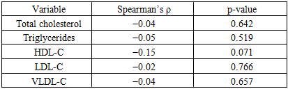
In this cohort of patients with stable IHD, circulating ADMA tracked most consistently with indices of insulin resistance and adiposity, while associations with standard lipid parameters were weak. ADMA showed small but statistically significant positive correlations with fasting insulin and HOMA-IR, as well as with BMI and waist circumference, indicating that higher ADMA levels co-occur with greater insulin resistance and central obesity. By contrast, correlations with triglycerides, LDL-C, VLDL-C and the atherogenic index were null, with only a borderline inverse trend for HDL-C. Taken together, these findings suggest that ADMA aligns more closely with the insulin-resistant/obesity phenotype than with conventional cholesterol fractions. Multivariable analyses reinforced this pattern: when BMI and HOMA-IR (or fasting insulin) were modeled together, BMI remained the sole independent predictor of ADMA, pointing to adiposity as the dominant correlate of circulating ADMA in this dataset. A plausible mechanistic interpretation is that excess adiposity-via low-grade inflammation, oxidative stress, and altered DDAH/NO signaling-elevates ADMA and impairs endothelial function, which in turn exacerbates insulin resistance.
Several considerations temper inference. The cross-sectional design precludes causal conclusions; medication use and unmeasured confounders (e.g., renal function, systemic inflammation) may attenuate lipid associations; and ELISA-based quantification can differ from chromatographic methods. Unit harmonization for ADMA (μmol/L vs nmol/L) should be verified across text and tables. Clinically, ADMA may have value as an adjunctive marker of cardiometabolic risk-particularly in patients with central obesity and insulin resistance-yet its incremental utility beyond established measures requires prospective validation and, ideally, interventional studies targeting weight reduction, insulin sensitivity, or the DDAH/NO axis.

4.1. Study Limitations

This analysis is cross-sectional, which precludes establishing causal relationships. It is therefore not possible to determine whether elevated ADMA levels cause insulin resistance and vascular dysfunction or arise secondarily in response to metabolic perturbations. Longitudinal studies are needed to clarify whether ADMA predicts the development of diabetes and atherosclerosis. In addition, the sample comprised a relatively heterogeneous group without strict clinical subclassification; future work should examine associations between ADMA and metabolic markers in more defined cohorts (e.g., patients with metabolic syndrome or with diabetes). Another limitation is the use of ELISA for ADMA measurement-while convenient, chromatographic methods (e.g., HPLC/LC–MS) provide greater analytical specificity. Nevertheless, our findings are broadly consistent with prior studies and are biologically plausible.

5. Conclusions

In this study, we identified a close relationship between circulating ADMA—an endogenous inhibitor of nitric oxide synthase—and metabolic disturbances characteristic of insulin resistance and dyslipidemia. Higher ADMA concentrations were associated with elevated fasting insulin and HOMA-IR, as well as with measures of adiposity (BMI and waist circumference), reflecting the coupling of endothelial dysfunction with insulin-resistant states. Although direct associations between ADMA and individual lipid parameters were weaker, we observed a trend toward an inverse relationship with HDL-C, consistent with the putative role of ADMA in atherogenesis. These findings add to a growing body of evidence that positions ADMA not merely as a passive marker but as a potential mediator of vascular injury in the context of metabolic disease. Mechanistically, elevated ADMA may contribute to reduced nitric oxide bioavailability, impaired vasodilation, and pro-inflammatory signaling, all of which accelerate atherosclerotic changes. The consistent associations across multiple indices of insulin resistance and adiposity suggest that ADMA could serve as an integrative indicator of cardiometabolic stress.
From a practical standpoint, ADMA may be considered a promising biomarker of cardiometabolic risk in patients with insulin resistance and related disorders such as type 2 diabetes or metabolic syndrome. Incorporating ADMA measurement into clinical risk stratification could help identify individuals at heightened risk for early vascular complications. Nevertheless, the observational nature of our study limits inference about causality. Definitive confirmation of a causal role for ADMA will require long-term prospective cohorts and randomized interventional trials aimed at lowering ADMA levels—through lifestyle modification, pharmacologic agents, or targeted enzyme modulation—and evaluating the downstream effects on metabolic and vascular outcomes. Future research should also explore whether genetic or epigenetic determinants of ADMA metabolism influence susceptibility to insulin resistance and cardiovascular disease, which could open avenues for precision-based prevention and therapy.

References

[1]  Di Pino A, DeFronzo RA. Insulin resistance and atherosclerosis: implications for insulin-sensitizing agents. Endocr Rev. 2019 Dec; 40(6): 1447-1467. doi:10.1210/er.2018-00141.
[2]  Ormazabal V, Nair S, Elfeky O, et al. Association between insulin resistance and the development of cardiovascular disease. Cardiovasc Diabetol. 2018; 17: 122. doi:10.1186/s12933-018-0762-4.
[3]  Amirkhizi F, Taghizadeh M, Hamedi-Shahraki S, Asghari S. Association of dietary phytochemical index with metabolic markers, serum asymmetric dimethylarginine and atherogenic indices in patients with polycystic ovary syndrome. Nutr Metab (Lond). 2025; 22(1): 1. doi:10.1186/s12986-025-00932-1.
[4]  Sibal L, Agarwal SC, Home PD, Boger RH. The role of asymmetric dimethylarginine (ADMA) in endothelial dysfunction and cardiovascular disease. Curr Cardiol Rev. 2010 May; 6(2): 82-90. doi:10.2174/157340310791162659. PMID: 21532773.
[5]  Boger RH, et al. Asymmetric dimethylarginine (ADMA): a novel risk factor for endothelial dysfunction in hypercholesterolemia. Circulation. 1998 Oct 27; 98(18): 1842-7. doi:10.1161/01.cir.98.18.1842.
[6]  Perticone F, Sciacqua A, Maio R, et al. Asymmetric dimethylarginine, L-arginine, and endothelial dysfunction in essential hypertension. J Am Coll Cardiol. 2005 Aug 2; 46(3): 518-523. doi:10.1016/j.jacc.2005.04.040.
[7]  Relationship between metabolical associated steatotic liver disease and ischemic heart disease: clinical aspects of comorbidity. (2024). CARDIOLOGY OF UZBEKISTAN, 1(4), 221-227. https://doi.org/10.70626/cardiouz-2024-1-00025.
[8]  Ragavan VN, Nair PC, Jarzebska N, et al. A multi-centric consortium study demonstrates that dimethylarginine dimethylaminohydrolase 2 is not a dimethylarginine dimethylaminohydrolase. Nat Commun. 2023 Jun 9; 14(1): 3392. doi:10.1038/s41467-023-38467-9. PMID: 37296100.
[9]  Nair PC, Mangoni AA, Rodionov RN. Redefining the biological and pathophysiological role of dimethylarginine dimethylaminohydrolase 2. Trends Mol Med. 2024 Jun; 30(6): 552-561. doi:10.1016/j.molmed.2024.03.001.
[10]  Németh B, Ajtay Z, Hejjel L, et al. The issue of plasma asymmetric dimethylarginine reference range: a systematic review and meta-analysis. PLoS One. 2017 May 11; 12(5): e0177493. doi:10.1371/journal.pone.0177493.
[11]  Nakhjavani M, et al. ADMA is a correlate of insulin resistance in early-stage diabetes independent of hs-CRP and body adiposity. Ann Endocrinol (Paris). 2010; 71(4): 303-308. doi:10.1016/j.ando.2010.02.026.
[12]  Taskinen MR, Borén J. New insights into the pathophysiology of dyslipidemia in type 2 diabetes. Atherosclerosis. 2015 Apr; 239(2): 483-495. doi:10.1016/j.atherosclerosis.2015.01.039.
[13]  Hirano T. Pathophysiology of diabetic dyslipidemia. J Atheroscler Thromb. 2018 Sep 1; 25(9): 771-782. doi:10.5551/jat.RV17023.
[14]  Xiao Y, Cai J, Su Y. Association between atherogenic index of plasma and cardiovascular disease in hypertension: a cross-sectional and longitudinal study. Heart Lung. 2025; 74: 49-56. doi:10.1016/j.hrtlng.2025.06.005.
[15]  Mendieta G, Pocock S, Mass V, Moreno A, Owen R, Garcia-Lunar I, et al. Determinants of progression and regression of subclinical atherosclerosis over 6 years. J Am Coll Cardiol 2023; 82: 2069–83. https://doi.org/10.1016/j.jacc.2023.09.814.
[16]  Wang J, Zhao X, Zhao Y, et al. Discordance of small dense LDL cholesterol beyond LDL cholesterol or non–HDL cholesterol and carotid plaque. JACC Asia. 2025; 5(8): 1012-1028. doi:10.1016/j.jacasi.2025.04.015.
[17]  El-salam MA, Abdelrahman T, Youssef M, et al. Evaluation of asymmetric dimethylarginine serum level and left ventricular function by 2D speckle tracking echocardiography in children on regular hemodialysis. Saudi J Kidney Dis Transpl. 2022 Mar-Apr; 33(2): 259-271. doi:10.4103/1319-2442.379024.
[18]  Nissen SE, Lincoff AM, Brennan D, et al. Bempedoic acid and cardiovascular outcomes in statin-intolerant patients. N Engl J Med. 2023; 388: 1353-1364. doi:10.1056/NEJMoa2215024.
[19]  Nicholls SJ, Lincoff AM, Garcia M, et al. Effect of high-dose omega-3 fatty acids vs corn oil on major adverse cardiovascular events in patients at high cardiovascular risk: the STRENGTH randomized clinical trial. JAMA. 2020; 324: 2268-2280. doi:10.1001/jama.2020.22258.
[20]  Fernandez-Friera L, Garcia-Alvarez A, Oliva B, et al. Association between subclinical atherosclerosis burden and unrecognized myocardial infarction detected by cardiac magnetic resonance in middle-aged low-risk adults. Eur Heart J Cardiovasc Imaging. 2024; 25: 968-975. doi:10.1093/ehjci/jeae044.
[21]  Peng AW, Dardari ZA, Blumenthal RS, et al. Very high coronary artery calcium (≥1000) and association with cardiovascular disease events, non-cardiovascular outcomes, and mortality: results from MESA. Circulation. 2021; 143: 1571-1583. doi:10.1161/CIRCULATIONAHA.120.050545.
[22]  Mach F, Koskinas KC, Roeters van Lennep JE, et al. 2025 Focused update of the 2019 ESC/EAS guidelines for the management of dyslipidaemias. Eur Heart J. 2025; ehaf190. doi:10.1093/eurheartj/ehaf190.
[23]  Raposeiras-Roubin S, Rossello X, Oliva B, et al. Triglycerides and residual atherosclerotic risk. J Am Coll Cardiol. 2021; 77: 3031-3041. doi:10.1016/j.jacc.2021.04.059.
[24]  D’Amario D, Laborante R, Bianchini E, et al. Statins as preventive therapy for anthracycline cardiotoxicity: a meta-analysis of randomized controlled trials. Int J Cardiol. 2023; 391: 131219. doi:10.1016/j.ijcard.2023.131219.
[25]  Felix N, Nogueira PC, Silva IM, et al. Cardio-protective effects of statins in patients undergoing anthracycline-based chemotherapy: updated meta-analysis of randomized controlled trials. Eur J Intern Med. 2024; 126: 43-48. doi:10.1016/j.ejim.2024.04.007.
[26]  Demonty I, Ras RT, van der Knaap HC, et al. Continuous dose-response relationship of the LDL-cholesterol-lowering effect of phytosterol intake. J Nutr. 2009; 139: 271-284. doi:10.3945/jn.108.095125.
[27]  Thavendiranathan P, Houbois C, Marwick TH, et al. Statins to prevent early cardiac dysfunction in cancer patients at increased cardiotoxicity risk receiving anthracyclines. Eur Heart J Cardiovasc Pharmacother. 2023; 9: 515-525. doi:10.1093/ehjcvp/pvad031.
[28]  Sherratt SCR, Libby P, Dawoud H, Bhatt DL, Mason RP. Eicosapentaenoic acid improves endothelial nitric oxide bioavailability via changes in protein expression during inflammation. J Am Heart Assoc 2024; 13: e034076. https://doi.org/10.1161/JAHA.123. 034076.