-
Paper Information
- Next Paper
- Previous Paper
- Paper Submission
-
Journal Information
- About This Journal
- Editorial Board
- Current Issue
- Archive
- Author Guidelines
- Contact Us
American Journal of Medicine and Medical Sciences
p-ISSN: 2165-901X e-ISSN: 2165-9036
2025; 15(9): 3121-3123
doi:10.5923/j.ajmms.20251509.60
Received: Aug. 17, 2025; Accepted: Sep. 12, 2025; Published: Sep. 29, 2025

Clinical Evaluation of the Oral Mucosa in Patients with Different Types of Removable Dentures
 Abstract
 Reference
 Full-Text PDF
 Full-text HTML
Ibragimova Feruza Ikromovna, Khakimov Farrukh Khakimovich
Bukhara State Medical Institute, Uzbekistan
Copyright © 2025 The Author(s). Published by Scientific & Academic Publishing.
This work is licensed under the Creative Commons Attribution International License (CC BY).
http://creativecommons.org/licenses/by/4.0/

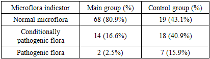
This article assessed the clinical condition of the oral mucosa in patients using various removable dentures. The study was conducted on 128 patients, who were divided into the main (84) and control (44) groups. The main group of patients used dentures made of modern materials and additionally used physiotherapeutic methods (laser therapy and ultrasound), while the control group used traditional dentures. Clinical examination, questionnaire, microbiological analysis and the effectiveness of physiotherapeutic methods were assessed. The results showed that in the main group, clinical and functional indicators of the mucosa were better preserved, signs of inflammation were reduced, and the level of comfort and adaptation of patients to the denture was higher. Microbiological examinations showed a high level of normal microflora and a significantly lower level of pathogenic flora. The study showed the importance of modern materials and physiotherapeutic approaches in maintaining the health of the oral mucosa and increasing the effectiveness of prosthetics.
Keywords: Removable dentures, Oral mucosa, Clinical assessment, Physiotherapeutic methods, Laser therapy, Ultrasound, Microbiological analysis, Denture adaptation
Cite this paper: Ibragimova Feruza Ikromovna, Khakimov Farrukh Khakimovich, Clinical Evaluation of the Oral Mucosa in Patients with Different Types of Removable Dentures, American Journal of Medicine and Medical Sciences, Vol. 15 No. 9, 2025, pp. 3121-3123. doi: 10.5923/j.ajmms.20251509.60.
Article Outline
1. Introduction
- The oral mucosa is of great importance in the prosthetics process, and its functional and clinical condition directly determines the patient's quality of life. Inflammation, atrophy, and discomfort of the mucous membrane are common when using removable dentures. According to statistics, various pathological changes in the mucous membrane are noted in 40–60% of patients wearing dentures [1,2]. Therefore, the use of modern materials and physiotherapeutic methods allows improving the clinical and functional indicators of the mucous membrane. The oral mucosa is one of the most important objects in dental practice. Assessment of the clinical condition of these tissues in patients using various removable dentures is of great importance in the treatment process. According to the latest data from the World Health Organization (WHO), about 3.5 billion people worldwide have various dental diseases, one of the most common of which is tooth loss [1]. It is estimated that 30–40% of the world's population has lost one or more teeth, and up to 70–80% of the population over the age of 65 has complete or partial edentulism [2,3]. Such epidemiological indicators indicate that prosthetics is a pressing global health issue.Removable dentures—complete dentures, partial dentures, clasp dentures, and options made of modern elastomeric materials—are widely used to meet the functional and aesthetic needs of patients [4,5]. However, as many scientists have noted, long-term exposure to dentures can lead to various pathological changes in the oral mucosa [6]. For example, a study by Budtz Jørgensen (1981) found that more than half of removable denture wearers developed denture stomatitis [7]. Recent observations have also confirmed that this figure remains in the range of 40–60% [8].Denture stomatitis is one of the most common complications of removable dentures, and fungi such as Candida albicans, as well as poor denture hygiene, mechanical stress, and biological inertness of materials play an important role in its development [9,10]. Gendreau and Loewy (2011) noted in their review that denture stomatitis has a high prevalence and significantly reduces the quality of life of patients [11]. Also, Arendorf and Walker (1987) found that inflammation and trophic disorders of the mucosa are common in patients with denture stomatitis [12].The changes observed in the mucosa vary depending on the type of removable prosthesis. In complete dentures, hyperemia and erosion are usually observed over a large area due to uneven pressure distribution and insufficient fixation [13]. Clip dentures partially reduce mechanical loading, but metal parts can cause local damage [14]. Although prostheses made of modern elastomeric materials are somewhat softer on the mucosa, they can also increase microbiological contamination if hygienic care rules are not followed [15].Changes in the oral mucosa during denture wear also depend on the general somatic condition of the patients. For example, in patients with diabetes, cardiovascular diseases, or immunodeficiency, it has been found that inflammatory processes and trophic disorders in the mucosa develop more quickly [16]. This situation indicates the need for an individual approach to each patient in clinical dentistry. Clinical observations have also confirmed that neglect of denture hygiene and care leads to changes in the oral microbiocenosis and an increased risk of caries and periodontitis [17].Therefore, assessing the clinical condition of the oral mucosa in patients using various removable prostheses is an important condition for increasing the effectiveness of prosthetics, early detection and prevention of complications, and improving the quality of life of patients. In this direction, modern clinical examination methods (clinical examination, cytological examinations, microbiological analyses, biomarker studies) are of particular importance and are widely used in scientific research [18,19].This study is to assess the clinical condition of the oral mucosa in patients using different types of removable dentures, to determine the effectiveness of modern approaches and to compare them with traditional methods.
2. Materials and Methods
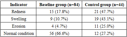
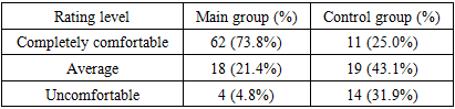
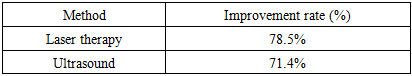
- This study was conducted on a total of 128 patients. All patients were divided into groups according to age, gender and clinical indicators. The study participants were divided into two groups. The first group was the main group, which included 84 patients. They were provided with modern removable prostheses, wore constructions made from new materials and underwent physiotherapeutic procedures (laser therapy, ultrasound). The second group was the control group, which included 44 patients. They used traditional removable prostheses and did not use physiotherapeutic methods.Several methods were used during the study. First, a clinical examination was performed, and pathological signs such as redness, swelling, and erosion of the mucous membrane were recorded. Then, using questionnaires, the patients' levels of comfort and discomfort in using the prosthesis were studied. Also, microbiological analyzes were performed, and the composition of the microflora in smears taken from the area under the prosthesis was determined. The effectiveness of physiotherapeutic procedures used in the main group was separately evaluated. The results were analyzed by comparing the main and control groups.
3. Research Results
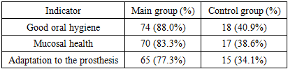
- According to the results of the study, the clinical indicators of the oral mucosa in the main group of patients were significantly improved compared to the control group. In the main group, there were fewer signs of inflammation, and the condition of the mucosa remained almost normal. In the control group, redness, swelling and erosion were much more common. In addition, the patients' adaptation to the prosthesis, the level of oral hygiene and the overall quality of life were higher in the main group.
|
|
|
|
|
4. Discussion
- It is very important to monitor the clinical condition of the oral mucosa when using removable dentures. The results of the study showed that when using dentures made of modern materials and physiotherapeutic methods (laser, ultrasound), the clinical indicators of patients, oral hygiene and adaptation to the prosthesis significantly improve. In the control group, the level of inflammation and discomfort was higher against the background of traditional dentures.From a scientific point of view, this study confirms the advantages of modern prosthetic approaches. From a social point of view, modern approaches play an important role in improving the quality of life of patients. From an economic point of view, better preservation of the mucosa and a shorter adaptation period to the prosthesis help reduce healthcare costs.
5. Conclusions
- As can be seen from the above study, the clinical condition of the oral mucosa varies significantly when using different removable prostheses. When prostheses made of modern materials and physiotherapeutic methods are used, clinical results are high, the condition of the mucosa is stable, and patients adapt to the prosthesis quickly. With traditional prostheses, inflammation, discomfort, and microflora disorders are common. Therefore, the widespread use of modern prosthetic materials and additional physiotherapeutic methods in dental practice increases clinical efficiency, improves the quality of life of patients, and allows for long-term preservation of the health of the oral mucosa.