-
Paper Information
- Next Paper
- Previous Paper
- Paper Submission
-
Journal Information
- About This Journal
- Editorial Board
- Current Issue
- Archive
- Author Guidelines
- Contact Us
American Journal of Medicine and Medical Sciences
p-ISSN: 2165-901X e-ISSN: 2165-9036
2025; 15(9): 3088-3091
doi:10.5923/j.ajmms.20251509.52
Received: Aug. 21, 2025; Accepted: Sep. 17, 2025; Published: Sep. 29, 2025

Improvement of Periodontal Disease Detection Strategies in Individuals with Epilepsy
 Abstract
 Reference
 Full-Text PDF
 Full-text HTML
Zoyirov Tulkin Elnazarovich1, Mardonova Dildora Kasimovna2
1Doctor of Medical Sciences (DSc), Professor, Samarkand State Medical University, Samarkand, Uzbekistan
2Independent Applicant for the Department of Therapeutic Dentistry, Samarkand State Medical University, Samarkand, Uzbekistan
Copyright © 2025 The Author(s). Published by Scientific & Academic Publishing.
This work is licensed under the Creative Commons Attribution International License (CC BY).
http://creativecommons.org/licenses/by/4.0/

Epilepsy is one of the most common chronic neurological diseases affecting approximately 65 million people worldwide. According to the World Health Organization, the prevalence of epilepsy is 4-10 cases per 1,000 population, with about 2.4 million new cases reported annually. In the Republic of Uzbekistan, more than 150,000 people suffer from epilepsy, which is 0.5% of the total population. This disease is characterized by repeated unprovoked epileptic seizures caused by abnormal electrical activity of the brain, and has a significant impact on the quality of life of patients and their social adaptation.
Keywords: Epilepsy, Periodontal diseases, Periodontitis, Gingivitis, Gum hyperplasia, Antiepileptic drugs, Phenytoin, Diagnosis, Periodontal examination, Indexes, Interdisciplinary approach, Personalized medicine
Cite this paper: Zoyirov Tulkin Elnazarovich, Mardonova Dildora Kasimovna, Improvement of Periodontal Disease Detection Strategies in Individuals with Epilepsy, American Journal of Medicine and Medical Sciences, Vol. 15 No. 9, 2025, pp. 3088-3091. doi: 10.5923/j.ajmms.20251509.52.
1. Introduction
- A growing number of scientific studies demonstrate a close relationship between epilepsy and oral health, especially in relation to periodontal diseases [1]. Patients with epilepsy are at increased risk of developing gingivitis, periodontitis and other inflammatory diseases of periodontal tissues. The prevalence of periodontal diseases among people with epilepsy varies from 38% to 95%, which is significantly higher than in the general population (20-50%). This pattern is due to a complex of factors, including direct and indirect effects of antiepileptic therapy, lifestyle features of patients, as well as systemic disorders associated with the underlying disease [2].The leading etiological factor in the development of periodontal diseases in patients with epilepsy is gum hyperplasia induced by taking antiepileptic drugs. The most pronounced gingival overgrowth is observed with the use of phenytoin (diphenylhydantoin), which causes gum hyperplasia in 50-100% of patients, depending on the dose and duration of use. The pathogenesis of phenytoin-induced hyperplasia is associated with impaired collagen metabolism in the connective tissue of the gum, increased synthesis of glycosaminoglycans, and decreased collagenase activity. Carbamazepine and valproic acid can also cause gingival growths, although with a lower frequency (6-20% of cases) [3].Hyperplastic gum tissue creates multiple retention zones for plaque accumulation, which contributes to the development of the inflammatory process and the progression of periodontitis. The altered architectonics of the gingival margin makes it difficult to conduct adequate oral hygiene, creating a vicious circle: drug hyperplasia → deterioration of hygiene → inflammation → further tissue overgrowth → progression of periodontitis [4].Additional risk factors for periodontal disease in patients with epilepsy include cognitive impairment, decreased manual skills, side effects of antiepileptic drugs (dry mouth, nausea, drowsiness), as well as socio-economic factors that restrict access to dental care. Epileptic seizures can be accompanied by traumatization of the soft and hard tissues of the oral cavity, which also contributes to the development of inflammatory processes in the periodontium [5].The systemic effects of epilepsy on the body include immune status disorders, changes in the blood coagulation system, endocrine dysfunctions and metabolic disorders that can modify the course of periodontal infection. Chronic stress associated with the underlying disease leads to activation of the hypothalamic-pituitary-adrenal system and an increase in cortisol levels, which negatively affects the reparative processes in periodontitis and reduces the effectiveness of the local immune response [6].Existing approaches to the diagnosis of periodontal diseases in patients with epilepsy are based on traditional methods of periodontal examination, including visual assessment of gum condition, probing of periodontal pockets, determination of hygiene and inflammation indices, as well as X-ray examination. However, these methods have a number of limitations when used in this category of patients. The altered anatomy of the gingival margin due to drug hyperplasia makes it difficult to accurately measure the depth of periodontal pockets and may lead to underestimation of the severity of periodontitis. Bleeding during probing can be caused not only by inflammation, but also by increased traumatization of hyperplastic tissues [7].X-ray diagnostics also presents certain difficulties in patients with epilepsy. The need for prolonged immobility can trigger an epileptic seizure, especially in patients with photosensitive epilepsy. In addition, traditional radiography does not always reveal early changes in the bone tissue of the alveolar process against the background of severe soft tissue hyperplasia [8].Special difficulties arise in the differential diagnosis of inflammatory and drug-induced gum hyperplasia. The clinical manifestations of these conditions may overlap, and their combination significantly complicates the assessment of the true condition of periodontitis and the planning of therapeutic measures. Standard clinical indexes do not take into account the specifics of the changes characteristic of patients with epilepsy, which can lead to an inadequate assessment of the severity of periodontal damage [9].Current trends in the development of periodontology are aimed at personalizing diagnostic approaches, taking into account individual risk factors and comorbid conditions. For patients with epilepsy, it is necessary to develop specialized diagnostic protocols that take into account the clinical manifestations of periodontal diseases against the background of antiepileptic therapy [10]. A promising area is the integration of modern instrumental diagnostic methods, including ultrasound examination of periodontal tissue, optical coherence tomography, confocal laser microscopy and molecular biological analysis methods. These technologies allow us to obtain objective information about the condition of periodontal tissues, minimizing the influence of subjective factors and anatomical features associated with drug hyperplasia [11]. An important component of optimizing diagnostic strategies is the development of specialized indexes and assessment scales adapted for patients with epilepsy. Such tools should take into account not only the traditional parameters of periodontal status, but also the specific manifestations of drug-induced changes, functional disorders and quality of life of patients [12].An interdisciplinary approach to the diagnosis of periodontal diseases in patients with epilepsy requires close collaboration between dentists, neurologists, pharmacologists and other specialists. Comprehensive assessment of neurological status, analysis of antiepileptic therapy, assessment of patient compliance and social factors are integral components of the diagnostic process. The cost-effectiveness of improved diagnostic strategies is due to the possibility of early detection of periodontal lesions, preventing their progression and reducing the need for complex surgical interventions. Timely diagnosis and treatment of periodontal diseases in patients with epilepsy can improve the control of the underlying disease, reduce the frequency of seizures and improve the quality of life [13].In the Republic of Uzbekistan, the problem of diagnosing periodontal diseases in patients with epilepsy is becoming particularly relevant due to the lack of awareness of medical personnel about the relationship between neurological and dental diseases, limited opportunities for interdisciplinary interaction and the lack of standardized examination protocols for this category of patients.The purpose of the study: to develop and scientifically substantiate a set of optimized diagnostic methods for detecting periodontal diseases in patients with epilepsy, taking into account the specifics of clinical manifestations and the management of this category of patients.
2. Research Materials and Methods
- Within the scope of the study, 149 individuals were taken between the ages of 18 and 60, making the main group of 67 patients diagnosed with epilepsy (29 men and 38 women). At the time of the examination, 59 patients were attending an outpatient appointment, while 8 patients were undergoing inpatient treatment.All patients with epilepsy were examined by an epileptologist, neurologist, psychiatrist and clinical psychologist. In the patients examined, parametric tests and functional research methods-EEG, reg, CT-were carried out. Data on identified neurological disorders have been documented in outpatient and stationary disease histories. Patients with epilepsy have been given traditional anti-epileptic seizure therapy. Diphenin has been used in 18 (26.9%) patients as part of complex treatment, and in 2 (3%) patients as monotherapy. The comparison group consisted of 52 patients (24 men and 28 women) with various mental disorders. There were 30 people (18 men and 12 women) in patients with schizophrenia and 22 people (8 men and 14 women) in patients with organic brain damage. At the time of the examination, all patients were in the stage of intragospital therapeutic remission.As a control group, 20 practically healthy individuals between the ages of 18 and 53 - volunteers without somatis pathology (8 men and 12 women) were examined, who visited the Samarkand Regional Dental Polyclinic.The study used Standard Indexes recommended by the World Health Organization. To assess the caries process, the distribution and intensity (GPU of the teeth) were calculated. The condition of the Parodont tissue was assessed using the general parodontal CPITN index.Thus, the study found that in patients with epilepsy, in general, the condition of the teeth is better than in patients with various mental disorders, since teeth with less tooth decay and treated and covered with coatings were more likely than in the comparison group. At the same time, the number of teeth removed in patients with epilepsy prevailed. This suggests that patients with epilepsy seek dental care more often than patients with various mental disorders, but in most cases, the visit of such patients to the dentist ends with the removal of problematic teeth. It is important to note that patients with epilepsy had better CPO index values in men and in the control group in women. In patients with various mental disorders, an excess of caries teeth is a hotbed of chronic odontogenic infection, which negatively affects the condition of the body as a whole in patients.CPITN index registration took into account bleeding when probed during the study, the presence of milkosti and milkusti tartar, and periodontal pockets of varying depths. To do this, the teeth were divided into 6 sextants, and the following teeth were examined: 17, 16 and, 26, 27, 31, 36, 37, 46, 47. If the sextant did not have Index teeth, all other teeth were examined and the heaviest code was recorded.
3. Results
- A comparative analysis of the data carried out showed that in patients with epilepsy and in the control group, pathological changes in the parodont were detected in approximately the same amount, in the main group, the changes in the parodont tissue were 1.3 times greater than in the control group. Hypertrophic gingivitis has been found in 11.3% of cases, with only anticonvulsants being observed in patients with receptive epilepsy. Patients with epilepsy were 2.2 times less likely to have catarrhal gingivitis than the control group (Figure 1).
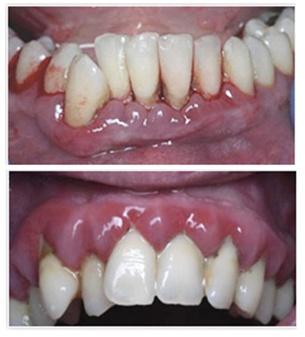 | Figure 1. The appearance of the oral cavity of a 34-year-old patient diagnosed with epilepsy |
4. Attitude
- The study found that in patients with epilepsy, in general, the condition of the teeth is better than in patients with various mental disorders, since teeth with less tooth decay and treated and covered with coatings were more likely than in the comparison group. At the same time, the number of teeth removed in patients with epilepsy prevailed. This suggests that patients with epilepsy seek dental care more often than patients with various mental disorders, but in most cases, the visit of such patients to the dentist ends with the removal of problematic teeth. It is important to note that patients with epilepsy had better CPO index values in men and in the control group in women. In patients with various mental disorders, an excess of caries teeth is a hotbed of chronic odontogenic infection, which negatively affects the condition of the body as a whole in patients.Inflammatory processes in Parodont tissues are associated with the development of pathogenic microflora, among which an important role is played by Bacteroides forsithus, Prevotella intermedia, Porphyromonas gingivalis, Actinobacillus actinomycetem-commitans, Treponema denticola, Veillonella recta, Peptostreptococcus micros, Str. intermedius, Actinomyces naeslundii, play Actinomyces israelii. Against the background of the obvious growth of pathogenic and conditional-pathogenic microorganisms, the concentration of representatives of the normative microflora is sharply reduced. Therefore, the development and application of means to help restore the normative microflora of the oral cavity and, in particular, the tissues of the parodont in the treatment of parodont inflammatory diseases is considered as a necessary condition for improving the effectiveness of the treatment of parodontitis. One of the promising directions is the use of biopreparations, the active part of which is affected is the representatives of the regulatory microflora of the oral cavity.
5. Conclusions
- 1. The study found a clear correlation relationship between dental indicators and the level of conformity in the control group. Whereas in patients with epilepsy and in patients with various mental disorders, correlation relationships are established only by some indicators. This is most likely due to personality changes.2. As you know, it is very difficult to change the habits of oral care in people of mature age from childhood (Chistova., 2001). Patients with epilepsy were found to have low hygiene. Therefore, only the formation of a specially oriented relationship of trust with the dentist makes it possible to increase the level of hygienic "re-education" and interaction of these patients. At the same time, the improvement of oral hygiene products makes it possible to solve the problem of optimizing hygienic care in hospitals in patients with epilepsy and patients with mental disorders. 3. In almost all cases, a fibrosis form of hypertrophic gingivitis has been observed. In 23 patients, hypertrophied milk suckers have a light pink color, and in one case, dark pink suckers with a cyanotic tone have been encountered. The gum suckers were slightly enlarged in size, densely fibrous, with a rough surface. During the examination, bleeding was recorded in one case. Under hypertrophied gum suckers, plaque and tartar buildup were observed on the tooth surfaces.4. Patients with epilepsy have been referred with the following complaints typical of hypertrophic gingivitis: 12 patients periodically complained to the blood of the gums while brushing their teeth, 55 patients to aesthetic defect. In 22 patients a deep prikus was detected, in 9 patients an orthognathic prikus was detected, in 13 patients a pathological absorption of dental hard tissue pathological absorption. When the prevalence of the hypertrophy process was studied in patients with epilepsy receiving diphenin, it was observed that the gums underwent hypertrophy, mainly in the frontal area of the lower and upper jaws. Two patients reported generalized hypertrophic gingivitis.