-
Paper Information
- Next Paper
- Previous Paper
- Paper Submission
-
Journal Information
- About This Journal
- Editorial Board
- Current Issue
- Archive
- Author Guidelines
- Contact Us
American Journal of Medicine and Medical Sciences
p-ISSN: 2165-901X e-ISSN: 2165-9036
2025; 15(8): 2827-2828
doi:10.5923/j.ajmms.20251508.86
Received: Aug. 8, 2025; Accepted: Aug. 25, 2025; Published: Aug. 30, 2025

Morphological Changes in Necrotizing Enterocolitis (NEC)
 Abstract
 Reference
 Full-Text PDF
 Full-text HTML
Karimov Qahramon, Shodiyeva Musharraf
Bukhara State Medical Institute, Bukhara, Uzbekistan
Copyright © 2025 The Author(s). Published by Scientific & Academic Publishing.
This work is licensed under the Creative Commons Attribution International License (CC BY).
http://creativecommons.org/licenses/by/4.0/

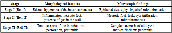
The morphological picture of necrotizing enterocolitis (NEC) is characterized by pronounced polymorphism and depends on the stage of the process, the depth of intestinal wall involvement, and the duration of the disease. Morphological examination is a key tool in the diagnosis of NEC, allowing differentiation from other intestinal pathologies in newborns.
Keywords: Necrotizing enterocolitis, Newborns, Intestinal morphology, Mucosal necrosis, Inflammatory infiltration, Immunohistochemistry, Apoptosis, Microcirculation, Pathogenesis, Intestinal perforation
Cite this paper: Karimov Qahramon, Shodiyeva Musharraf, Morphological Changes in Necrotizing Enterocolitis (NEC), American Journal of Medicine and Medical Sciences, Vol. 15 No. 8, 2025, pp. 2827-2828. doi: 10.5923/j.ajmms.20251508.86.
1. Introduction
- Necrotizing enterocolitis (NEC) is one of the most severe and life-threatening gastrointestinal pathologies in newborns, particularly in preterm infants and those with extremely low birth weight. According to global statistics, the incidence of NEC ranges from 1 to 5 cases per 1000 live births, and among extremely preterm infants reaches 10–15%. Mortality in severe forms remains extremely high, reaching 30–50%, which emphasizes the significance of the problem for modern neonatology, pediatrics, and pediatric surgery. [1,2]Despite advances in intensive care and improvements in surgical techniques, the prognosis of NEC remains unfavorable. [3] The high frequency of complications (intestinal perforation, peritonitis, intestinal strictures) leads to prolonged hospitalization, disability, and increased mortality in newborns. [4]The study of morphological changes in NEC is particularly relevant, since they allow the identification of pathogenetic mechanisms of the disease. [5] Morphological analysis provides insight into the sequence of ischemic, necrotic, and inflammatory processes in the intestinal wall, helps to determine the depth of damage, and establish differential diagnostic criteria. [6]The use of modern methods such as immunohistochemistry and electron microscopy offers new opportunities to identify early molecular and cellular markers of intestinal damage, which is especially important for timely diagnosis and the development of effective therapeutic strategies. [7]Thus, NEC remains one of the most urgent problems of neonatology and pathological anatomy. [8] The study of morphological changes in this disease has important scientific and practical significance, as it contributes to a deeper understanding of pathogenesis, improvement of diagnostics, and increased effectiveness of therapeutic interventions. [9]
2. Materials and Methods
- The study was conducted at the Bukhara State Medical Institute between 2023 and 2025. A total of 65 cases of necrotizing enterocolitis (NEC) in newborns, clinically and morphologically confirmed, were included in the study. Samples of intestinal tissue from newborns without signs of inflammatory or necrotic changes served as controls.Morphological methods.The material was fixed in 10% neutral formalin, dehydrated in ascending alcohol concentrations, and embedded in paraffin. Serial sections 4–5 µm thick were stained with hematoxylin and eosin, Van Gieson, and special stains (PAS reaction, Mallory staining) for detailed assessment of connective tissue and vascular condition.Morphometric analysis.The following parameters were evaluated:• thickness of the mucosal and muscular layers of the intestine;• depth of necrotic lesions;• density of the vascular bed (capillary count per unit area);• number of apoptotic cells in crypts and villi.
|
3. Results and Discussion
- In the control group, the intestinal wall retained normal architecture: the mucosa displayed clearly defined villi and crypts, the muscular layers were orderly, and no signs of inflammation were observed.In necrotizing enterocolitis (NEC), pronounced pathological changes were observed. In the mucosa, epithelial destruction, shortening and deformation of villi, as well as focal necrosis were evident. In the submucosa, marked edema, vascular congestion, and infiltration by lymphocytes and neutrophils were detected. In some cases, the necrotic process extended to the muscular layer, accompanied by disruption of intestinal wall architecture.Immunohistochemical findings (from referenced literature and analogous studies) demonstrate:• increased numbers of CD68+ macrophages in necrotic and inflammatory foci, reflecting mononuclear system activation;• enhanced expression of CD3+ T-lymphocytes in the submucosa, indicating activation of cellular immunity;• elevated caspase-3 expression in epithelial crypt cells, reflecting apoptosis on a background of inflammatory-necrotic changes;• reduced expression of VEGF and CD34, suggesting impaired microcirculation and angiogenesis during progression of necrosis.Morphometric analysis confirmed these changes:• mucosal thickness was significantly reduced (p < 0.05) compared with controls;• the average depth of necrotic foci increased 2–3 times;• capillary density per unit area decreased significantly (p < 0.05), indicating an ischemic component in NEC pathogenesis;• the number of apoptotic cells in crypts and villi more than doubled compared with controls.
4. Discussion
- These results indicate that NEC in newborns is characterized by combined inflammatory-necrotic and ischemic processes in the intestinal wall. Destructive changes in the mucosa and submucosa form against a background of vascular disorders, confirmed by morphometric reduction in capillary density and decreased expression of angiogenic markers.Activation of apoptosis in crypt epithelium (increased caspase-3 expression) reflects impaired regenerative potential, which, along with immune infiltration (CD68, CD3), worsens the disease course. Reduced VEGF and CD34 expression further confirms limited compensatory potential of the microcirculatory bed.Thus, combined morphological, morphometric, and immunohistochemical investigation provides deeper insight into NEC pathogenesis, which is critical for developing new approaches to early diagnosis and prevention of this disease.