-
Paper Information
- Next Paper
- Previous Paper
- Paper Submission
-
Journal Information
- About This Journal
- Editorial Board
- Current Issue
- Archive
- Author Guidelines
- Contact Us
American Journal of Medicine and Medical Sciences
p-ISSN: 2165-901X e-ISSN: 2165-9036
2025; 15(8): 2734-2739
doi:10.5923/j.ajmms.20251508.66
Received: Aug. 2, 2025; Accepted: Aug. 26, 2025; Published: Aug. 30, 2025

Clinical Significance of Early Detection of Eye Burn Sequelae Using AS-OCT and Corneal Densitometry
 Abstract
 Reference
 Full-Text PDF
 Full-text HTML
Kamilov Khalidzhan Makhamadzhanovich1, Maksudova Laylo Maskhutovna2, Inagamdjanova Shakhzoda Bobirovna3
1DSc, Professor, Head of the Department of “Ophthalmology” of the Center for the Development of Professional Qualifications of Medical Personnel, Tashkent, Uzbekistan
2DSc, Associate Professor of the Department of “Ophthalmology”, Center for Advanced Medical Personnel Training, Tashkent, Uzbekistan
3Freelance Applicant for the Department of Ophthalmology of the Center for Advanced Medical Personnel Training, Tashkent, Uzbekistan
Copyright © 2025 The Author(s). Published by Scientific & Academic Publishing.
This work is licensed under the Creative Commons Attribution International License (CC BY).
http://creativecommons.org/licenses/by/4.0/

The aim of this study was to evaluate the dynamics of corneal epithelial regeneration, stromal edema, and recovery of optical transparency in patients with grade I–III eye burns using corneal densitometry (Pentacam) and anterior segment optical coherence tomography (AS-OCT), as well as to analyze the cost-effectiveness of this approach.
Keywords: Ophthalmocombustiology, Corneal densitometry, AS-OCT, Cornea, Epithelial regeneration, Contact lens, Modern diagnostics and therapy
Cite this paper: Kamilov Khalidzhan Makhamadzhanovich, Maksudova Laylo Maskhutovna, Inagamdjanova Shakhzoda Bobirovna, Clinical Significance of Early Detection of Eye Burn Sequelae Using AS-OCT and Corneal Densitometry, American Journal of Medicine and Medical Sciences, Vol. 15 No. 8, 2025, pp. 2734-2739. doi: 10.5923/j.ajmms.20251508.66.
Article Outline
1. Introduction
- Eye burns are among the dangerous ophthalmological conditions for the visual organ that require immediate treatment. They result in deep disturbances in the epithelial tissues, stroma, and optical transparency of the cornea. Chemical eye burns are a frequent, complex, and vision-impairing pathological condition in ophthalmological practice [1]. Such injuries, especially those caused by contact with alkaline or acidic substances, can damage the cornea from its epithelial layer to its deep stromal layers. The effectiveness of the treatment and rehabilitation process largely depends on early diagnosis, accurate clinical staging, and modern treatment approaches [2]. Modern diagnostic tools – corneal densitometry (Pentacam) and anterior segment optical coherence tomography (AS-OCT) — play an important role in identifying and dynamically monitoring this condition.One of the main consequences of corneal burns is epithelial and stromal edema. The swelling occurs as a result of inflammation, vascular penetration, and fluid accumulation, and can lead to a decrease in the optical clarity of the cornea. Optical coherence tomography (OCT) is a non-invasive diagnostic method that allows for accurate assessment of microstructural changes in corneal tissues. This method is used to visualize and dynamically monitor corneal edema after burns [3].Restoring the optical transparency of the cornea is crucial in eye burn cases, as changes in corneal density lead to a decrease in vision quality. In this regard, our study thoroughly analyzed the process of corneal transparency recovery using densitometric parameters obtained with the Pentacam HR (Oculus, Germany) device.
2. Materials and Methods
- Diagnostic and clinical studies were conducted on patients presenting with I and III degree eye burns at the Republican Clinical Ophthalmological Hospital of the Ministry of Health of the Republic of Uzbekistan during the period from 2021 to 2024.As the object of clinical studies, 112 patients (149 eyes) with I, II, and III degree eye burns, examined and treated at the Republican Clinical Ophthalmological Hospital of the Ministry of Health of the Republic of Uzbekistan, and subsequently under dispensary observation, were included. Industrial burns accounted for 65%, and domestic burns for 35% (Figure 1).
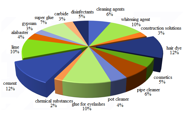 | Figure 1. Most common chemical substances causing burns |
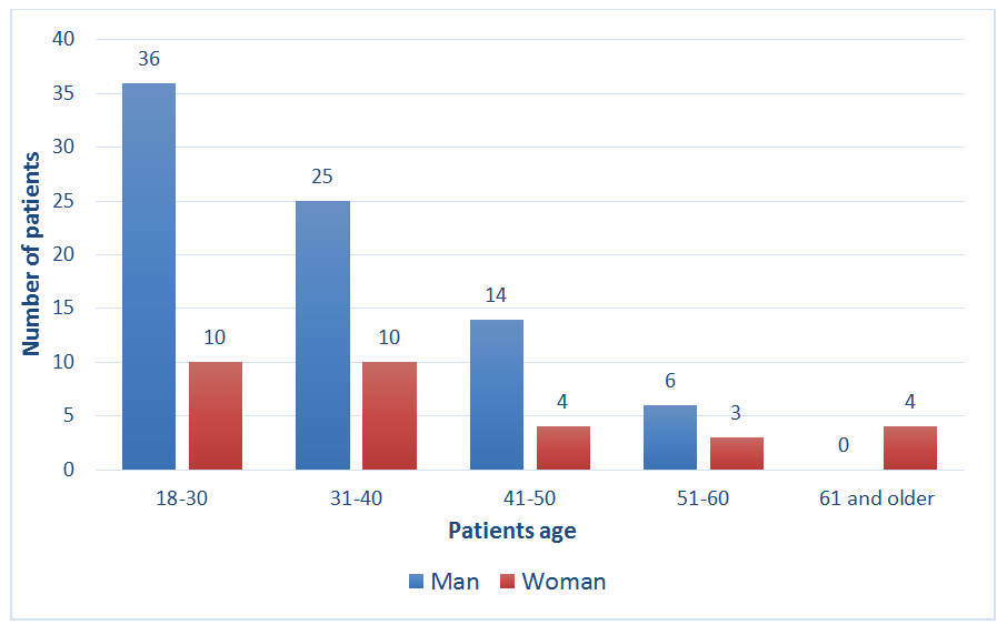 | Figure 2. Age distribution of patients participating in the study |
|

3. Methodology for Assessing Corneal Edema Using AS-OCT
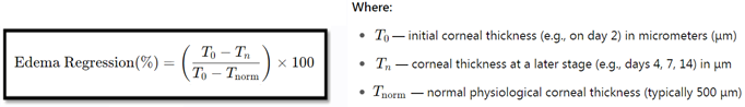
- One of the main consequences of corneal burns is epithelial and stromal edema. Optical coherence tomography (OCT) is a non-invasive diagnostic method that allows for accurate assessment of microstructural changes in corneal tissues [6]. This method is used to visualize and dynamically monitor corneal edema after burns. OCT technology is based on infrared light interferometry, capable of highly accurately displaying changes in the cornea and anterior segment (5 – 10 µm) [7].To monitor the corneal edema recovery process using OCT, the examination is repeated every 48 – 72 hours. To assess the effectiveness of the treatment strategy, we applied the following evaluation criteria:- Day 1: Cornea with significant edema, optical clarity of the stroma disturbed (OCT: 650 – 700 µm).- Day 3: Fluid decreased in the lower layers of the stroma, epithelial regeneration beginning (OCT: 550 – 600 µm).- Day 7: Epithelial and stromal thickness approaching normal, transparency recovering (OCT: 500 – 550 µm).- Day 14: Corneal transparency restored, edema almost absent (OCT: 480 – 520 µm, normal state).According to the analysis of initial (Day 2) indicators, the average corneal density in the main group was 67±6 GSU, and in the control group, it was 70±5 GSU. At this stage, the difference between the groups was not statistically significant (p>0.05). This confirms that the initial state of the patients was almost identical in both groups.By Day 4, corneal density in the main group significantly decreased to 43±5 GSU, while in the control group, this indicator was 54 ±4 GSU (p<0.05). During this period, an improvement in optical transparency was observed in patients of the main group, indicating the antioxidant and regenerative effects of the active substances in Keraton AC and Vita-POS ointments.According to the evaluation results on Day 7, corneal density in patients of the main group decreased to 29±4 GSU, while in the control group, the density remained at 39±5 GSU. At this stage, the difference between the two groups became even more pronounced (p<0.01). Clinically, a significant improvement in visual comfort and quality of vision was noted in patients of the main group.In the final analysis on Day 14, the average corneal density in patients of the main group decreased to 18±3 GSU, approaching normal. In the control group, this indicator remained higher, averaging 27±4 GSU. These results indicated that the difference between the groups was statistically highly significant (p<0.001).
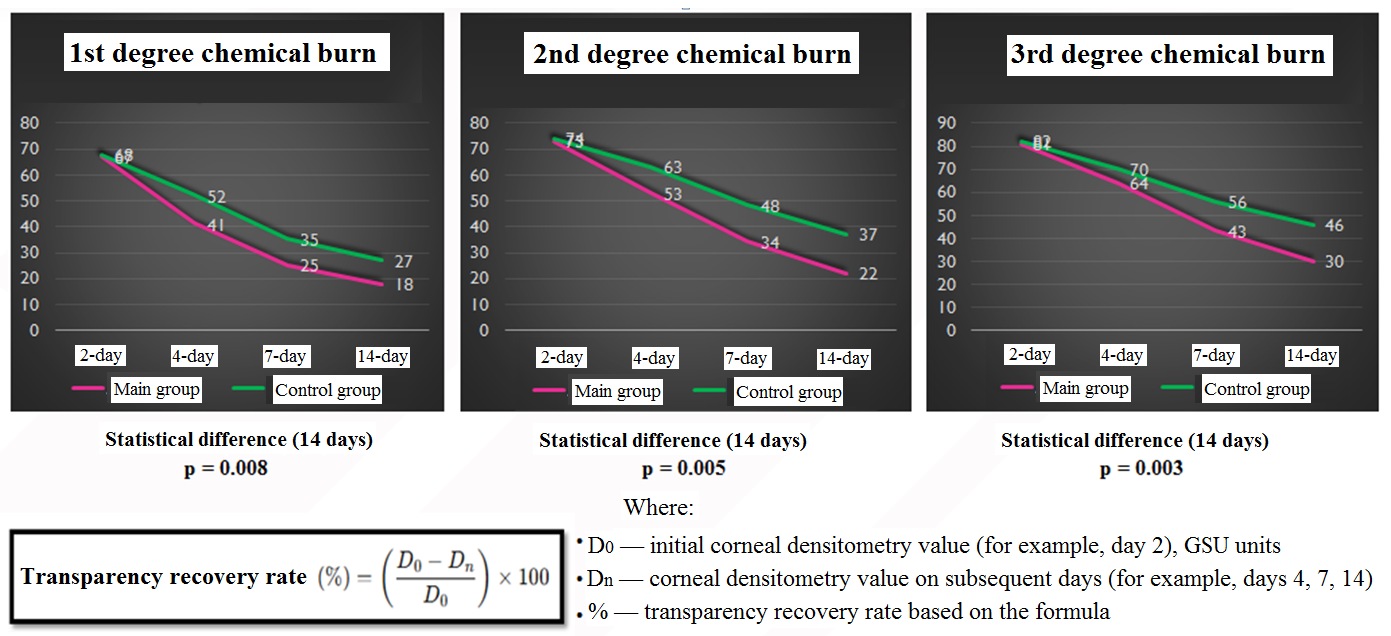 | Figure 3. Evaluation of the effectiveness of corneal transparency restoration from chemical burns (in the Corneal Densitometry module) |
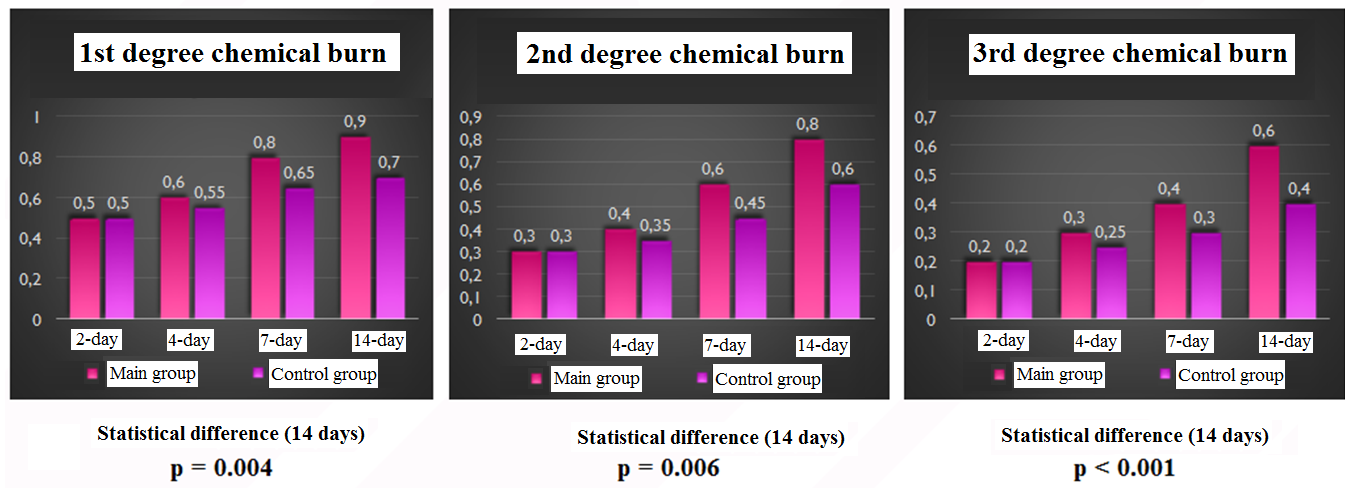 | Figure 4. Evaluation of the effectiveness results in restoring visual acuity |
4. Economic Efficiency of the Recommended Complex Treatment by Disease Severity
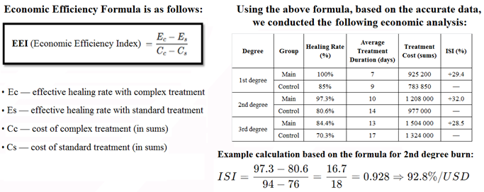 As a result of the analysis, we can conclude that although complex therapy is slightly more expensive in terms of initial costs, it increases overall economic efficiency due to a shorter treatment period, higher recovery rate, and more efficient use of hospital resources. Especially in II- and III-degree burns, the economic efficiency index (EEI) was over 90%, which allows recommending complex therapy as a non-invasive, yet cost-effective approach for the healthcare system.AS-OCT is a high-resolution non-contact diagnostic method through which inter-layer structural changes in the cornea can be assessed with high accuracy. In our study, Central Corneal Thickness (CCT) was measured and thoroughly analyzed in both groups (main and control) on days 2, 4, 7, and 14.On Day 2, the indicators in both groups were similar, and no statistically significant difference was recorded. This indicates that the initial conditions were comparable for mutual comparison.Starting from Day 4, corneal thickness in the main group decreased statistically significantly, and edema regression proceeded rapidly and stably.On Day 7 and especially on Day 14, edema significantly decreased in the main group, approaching normal indicators.
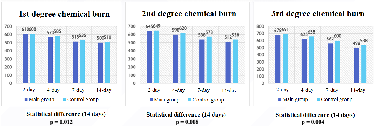 | Figure 5. Assessment of the effectiveness of corneal edema reduction using AS-OCT results |
5. Conclusions
- 1. Corneal regeneration and edema dynamics were accurately assessed using corneal densitometry and AS-OCT.2. The innovative treatment method accelerated corneal epithelization, reduced the degree of edema, and significantly improved transparency.3. Modern methods (Corneal densitometry, AS-OCT) play an important role in assessing corneal burns and are recommended for use in practical ophthalmology.