
-
Paper Information
- Previous Paper
- Paper Submission
-
Journal Information
- About This Journal
- Editorial Board
- Current Issue
- Archive
- Author Guidelines
- Contact Us
American Journal of Medicine and Medical Sciences
p-ISSN: 2165-901X e-ISSN: 2165-9036
2025; 15(8): 2621-2625
doi:10.5923/j.ajmms.20251508.43
Received: Jul. 16, 2025; Accepted: Aug. 3, 2025; Published: Aug. 7, 2025

Comparative Evaluation of Structural and Geometrical Parameters of the Heart in Patients with Mitral Valve Stenosis after Balloon Mitral Valvuloplasty Depending on the Type of Imaging
 Abstract
 Reference
 Full-Text PDF
 Full-text HTML
Zufarov M. M., Khamdamov S. K., Im V. M.
Republican Specialized Scientific and Practical Medical Center of Surgery named after Academician V. Vakhidov, Uzbekistan
Copyright © 2025 The Author(s). Published by Scientific & Academic Publishing.
This work is licensed under the Creative Commons Attribution International License (CC BY).
http://creativecommons.org/licenses/by/4.0/

Introduction. At present, echocardiography is an effective non-invasive method for assessing the degree of heart chamber remodeling and prognosis during balloon mitral valvuloplasty (BMV). Objective. The aim of our study is to analyze the structural and geometrical parameters of the heart in a comparative aspect with angiocardiographic examination during BMV in patients with mitral valve stenosis. Materials and Methods. The results of the examination of 87 patients with mitral stenosis are presented. The patients were divided into two groups: Group I consisted of 39 patients who underwent BMV under fluoroscopic guidance using the Inoue technique under local anesthesia; Group II included 48 patients who underwent BMV under echocardiographic guidance, with fluoroscopic control minimized and used mainly in technically difficult situations. Results. After BMV, both groups showed positive dynamics in the restoration of atrial geometry, which significantly lagged behind the recovery of the ventricles. In the postoperative period, the ventricles returned to normal values, whereas atrial parameters, despite positive remodeling dynamics, remained significantly above the normal range. As a result, the heart chamber ratio improved, but the index of chamber proportion did not return to normal. The remodeling index increased significantly, but its values remained reliably below the lower limit of normal. Conclusion. The above allows us to conclude that echocardiographic guidance during BMV in patients with mitral valve stenosis is a promising technique.
Keywords: Echocardiography, Mitral stenosis, Balloon mitral valvuloplasty, Cardiac chamber remodeling, Angiography
Cite this paper: Zufarov M. M., Khamdamov S. K., Im V. M., Comparative Evaluation of Structural and Geometrical Parameters of the Heart in Patients with Mitral Valve Stenosis after Balloon Mitral Valvuloplasty Depending on the Type of Imaging, American Journal of Medicine and Medical Sciences, Vol. 15 No. 8, 2025, pp. 2621-2625. doi: 10.5923/j.ajmms.20251508.43.
1. Introduction
- Structural and geometrical changes in the heart chambers include processes of hypertrophy and dilation, leading to impaired systolic and diastolic function. Pathological alterations in the structure and geometry of the heart chambers, referred to as “remodeling” of the heart in response to overload or loss of viable myocardium, often precede the clinical manifestation of heart failure. These changes can independently exacerbate cardiac dysfunction and negatively affect the quality of life and prognosis in diseases of the mitral valve of rheumatic origin.Modern non-invasive diagnostic methods make it possible to assess morphological, structural-geometrical, and hemodynamic parameters of cardiac function with high reliability. Among these modern methods, echocardiography stands out, providing high-quality imaging of the heart chambers and intracardiac hemodynamics [1-4].Currently, echocardiography (EchoCG) is the most commonly used and widely accessible method for diagnosing valvular heart disease. It is more cost-effective than magnetic resonance imaging and offers better assessment of the valvular apparatus. An important advantage is its non-invasive nature, in contrast to cardiac catheterization [4-7]. Among effective minimally invasive methods for treating mitral stenosis (MS) is balloon mitral valvuloplasty (BMV) using the Inoue technique [6,8-12]. This procedure is performed with the aid of catheters and specialized intravascular tools introduced via the femoral vessels, far from the target site. Visualization is achieved via fluoroscopy, which displays the instruments over the outline of the heart but does not provide a precise spatial representation of catheter position relative to cardiac chambers. Therefore, intervention effectiveness is assessed indirectly—mainly by measuring pressure in the heart chambers. During fluoroscopic imaging, the interatrial septum (IAS), particularly the region of the fossa ovalis, is not clearly visible. Anatomical changes caused by mitral stenosis and other concomitant conditions (e.g., other valvular defects, atrial fibrillation) further complicate orientation during the procedure. Echocardiography allows for precise identification of catheter position, instrument relationships, guidance during transseptal puncture, and placement of the balloon catheter and its dilation [9,11,13,14].The aim of our study is to assess the structural and geometrical parameters of the heart in a comparative context using echocardiographic versus angiocardiographic guidance during BMV in patients with mitral valve stenosis.
2. Materials and Methods
- This study presents the results of an examination of 87 patients with rheumatic mitral stenosis (MS) who underwent balloon mitral valvuloplasty (BMV) using the Inoue technique at the Department of Interventional Cardiology, Arrhythmology, and Endovascular Surgery of the Republican Specialized Scientific and Practical Medical Center of Surgery named after Academician V. Vakhidov.Patients were divided into two groups:• Group I included 39 patients who underwent BMV under fluoroscopic guidance using the Inoue technique under local anesthesia.• Group II included 48 patients who underwent BMV under echocardiographic guidance, with fluoroscopy minimized and used only in cases of technical difficulty.The initial stages of BMV involved catheterization of the right heart chambers and transseptal puncture of the interatrial septum. Right heart catheterization was performed for diagnostic purposes and did not differ from standard intracardiac catheterization techniques. However, the transseptal puncture had to be performed precisely at the fossa ovalis, as it served as the access point for surgical catheter intervention. Transseptal puncture was performed using a J. Endrys (USA) transseptal needle inserted into the catheter and advanced to the opening of the superior vena cava. It was then directed into the right atrium, with the proximal control flag of the needle rotated dorsally at 45° relative to the sagittal plane of the patient. This maneuver oriented the needle tip toward the left atrium. Continuing advancement of the catheter and needle allowed engagement with the fossa ovalis after sliding off the muscular ridge defining its superior edge. A quick, forward piercing motion enabled puncture of the interatrial septum, followed by a “give” into the left atrial cavity. The catheter was then advanced into a stable position within the left atrium.An ultrasound system (GE VIVID S70) was positioned to the patient’s left, at the head of the operating table. The ultrasound specialist stood next to the machine and manipulated the probe with the left hand, allowing repeated imaging without interfering with the surgeon, who stood on the patient’s right.Prior to intracardiac intervention, baseline intracardiac hemodynamic parameters were established. Determining the catheter position during transseptal puncture was a mandatory step. The surgeon and imaging specialist worked simultaneously. Standard parasternal imaging in the second to fourth intercostal spaces was not feasible during fluoroscopy, as the probe obstructed the field of view. Therefore, echocardiographic imaging was performed from the apical window in a four-chamber view. It was crucial to help the surgeon guide the needle tip to the fossa ovalis, adjusting maneuvers based on ultrasound imaging.Once the transseptal puncture was completed, the catheter position was confirmed. If the catheter could not be visualized from the apical window, the procedure was temporarily paused and imaging was switched to the standard parasternal position. Immediately afterward, the pericardial cavity was examined to exclude hemopericardium. If fluid was detected, serial assessments were performed every 3–5 minutes to monitor the rate of blood accumulation and signs of cardiac tamponade. Parameters such as chamber dimensions, interventricular septal motion, respiratory variations in atrioventricular flow, and ventricular wall motion were analyzed. Accumulation of ≥200 mL of blood or evidence of tamponade necessitated emergency percutaneous drainage. If no complications occurred, pericardial control was repeated after 15 minutes [9,13,14].In cases of difficulty advancing the catheter through the narrowed mitral orifice (MO), the EchoCG specialist assisted the surgeon by monitoring catheter position in real time. These assessments were also performed from the apical view to avoid interrupting the procedure [6,8,13].Mitral orifice dilation was performed gradually by stepwise balloon inflation. After each series of balloon inflations, the balloon was retracted into the left atrium, and transmitral blood flow was evaluated using Doppler echocardiography. When the peak transmitral pressure gradient decreased to 5–9 mmHg, the procedure was concluded. The mitral orifice area was calculated based on the pressure half-time using an apical view aligned with the transmitral flow. From this same view, the dynamics of mitral regurgitation were assessed. The emergence or worsening of regurgitation to grade 2 was considered an indication to stop the procedure. Final mitral orifice area measurements were performed in the short-axis view at the end of the procedure.The effectiveness of mitral valve dilation was confirmed by intraoperative echocardiographic findings: reduction of the transmitral pressure gradient, increase in the mitral orifice area, and absence of worsening mitral regurgitation [6,13,14].Throughout the procedure, both standard and specific echocardiographic parameters were analyzed to assess structural and geometrical remodeling using established remodeling index calculations.Statistical analysis was conducted using Microsoft Excel, calculating arithmetic means and standard errors. Statistical significance was determined using Student’s t-test, with a significance level of p<0.05 considered statistically meaningful. Differences were interpreted as significant at p<0.05 (95% confidence), p<0.01 (99% confidence), and p<0.001 (99.9% confidence).
3. Results and Discussion
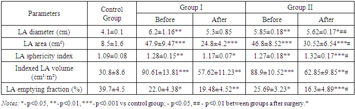
- To evaluate the outcomes of structural and geometrical heart parameters, patients were divided into two groups:• Group I: 39 patients who underwent BMV under fluoroscopic guidance.• Group II: 48 patients who underwent BMV under echocardiographic guidance, with minimal use of fluoroscopy.A control group of 37 healthy individuals was used for comparison.In addition to structural and geometrical parameters, we also performed a comparative analysis of radiation exposure received during BMV.The patient groups were comparable in terms of disease severity and duration.As shown in the presented data (Table 1), the left atrial volume (cm) before surgery was 6.2±1.16 in Group I and 5.85±0.18 in Group II. After surgery, the values decreased to 5.3±0.85 in Group I and 5.62±0.17 in Group II. In the control group, the left atrial volume was 8.5±1.6 (P<0.05). The left atrial area in Group I was 47.9±9.47, and in Group II, it was 46.8±8.52. After surgery, the values were 24.8±4.2 in Group I and 30.52±6.54 in Group II, compared to 8.5±1.6 in the control group (P<0.01). The left atrial pressure (mmHg) in Groups I and II before surgery was 1.28±0.15 and 1.27±0.18, respectively, while after surgery the values were 1.17±0.07 in Group I and 1.32±0.11 in Group II. The values of the indexed left atrial pressure (cm/m²) were 90.61±13.81 in Group I and 88.9±10.52 in Group II. After surgery, these values decreased to 57.62±11.23 in Group I and 62.85±9.85 in Group II, which were significantly higher (P<0.001 compared to the control group). The fraction of left atrial volume change (%) before surgery was 22.0±4.38 in Group I and 25.69±3.23 in Group II; after surgery, these values changed to 19.48±4.52 in Group I and 16.3±4.89 in Group II.
|
|
|
|

4. Conclusions
- 1. Structural and geometrical parameters of the heart, as assessed by echocardiographic examination, reflect the severity of the pathological process in mitral valve stenosis.2. The remodeling outcomes observed using traditional angiographic and echocardiographic assessments were found to be comparable.3. The degree of geometric recovery of the heart depends on its baseline structural state prior to the intervention, which in turn influences the improvement of intracardiac hemodynamics in the postoperative period.4. A comparative analysis of the two patient groups demonstrated that performing BMV under echocardiographic guidance significantly reduces patient radiation exposure.