
-
Paper Information
- Next Paper
- Previous Paper
- Paper Submission
-
Journal Information
- About This Journal
- Editorial Board
- Current Issue
- Archive
- Author Guidelines
- Contact Us
American Journal of Medicine and Medical Sciences
p-ISSN: 2165-901X e-ISSN: 2165-9036
2025; 15(8): 2607-2611
doi:10.5923/j.ajmms.20251508.40
Received: Jul. 14, 2025; Accepted: Aug. 3, 2025; Published: Aug. 7, 2025

Histological Characteristics of Gestational Pyelonephritis
 Abstract
 Reference
 Full-Text PDF
 Full-text HTML
Tojiboev Timur
Assistant, Department of Pathological Anatomy, Andijan State Medical Institute, Uzbekistan
Correspondence to: Tojiboev Timur, Assistant, Department of Pathological Anatomy, Andijan State Medical Institute, Uzbekistan.
| Email: |  |
Copyright © 2025 The Author(s). Published by Scientific & Academic Publishing.
This work is licensed under the Creative Commons Attribution International License (CC BY).
http://creativecommons.org/licenses/by/4.0/

Gestational pyelonephritis, which develops during pregnancy, is a common infectious-inflammatory disease of the upper urinary tract in obstetric practice. Its histological characteristics are of great importance for identifying the morphological features of inflammatory processes in renal tissue. This article analyzes the histological changes observed in gestational pyelonephritis, including interstitial lymphocytic and neutrophilic infiltration, epithelial cell damage, tissue dystrophy, and fibrosis. Additionally, immunohistochemical studies provide insights into the activity of immune cells and cytokines involved in the inflammatory response. Physiological immunomodulation and hormonal changes during pregnancy influence the pathogenetic features of pyelonephritis. Histological analysis serves as a crucial tool for understanding the clinical manifestations and complications of the disease. This study aims to improve diagnostic and therapeutic strategies for gestational pyelonephritis, offering valuable information for obstetricians, gynecologists, and nephrologists.
Keywords: Gestational pyelonephritis, Pregnancy, Histology, Inflammation, Immunohistochemistry, Renal tissue, Epithelial damage, Fibrosis
Cite this paper: Tojiboev Timur, Histological Characteristics of Gestational Pyelonephritis, American Journal of Medicine and Medical Sciences, Vol. 15 No. 8, 2025, pp. 2607-2611. doi: 10.5923/j.ajmms.20251508.40.
Article Outline
1. Introduction
- Pyelonephritis during pregnancy is a common upper urinary tract infections-inflammatory disease in obstetric practice, posing significant medical risks to both the mother and the fetus [1,2]. This condition develops in pregnant women due to physiological immunosuppression, hormonal changes, and mechanical alterations in the urinary tract, which complicate the process of infection spread and tissue damage [3,4,9]. The gestational form of pyelonephritis often presents with severe clinical manifestations and can lead to impaired renal function, sepsis, subsequent gestational complications including abnormal labor contractions, and neonatal problems [5,6].Physiological and immunological changes during pregnancy influence the pathogenetic mechanisms of the disease, resulting in various microscopic alterations in the kidney tissue during pyelonephritis, such as interstitial lymphocytic and neutrophilic infiltration, epithelial cell dystrophy, necrosis, and fibrosis [7,10,12]. Histological analysis plays a critical role in identifying these changes as it helps determine the stage, severity, and pathogenesis of the disease [11].Immunohistochemical methods allow for the detection of cytokines, chemokines, and immune cells involved in the inflammatory process, providing essential information for understanding the immunopathogenesis of the disease [9,12]. These insights are crucial for predicting clinical outcomes of gestational pyelonephritis and for developing effective treatment strategies.Moreover, histomorphological and immunohistochemical research findings offer obstetricians, gynecologists, and nephrologists the opportunity to develop new diagnostic and therapeutic approaches, which are vital for preserving the health of both the mother and the newborn [8].Therefore, studying the histological features of pyelonephritis developing during pregnancy remains a pressing scientific and clinical issue in modern medicine [1,2,4].
2. Purpose of the Research
- The purpose of this study is to investigate the histological and immunohistochemical characteristics of pyelonephritis developing during pregnancy. The research aims to deepen the understanding of the disease pathogenesis by identifying inflammatory processes and tissue changes in the renal parenchyma, thereby enabling the early prediction of clinical complications.Additionally, analyzing the activity of immune cells and cytokines involved in the inflammatory response through immunohistochemical methods is a key objective. These findings will provide a scientific basis for obstetricians and nephrologists to improve the diagnosis and develop more effective treatment strategies for gestational pyelonephritis.
3. Materials and Methods
- The subject of the study comprised kidney tissues from 40 pregnant women who died due to renal pathologies, examined at the Republican Center of Pathological Anatomy between 2020 and 2023.The research methods included the following.Morphological method:Kidney tissues were stained with hematoxylin-eosin and their microscopic structures were examined.Morphometric method:Quantitative analyses of the tissue components were performed which helped identify changes in the kidney tissue.Immunohistochemical method:The expression levels of CD34, p53, and BCL-2 markers in the kidney tissue were determined this method allowed the study of cell proliferation, apoptosis processes, and vascular characteristics within the tissue.
4. Results and Discussion
- In our study, the pathomorphology of gestational pyelonephritis developing at various stages of pregnancy was thoroughly investigated. According to the results, gestational pyelonephritis occurring in the first trimester is primarily associated with impaired urodynamics, leading to urine stasis in the urinary tract and renal pelvis. This was accompanied by the formation of desquamative inflammatory foci in the mucosal lining of the renal pelvis and calyces. Additionally, pyeloectasia, hyperplastic foci in the epithelium covering the renal pyramids, dystrophic and necrobiotic changes in the distal tubules, as well as neutrophilic infiltration were identified. These changes confirm the acute nature of the pathological process.Moreover, damage to the renal parenchyma was observed in first-trimester gestational pyelonephritis, specifically morphological alterations in the tubules and collecting ducts. These pathological changes manifested clinically as nephrotic syndrome and, from a morphological standpoint, indicated a high likelihood of progression to renal failure.Macroscopic analysis revealed that kidneys affected by gestational pyelonephritis were enlarged on average by 1.25 times compared to normal size. The primary route of infection was through the urinary tract, with hematogenous or lymphogenous spread occurring rarely. In 80% of cases, Escherichia coli was the main pathogen, normally part of intestinal flora, which can enter the urinary tract when hygiene is compromised, leading to infection. Consequently, immune function in the urinary tract is impaired, hormonal changes occur, and infections progress rapidly, especially under conditions such as diabetes mellitus.The infectious process began in the renal calyces and extended through the collecting ducts, calyces, and renal pelvis, with urine stasis causing kidney enlargement. Histological examination showed multifocal desquamation of the epithelial lining of the pelvis and calyces, congestion in blood vessels, hydropic dystrophy in distal tubules, massive hydropic and hyaline droplet degeneration in the epithelium of terminal collecting tubules, multifocal necrosis, and numerous hemorrhagic foci. Developed congestion in blood vessels and hyperplastic foci in the epithelium lining the pelvis and calyces were also confirmed (see figure 1 and 2).
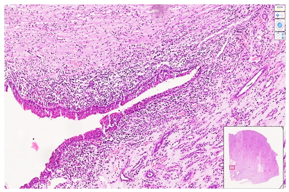 | Figure 1. Kidney tissue from a 38-week pregnant woman obtained at autopsy (first trimester pregnancy). Mucosal layer of the renal pelvis. Hyperplasia of the mucosal surface cells with foci of lymphocytic infiltration beneath the mucosa are observed. Staining: H&E. Magnification: 4x10 |
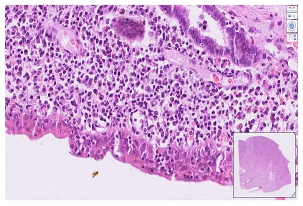 | Figure 2. Renal pelvis tissue from a 37-week pregnant woman obtained at autopsy (first trimester pregnancy). The mucosal layer consists of multilayered pseudostratified epithelium. Foci of lymphocytic infiltration are observed in the submucosal layer. Blood vessels are dilated with perivascular edema. Staining: H&E. Magnification: 20x10 |
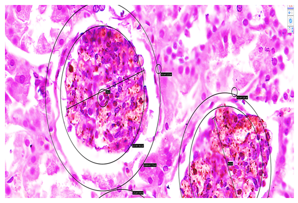 | Figure 3. Kidney glomerulus with Bowman’s space showing the epimetry and perimeter; the diameter is measured along the segmental diagonal. Scanned using NanoZoomer (REF C13140-21, S/N000198, HAMAMATSU PHOTONICS, Japan). Staining: H&E. Magnification: 20x10 |
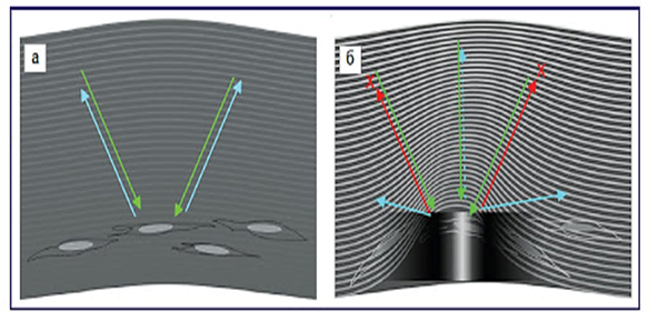 | Figure 4. To create a three-dimensional phase shape from a two-dimensional flat tissue, the flat surface is stretched vertically from a central point, generating intersecting surfaces perpendicular to the axes around it. This process enables the formation of confocal images, which are then used by specialized software to produce 3D morphogram images |
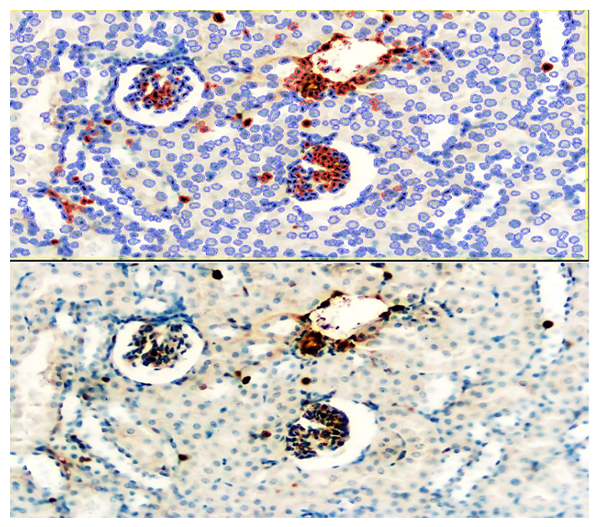 | Figure 5. Kidney tissue during first pregnancy. Moderate positive expression of the P53 marker observed in the glomerular capillary network and peritubular blood vessels of the renal tissue. Magnification: ×40. Scanned using QuPath-0.4.0.ink software. Positively expressed cells are shown in dark red. Staining: DAB chromogen and hematoxylin. Image size: 20×10 |
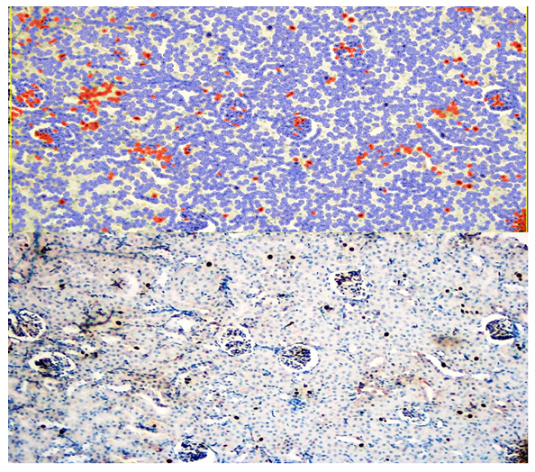 | Figure 6. Kidney tissue in the second pregnancy. Mild positive expression of the BcL-2 marker is observed in the glomerular capillaries and peritubular blood vessels of the kidney tissue. Scanned using QuPath-0.4.0.ink software. Positively expressed cells are marked in dark red. Staining: DAB chromogen with hematoxylin. Scale: 20x10 |
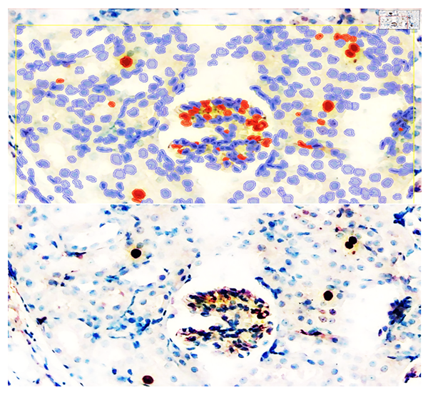 | Figure 7. Kidney tissue in the third pregnancy. Low positive expression of the P53 marker is observed in the glomerular capillaries and peritubular blood vessels of the kidney tissue. Scanned and expression level analyzed using QuPath-0.4.0.ink software. Positively expressed cells are marked in dark red. Staining: DAB chromogen with hematoxylin. Scale: 20x10 |
5. Conclusions
- The pathomorphological and immunohistochemical study of gestational pyelonephritis at different stages of pregnancy demonstrated that the most severe kidney damage occurs during the first trimester. In this period, significant disturbances in urodynamics, tissue injury, and degenerative changes were observed. Morphofunctional alterations, including epithelial hyperplasia, necrobiotic signs, and damage to the renal tubules and blood vessels, indicate a pronounced acute pathological process. The average kidney size increased by 1.25 times, accompanied by significant enlargement of glomeruli and vascular congestion, suggesting a high risk of renal failure.Immunohistochemical markers CD34, Ki-67, P53, and BcL2 revealed significant cellular damage and functional impairment of kidney tissue. Notably, the high expression of P53 in the first trimester indicated active hypoxia, apoptosis, and increased cell death, which may occur even in the absence of infection, contributing to the deterioration of renal function.In the second and third trimesters, morphological changes in the kidneys were predominantly atrophic and sclerotic, accompanied by a decrease in P53 expression. These findings reflect a morphological adaptation and a reduction in the acute phase of tissue injury. The low positive expression of P53 and the presence of chronic interstitial nephritis in the third trimester further confirm this trend.Overall, gestational pyelonephritis leads to the most pronounced renal damage in early pregnancy. Timely and appropriate treatment significantly improves the prognosis and increases the chances of maternal survival. Therefore, early diagnosis and effective therapeutic intervention are crucial in clinical practice to prevent severe complications associated with gestational pyelonephritis.