
-
Paper Information
- Next Paper
- Paper Submission
-
Journal Information
- About This Journal
- Editorial Board
- Current Issue
- Archive
- Author Guidelines
- Contact Us
American Journal of Medicine and Medical Sciences
p-ISSN: 2165-901X e-ISSN: 2165-9036
2025; 15(8): 2603-2606
doi:10.5923/j.ajmms.20251508.39
Received: Jun. 25, 2025; Accepted: Aug. 1, 2025; Published: Aug. 7, 2025

Method for Determining the Viability of Echinococcus Germ Cells in an in Vitro Experiment
 Abstract
 Reference
 Full-Text PDF
 Full-text HTML
Shamsiev Azamat Mukhitdinovich, Muxitdinov Sherzod Shavkatovich, Shamsiyev Jamshid Azamatovich, Suvonkulov Uktamjon Toirovich, Shamsiev Shokhzod Jamshidovich
Specialized Pediatric Surgical Clinic of Samarkand State Medical University, Samarkand, Uzbekistan
Correspondence to: Shamsiev Azamat Mukhitdinovich, Specialized Pediatric Surgical Clinic of Samarkand State Medical University, Samarkand, Uzbekistan.
| Email: |  |
Copyright © 2025 The Author(s). Published by Scientific & Academic Publishing.
This work is licensed under the Creative Commons Attribution International License (CC BY).
http://creativecommons.org/licenses/by/4.0/

Echinococcosis of the liver in children remains an urgent problem in endemic regions, including Uzbekistan, with a high incidence of postoperative recurrences. Prevention of recurrences requires effective intraoperative antiparasitic treatment of the residual cyst cavity. The article presents a new method of express diagnostics of echinococcal protoscolex viability based on the assessment of cuticular layer motility and morphostructure of parenchyma. The method allows reducing the diagnostic time to 1-2 min, increasing the safety of echinococcectomy and reducing the risk of recurrences.
Keywords: Echinococcosis, Children, Echinococcectomy, Protoscolexes, Antiparasitic treatment, Rapid diagnosis
Cite this paper: Shamsiev Azamat Mukhitdinovich, Muxitdinov Sherzod Shavkatovich, Shamsiyev Jamshid Azamatovich, Suvonkulov Uktamjon Toirovich, Shamsiev Shokhzod Jamshidovich, Method for Determining the Viability of Echinococcus Germ Cells in an in Vitro Experiment, American Journal of Medicine and Medical Sciences, Vol. 15 No. 8, 2025, pp. 2603-2606. doi: 10.5923/j.ajmms.20251508.39.
1. Relevance
- To date, the main treatment option for hepatic echinococcosis in children is organ-preserving echinocolectomy [1]. However, the frequency of postoperative recurrences, according to different authors, remains high 17 -41% [2,3]. In order to prevent recurrences of the disease during surgery, the residual cavity of the echinococcal cyst is treated with germicidal drugs, but the effectiveness of many of them has not been proven to date and is questionable [4]. To assess the effectiveness of antiparasitic treatment, it is important to determine the viability of echinococcal germ cells, which is associated with certain difficulties [5].In addition, existing methods for assessing the viability of protoscolexes often require considerable time, which increases the duration of surgery and may affect its outcome [6]. In endemic regions such as Uzbekistan, where echinococcosis remains a common disease, the development of a rapid and reliable method of intraoperative diagnosis is a priority to improve the effectiveness of surgical treatment and reduce the risk of recurrence [7].Purpose of the research: development of a fast and reliable method of intraoperative express-diagnostics of antiparasitic treatment efficiency of echinococcosis germinal elements.
2. Materials and Methods
- The material for in vitro studies were germinal elements of cystic echinococcosis obtained during surgery - echinococcectomy in children with hepatic echinococcosis who were hospitalized at the Specialized Children's Surgical Clinic of SamSMU.Earlier, a method for assessing the viability of protoscolexes during echinococcectomy in children was developed in the clinic (invention patent No. IDP 05334). The method is based on the assessment of the motor activity of echinococcal protoscolexes - the ability of protoscolexes to fold and evert [8]. When folding, protoscolexes acquire an oval shape; when eversion takes place, they acquire the shape of a fungus (Fig. 1). The essence of the technique: after treatment of the echinococcal cyst and fibrous capsule with a germicidal drug, a flush is taken for sampling. In order to activate the motor function of protoscolexes, the sample is placed in nutrient medium at pH 7.5, incubated in the thermostat for 10 minutes, then microscoped. In the absence of motor activity, reduction in size, deformation and brightening of the protoscolex parenchyma, the loss of their viability is diagnosed (Fig. 2).
 | Figure 1. Protoscolexes unerupted and erupted, alive. Before antiparasitic treatment. Zoom 10×10 |
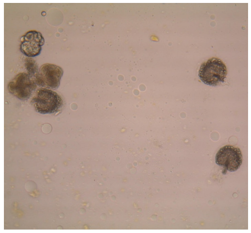 | Figure 2. Protoscolexes unerupted and erupted, dead. After anti-parasitic treatment. Zoom 10×10 |
 | Figure 3. Videophotomicroscopic study of the motor activity of the protoscolex. a-Folded protoscolex; b-Incomplete eversion; c-Full eversion. The duration of the process is 3-5 min |
 | Figure 4. Videophotomicroscopy: on a series of photographs (a, b, c, d) we tried to show the motor activity of the cuticular layer edge and protoscolex parenchyma in the form of insignificant “breathing” movements |
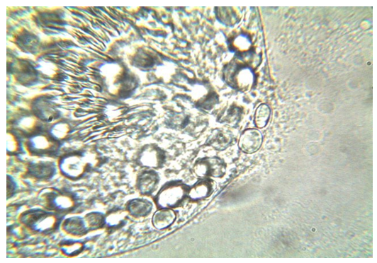 | Figure 5. Live protoscolex. Prior to antiparasitic treatment. Zoom 10×100. Preservation of clear structure of parenchyma is noted |
3. Results
- Complete loss of motility of cuticular layer and parenchyma of protoscolexes, destruction of structural elements and smoothing of parenchyma indicate irreversible destructive changes and death of germinal elements of the parasite (Fig. 6).
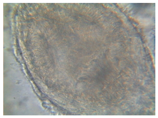 | Figure 6. Deceased protoscolex after antiparasitic treatment. Zoom 10×100. Complete absence of motor activity of cuticular layer and parenchyma, smoothness of parenchyma structure, margins of protoscolex are festooned |
4. Discussion
- Determination of the above features is simple and does not require a specially trained laboratory parasitologist. Comparison with the previous method based on the assessment of the ability of protoscolexes to curl and evert shows that the new method is significantly faster (1-2 minutes vs. 20-30 minutes) and does not require complex sample preparation [10]. This makes it more convenient for intraoperative application, which is especially important when time is limited during surgery.The effectiveness of antiparasitic treatment has a direct impact on reducing the risk of recurrence of echinococcosis, and our method can promptly confirm the death of protoscolexes, which increases the safety of surgery [11]. Unlike other methods such as staining of protoscolexes or the use of biochemical markers, our approach does not require additional reagents and sophisticated equipment, making it affordable to apply [12].
5. Conclusions
- Thus, the developed new method of determining the viability of Echinococcus protoscolexes after treatment of the echinococcal cyst and fibrous capsule cavity with an antiparasitic agent of contact action is reliable, fast and simple, which is important in echinococcosis surgery.Information about the source of support in the form of grants, equipment, and drugs. The authors did not receive financial support from manufacturers of medicines and medical equipment.Conflicts of interest. The authors have no conflicts of interest.