
-
Paper Information
- Previous Paper
- Paper Submission
-
Journal Information
- About This Journal
- Editorial Board
- Current Issue
- Archive
- Author Guidelines
- Contact Us
American Journal of Medicine and Medical Sciences
p-ISSN: 2165-901X e-ISSN: 2165-9036
2025; 15(8): 2517-2519
doi:10.5923/j.ajmms.20251508.20
Received: Jul. 6, 2025; Accepted: Jul. 29, 2025; Published: Aug. 4, 2025

Evaluation of IL-1β and IL-33 Levels Results in Children with Congenital Heart Defects
 Abstract
 Reference
 Full-Text PDF
 Full-text HTML
Usmanova D. D.1, Khaydarov K. I.2, Xamraeva Gulchekhra Shakhabovna2
1Tashkent Pediatric Medical Institute, Uzbekistan
2Center for the Development of Professional Qualifications of Medical Workers, Uzbekistan
Copyright © 2025 The Author(s). Published by Scientific & Academic Publishing.
This work is licensed under the Creative Commons Attribution International License (CC BY).
http://creativecommons.org/licenses/by/4.0/

Congenital heart disease (CHD) is the leading cause of birth defect-related morbidity and mortality, with 48.5% of cardiovascular deaths in children caused by CHD. Although simple CHD (e.g. ventricular septal defect) can be treated completely, complex CHD requires surgical intervention in early infancy and has a high mortality rate. The first year of life is critical for postnatal heart development. During this period, the structure and function of the infant heart change rapidly, with the heart rate declining from 150 beats per minute to 80 beats per minute and the systolic blood pressure rising from 55 mmHg to 100 mmHg. Almost 55.3% of CHD deaths occur in the first year of life. Although cardiomyocyte cell cycle activity is critical for heart development and damage repair, information on this process is limited. For example, data supporting that the average percentage of M-phase cardiomyocytes is 0.012 ± 0.003% is obtained from two samples only.
Keywords: Congenital heart defects, Cytokines, Interleukin-1β, Interleukin-33, Serum
Cite this paper: Usmanova D. D., Khaydarov K. I., Xamraeva Gulchekhra Shakhabovna, Evaluation of IL-1β and IL-33 Levels Results in Children with Congenital Heart Defects, American Journal of Medicine and Medical Sciences, Vol. 15 No. 8, 2025, pp. 2517-2519. doi: 10.5923/j.ajmms.20251508.20.
1. Introduction
- Interleukin-1β (IL-1β) is a key proinflammatory cytokine that plays a central role in the activation of the innate immune response, the development of systemic inflammation and tissue remodeling [2,7,9]. Its concentration in the blood serum often serves as an indicator of the presence and severity of the inflammatory process, including in cardiovascular diseases [1,4,10]. In children with congenital heart defects, the IL-1β level can act not only as an inflammation marker, but also as a prognostic indicator of the risk of complications in the perioperative period. Interleukin-33 (IL-33) is a member of the IL-1 cytokine family and functions as an alarmin, a signaling molecule released during cellular damage and playing a key role in triggering the immune and inflammatory response [3,6,10]. IL-33 is particularly active in the cardiovascular system, where it is expressed in endothelial cells and fibroblasts, participating in the formation of inflammation, myocardial remodeling and the development of heart failure [5,8].The aim of the study: determine the concentration of proinflammatory cytokines interleukin-1β (IL-1β) and interleukin-33 (IL-33) in blood serum in children with congenital heart defects.
2. Material and Methods of the Study
- Clinical studies were conducted in the cardiac surgery departments of the Tashkent Pediatric Medical Institute and the Era Med Multidisciplinary Medical Center from 2021 to 2024. The study involved 232 children, including 202 patients with a confirmed diagnosis of congenital heart disease. These patients were divided into two groups.The first group included 92 children (47.4%) with cyanotic forms of CHD, which were divided into 2 subgroups. Subgroup 1a consisted of 47 children who underwent CPB with classical ultrafiltration. Subgroup 1b consisted of 45 children who underwent CPB with modified ultrafiltration. The second group consisted of 110 children (39.7%) with acyanotic forms of CHD, which were also divided into 2 subgroups. Subgroup 2a consisted of 57 children who underwent CPB with classical ultrafiltration. Subgroup 2b consisted of 53 children who underwent CPB with modified ultrafiltration.The control group consisted of 30 (12.9%) practically healthy children.The criteria for exclusion from the study were: children with severe genetic diseases and stigmas of dysembryogenesis, the presence of infectious and inflammatory diseases.The diagnosis of congenital heart disease and the type of defect were established based on echocardiography data.Kits for quantitative determination of IL-1β and IL-33 by ELISA (Human IL-1β ELISA Kit, Human IL-33 ELISA Kit - manufactured by R&D Systems).
3. Research Results
- The study revealed a clear dependence of the IL-1β level on the presence and nature of congenital heart disease. In children in the control group who did not have cardiovascular pathology, the IL-1β level was 2.9±0.4 pg/ml, which corresponds to normal physiological values and reflects the absence of an active inflammatory process (Fig. 1).
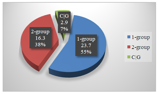 | Figure 1. Average levels of proinflammatory cytokine IL-1 (pg/ml) in children with CHD |
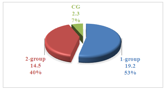 | Figure 2. Average levels of proinflammatory cytokine IL-33 (pg/ml) in children with CHD |
4. Conclusions
- Thus, the integrated assessment of these cytokines allows for more accurate risk stratification, monitoring of the child's condition dynamics and predicting the outcomes of surgical treatment of congenital heart disease. Inclusion of these indicators in clinical practice can significantly improve the effectiveness of diagnostics, prognosis and individualization of therapy.