
-
Paper Information
- Next Paper
- Previous Paper
- Paper Submission
-
Journal Information
- About This Journal
- Editorial Board
- Current Issue
- Archive
- Author Guidelines
- Contact Us
American Journal of Medicine and Medical Sciences
p-ISSN: 2165-901X e-ISSN: 2165-9036
2025; 15(8): 2513-2516
doi:10.5923/j.ajmms.20251508.19
Received: Jul. 10, 2025; Accepted: Jul. 30, 2025; Published: Aug. 4, 2025

Predictive Significance of Biochemical Markers in Secondary Restless Legs Syndrome
 Abstract
 Reference
 Full-Text PDF
 Full-text HTML
Raimova Malika Mukhamedjanovna, Mukhamedsaidova Iroda Abduvakhob kizi
Tashkent State Dental Institute, Tashkent, Uzbekistan
Copyright © 2025 The Author(s). Published by Scientific & Academic Publishing.
This work is licensed under the Creative Commons Attribution International License (CC BY).
http://creativecommons.org/licenses/by/4.0/

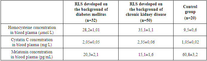
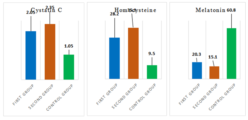
The article studies the clinical and biochemical indicators of the disease in 82 patients with secondary restless legs syndrome, which developed on the background of diabetes mellitus and chronic kidney disease. The biochemical indicators studied included homocysteine, cystatin C, and melatonin. The study's results showed that the severity and duration of the disease have a strong positive correlation with elevated levels of homocysteine and cystatin C. At the same time, there is a moderate negative correlation with decreased melatonin levels. Based on the obtained results, it was concluded that the investigated laboratory markers (homocysteine, cystatin C, and melatonin) can be used to identify the severity grades of secondary restless legs syndrome and to predict its course.
Keywords: Secondary restless legs syndrome, Homocysteine, Melatonin, Cystatin C
Cite this paper: Raimova Malika Mukhamedjanovna, Mukhamedsaidova Iroda Abduvakhob kizi, Predictive Significance of Biochemical Markers in Secondary Restless Legs Syndrome, American Journal of Medicine and Medical Sciences, Vol. 15 No. 8, 2025, pp. 2513-2516. doi: 10.5923/j.ajmms.20251508.19.
1. Introduction
- Restless Leg Syndrome (RLS) is one of the most common diseases of the nervous system, one of the main manifestations of which is circadian sensorimotor disorders. They are characterized by unpleasant sensations in the legs that occur during rest (often in the evening and at night). These sensations are relieved by movement, which often forces the person to move and frequently results in sleep disturbances. According to several researchers, RLS is the fourth most common cause of insomnia. In the International Classification of Diseases X, RLS is included in the section "other extrapyramidal and motor diseases," in the section "other specified extrapyramidal and motor diseases" (G 25.8). Additionally, RLS is also considered within the section of "sleep disorders" in the International Classification of sleep disorders. [2,4,7]Restless legs syndrome is considered a serious clinical problem. Restless legs syndrome is divided into primary (idiopathic) and secondary restless legs syndrome. Both occur with approximately the same frequency. Primary RLS develops without any identifiable cause and is considered an independent disease. According to various authors, a positive family history is observed in 25-75 percent of primary RLS, which indicates the role of the genetic factor. Primary RLS often has an early onset (up to 45 years) and symptoms usually persist throughout life, however, during life, their intensity can change significantly during stress, severe physical exertion, and pregnancy. In most cases, there is a tendency towards a gradual increase in symptoms over time, and periods of stabilization or remission (which can last up to several years) may occur. Secondary RLS typically arises after the age of 50 and occurs on the background of somatic or neurological disorders. The clinical features of secondary RLS progress relatively rapidly, and generally, periods of remission do not occur. The course of secondary RLS is determined by the progression of the underlying disease and the success of its treatment. Some researchers believe that the term “secondary” should not be used and propose the term “comorbid” RLS.Restless legs syndrome may arise from various illnesses and conditions. The most common etiological factors are iron deficiency anemia, pregnancy, renal failure, and polyneuropathies. [9,11,12] Among the factors in the development of RLS, iron deficiency is one of the most important. It is important to consider that iron deficiency may not necessarily present with clinically manifest anemia, and among the laboratory parameters, ferritin concentration serves as the most reliable indicator. [5,7]During pregnancy, RLS can develop more frequently, which is often associated with a decrease in iron and folic acid reserves. Epidemiological studies show that symptoms of RLS develop in 20-25% of pregnant women, and most often, the symptoms of this disease disappear in the third trimester of pregnancy. Furthermore, RLS was found to be more common in patients with uremia (15-70%). The pathogenesis of the development of this syndrome on the background of severe renal failure is not yet fully clear.Secondary RLS has various causes, including acquired conditions (such as alcoholic, diabetic, amyloid, porphyria, and others) as well as hereditary polyneuropathies. [1,8,9] For the early diagnosis of secondary RLS, it is important to determine the levels of biomarkers - melatonin, cystatin C, and homocysteine. Melatonin is the central regulator of the sleep-wake biorhythm, and its deficiency is associated with a deterioration in sleep quality. Cystatin C is a low molecular weight protein synthesized by all cells of the body and is used as an indicator of metabolic status, kidney function, and inflammatory processes. Cystatin C is considered a sensitive marker for assessing glomerular filtration and allows a comprehensive evaluation of the pathogenesis associated with chronic kidney disease. Cystatin C is also considered as an indicator reflecting systemic inflammation and possible involvement of other organs. High levels of homocysteine exacerbate oxidative stress and toxic effects on nerve tissue. Elevated homocysteine levels may be associated with restless legs syndrome, however, they do not represent the sole or primary etiological factor. Homocysteine is an amino acid whose increased concentration can contribute to a range of health complications, including neurological disorders. The elevation of homocysteine is frequently linked to deficiencies in vitamins B6, B12, and folic acid, which are essential cofactors in homocysteine metabolism. Deficiency of these vitamins can lead to the accumulation of homocysteine in the bloodstream, potentially exacerbating the clinical manifestations of RLS. [1,3,4] Sometimes RLS is the result of other diseases or conditions such as kidney disease, iron deficiency anemia, or neurological disorders. Elevated homocysteine can be one of the factors contributing to the development of secondary RLS. Purpose of the study: to study the clinical and biochemical features of secondary RLS, developed on the background of diabetes mellitus and chronic kidney diseases.
2. Materials and Methods
- A total of 82 patients diagnosed with RLS developed on the background of diabetes mellitus and chronic kidney disease were examined. Of these, 32 patients with BOS developed on the background of diabetes mellitus and 50 patients with RLS on the background of chronic kidney diseases. The age of the patients ranged from 35 to 58 years, and the average age was 45.4±7.8 years. Among the patients, 45 (54,8%) were women and 37 (45,2%) were men.Biochemical tests were performed on blood serum. The levels of homocysteine, cystatin C, and melatonin were determined in the patients and compared with the course and severity of the disease. For analysis, blood serum was taken on an empty stomach in the morning, performed by enzyme-linked immunosorbent assay (ELISA) using a semi-automated RAYTO RT2100C analyzer. During the analysis, a certified reagent kit from Elabscience was used, which was quantitatively optimized to detect specific antigens in blood serum. All studies were conducted in accordance with the manufacturer’s instructions. The results were recorded photometrically at a wavelength of 450 nm. Quantitative interpretation was carried out based on the calibration line: for cystatin C, the results were calculated in mg/l (milligrams/litre), for homocysteine in mmol/l (millimol/litre), for melatonin in pg/ml (picograms/millilitres). An increase in the level of cystatin C indicates inflammatory processes and a worsening of the general condition of the body.
3. Results
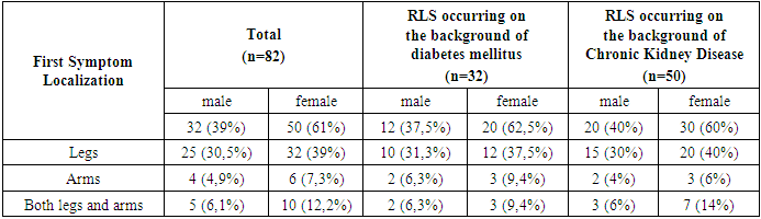
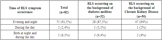
- During the anamnesis analysis, patients with RLS more frequently reported unpleasant sensations in the legs. Among patients with RLS on the background of diabetes mellitus, 22 out of 32 patients (68.8%) experienced such symptoms, while among those with RLS on the background of chronic kidney disease, 35 out of 50 patients (70%) reported similar complaints.
|
|
|
 | Figure 1 |