
-
Paper Information
- Next Paper
- Previous Paper
- Paper Submission
-
Journal Information
- About This Journal
- Editorial Board
- Current Issue
- Archive
- Author Guidelines
- Contact Us
American Journal of Medicine and Medical Sciences
p-ISSN: 2165-901X e-ISSN: 2165-9036
2025; 15(8): 2510-2512
doi:10.5923/j.ajmms.20251508.18
Received: Jun. 29, 2025; Accepted: Jul. 26, 2025; Published: Aug. 4, 2025

Criteria for Selecting Surgical Approaches for Perforated Gastroduodenal Ulcers Based on Laparoscopic and Video-Assisted Combined Techniques
 Abstract
 Reference
 Full-Text PDF
 Full-text HTML
Davronov Alisher Uktamovich, Kurbaniyazov Zafar Babajanovich
Samarkand State Medical University, Samarkand, Uzbekistan
Copyright © 2025 The Author(s). Published by Scientific & Academic Publishing.
This work is licensed under the Creative Commons Attribution International License (CC BY).
http://creativecommons.org/licenses/by/4.0/

This study is based on the surgical outcomes of 116 patients treated for perforated gastroduodenal ulcers. Closure of the ulcer using minimally invasive techniques, in combination with modern postoperative anti-ulcer therapy, resulted in favorable clinical outcomes and a relapse-free course of peptic ulcer disease in 91.6% of cases. Among patients who underwent laparoscopic or video-assisted combined closure, the Visick score revealed predominantly excellent (88.6% and 85%) and good (4.5% and 7.5%) outcomes, respectively. These findings underscore the effectiveness of mini-access ulcer closure in cases where laparoscopic repair is not feasible.
Keywords: Perforated gastroduodenal ulcer, Ulcer closure, Minimally invasive techniques
Cite this paper: Davronov Alisher Uktamovich, Kurbaniyazov Zafar Babajanovich, Criteria for Selecting Surgical Approaches for Perforated Gastroduodenal Ulcers Based on Laparoscopic and Video-Assisted Combined Techniques, American Journal of Medicine and Medical Sciences, Vol. 15 No. 8, 2025, pp. 2510-2512. doi: 10.5923/j.ajmms.20251508.18.
1. Introduction
- Perforated peptic ulcer remains one of the leading causes of mortality in patients with peptic ulcer disease, with fatality rates consistently ranging from 5% to 17% over the past decades. Surgical approaches to the management of perforated gastroduodenal ulcers vary significantly depending on the clinical context. The indications for surgery, as well as the choice of operative technique and method, are still a matter of ongoing debate.In Uzbekistan, there is a broad spectrum of surgical strategies employed in treating perforated gastroduodenal ulcers. The most frequently performed procedure is simple suture closure of the perforation, favored for its technical simplicity and reliability in preserving patient survival. More radical interventions, such as truncal vagotomy combined with pyloroplasty or distal gastrectomy, are reserved for selected cases and are often constrained by the time elapsed since perforation, the extent of peritonitis, and the overall condition of the patient. These advanced procedures are associated with higher rates of postoperative complications and mortality.Challenges arise when the perforation exceeds 2 cm in diameter or when the surrounding tissues are friable and inflamed, making primary closure technically difficult and potentially unsafe. In such cases, if the initial attempt at closure fails, a second attempt may also be ineffective. Under these circumstances, proceeding with gastric resection may offer a safer and more definitive solution.The introduction of innovative surgical technologies has expanded the treatment arsenal for perforated ulcers. One such advancement is the hybrid approach, which integrates endovideoscopic techniques for abdominal exploration and lavage with a mini-laparotomy to perform definitive closure of the perforation. These video-assisted procedures can achieve therapeutic outcomes comparable to traditional surgeries, while retaining the benefits of minimally invasive techniques. Nonetheless, clinical guidelines for patient selection and procedural indications remain to be clearly established.Objective of the Study. To enhance the clinical outcomes of surgical treatment in patients with perforated gastroduodenal ulcers by prioritizing the application of minimally invasive surgical technologies.
2. Materials and Methods
- The present study is based on the surgical treatment outcomes of 116 patients with perforated gastroduodenal ulcers who underwent emergency procedures at the Navoi branch of the Republican Scientific Center for Emergency Medical Care between 2019 and 2023. The age of the patients ranged from 18 to 67 years, with a mean age of 35.7 ± 15.4 years. The majority of patients (90%) were of working age. Males predominated in the cohort, comprising 83.4% of cases, while females accounted for 16.5%. All patients were admitted on an emergency basis.The comparison group included 61 patients (52.6%) who underwent laparoscopic suturing of the perforated ulcer. In the main group, which consisted of 55 patients, the ulcer closure was performed via a mini-access approach. Several intraoperative challenges limited the feasibility of proceeding with laparoscopic treatment in these patients.In 15 patients (27.2%), the ulcer was located in an anatomically unfavorable position for laparoscopic access, particularly in close proximity to the lesser curvature of the stomach. In 14 cases (25.4%), the diameter of the perforation exceeded 10 mm. In 8 patients (16.3%), dense inflammatory infiltration was observed in the perforated area, with a defect measuring more than 1.0 cm, necessitating wide sutures that partially incorporated the inflammatory mass, a task not achievable via laparoscopy.
3. Results and Discussion
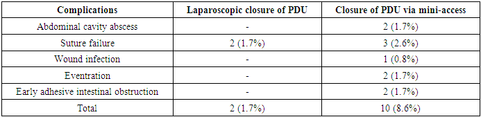
- The duration of laparoscopic closure for perforated gastroduodenal ulcers ranged from 40 to 150 minutes, with an average operative time of 62.8 ± 3.9 minutes. Within the first 24 hours after surgery, the severity of postoperative pain assessed using the Wong-Baker scale varied between 2 and 7 points, averaging 4.3 ± 0.5 points.The length of hospital stay in the control group ranged from 3 to 12 days, with a mean of 7.5 ± 0.3 days. At the early stage of adopting the mini-access video-assisted technique, the operation duration ranged between 70 and 110 minutes. However, with increased surgical experience, this time was reduced to 40–60 minutes. On average, the duration of ulcer closure performed via mini-access was 45.4 ± 3.2 minutes.In patients who underwent mini-access repair of the perforation, postoperative pain scores on the Wong-Baker scale ranged from 3 to 8, with a mean of 6.3 ± 0.7 points. In the early postoperative period, complications developed in 12 out of 116 patients (10.3%) who underwent minimally invasive ulcer repair.Intra-abdominal abscesses were identified in two patients (1.7%), all of whom were successfully treated via ultrasound-guided aspiration and drainage. Dehiscence of the sutured ulcer site occurred in five cases (4.3%): three patients (2.6%) from the mini-access group and two patients (1.7%) from the laparoscopic group. Patients from the main group underwent relaparotomy followed by gastric resection using the Billroth II method. In the control group, reoperation involved ulcer excision with Jaboulay pyloroplasty.Surgical site infections were observed in three patients (2.6%) in the main group and in one patient (0.8%) in the control group. Omental eventration through the trocar site occurred in two patients (1.7%) and was managed with intravenous sedation and surgical repair. All patients were discharged in stable condition with clinical improvement (see Table 1).
|
4. Conclusions
- 1. The choice of surgical technique for managing perforated gastroduodenal ulcers should be guided by the severity of the patient's clinical condition, the size of the perforation, and the extent of local inflammatory changes. Implementation of this diagnostic and therapeutic algorithm has enabled the avoidance of conversion from laparoscopic to open surgery.2. Laparoscopic closure was performed in 52.6% of patients and was deemed appropriate for ulcer perforations less than 1 cm in diameter and with surrounding inflammatory infiltration not exceeding 0.5 cm. Video-assisted combined procedures were used in 47.4% of patients as a minimally invasive alternative to wide laparotomy, particularly in cases with larger perforations (over 10 mm), significant inflammatory changes around the defect (with risk of suture tearing), difficult anatomical access (e.g., posterior gastric wall, low duodenal ulcers), or combined complications (such as simultaneous perforation with bleeding or stenosis).3. Minimally invasive repair of perforated gastroduodenal ulcers, when combined with modern postoperative anti-ulcer therapy, resulted in favorable clinical outcomes and a relapse-free course of peptic ulcer disease in 91.6% of cases. According to the Visick grading system, excellent and good long-term results were observed in the majority of patients who underwent laparoscopic or video-assisted mini-access closure (88.6% and 85% excellent outcomes; 4.5% and 7.5% good outcomes, respectively). These findings support the clinical value of mini-access ulcer closure in situations where laparoscopic intervention is not feasible.