Umidjon Donakuziyevich Usmonov1, 2
1PhD, Associate Professor, Department of Surgical Diseases, Andijan State Medical Institute, Uzbekistan
2Doctoral Researcher at the Military Medical Academy of the Armed Forces of the Republic of Uzbekistan, Tashkent, Uzbekistan
Correspondence to: Umidjon Donakuziyevich Usmonov, PhD, Associate Professor, Department of Surgical Diseases, Andijan State Medical Institute, Uzbekistan.
| Email: |  |
Copyright © 2025 The Author(s). Published by Scientific & Academic Publishing.
This work is licensed under the Creative Commons Attribution International License (CC BY).
http://creativecommons.org/licenses/by/4.0/

Abstract
The aim of this work was to conduct a comprehensive assessment of the clinical efficiency of a new method of primary pre-hospital care for non-gunshot wounds of soft tissues based on the multi-stage application of the developed antiseptic gel, with the analysis of the dynamics of pain syndrome, local inflammatory reaction and the frequency of postoperative complications.
Keywords:
Non-gunshot wounds of soft tissues, First aid, Antiseptic gel, Inflammatory reaction, Prevention of wound complications, Wound treatment
Cite this paper: Umidjon Donakuziyevich Usmonov, Analysis of the Clinical Effectiveness of a New Method for Primary Treatment of Non-Gunshot Soft Tissue Wounds in Pre-Medical Care, American Journal of Medicine and Medical Sciences, Vol. 15 No. 8, 2025, pp. 2479-2485. doi: 10.5923/j.ajmms.20251508.11.
1. Introduction
In contemporary armed conflicts, the primary causes of injuries are firearms, artillery shells, landmines, and explosive devices. However, despite the lower incidence of non-gunshot injuries, their nature and consequences remain highly significant. Among non-gunshot injuries, soft tissue damage represents one of the most prevalent types of trauma both in wartime and peacetime. These injuries include stab and cut wounds caused by knives or shrapnel, blunt trauma from impacts or falls, burns of varying severity, and others [1,9].In military conflicts, soft tissue injuries constitute a substantial proportion of combat-related trauma, with various studies reporting incidences of up to 60% of all cases. In modern military engagements, non-gunshot soft tissue injuries among service members typically result from road traffic accidents, falls from heights, collisions with military equipment, and other mechanical forces. These injuries may be either open or closed and can resemble gunshot wounds in their nature and severity. In peacetime, soft tissue injuries are also widespread: according to medical institution statistics, they account for up to 40% of visits to emergency departments, underscoring their significance for the healthcare system [2,4,16].The organization of surgical care for non-gunshot soft tissue injuries among military personnel at the initial stages of medical evacuation is a crucial aspect in ensuring timely treatment, preserving lives, and increasing the likelihood of full recovery. This process encompasses a range of organizational, surgical, and evacuation measures aimed at minimizing complications and preventing disability. Timely surgical care for non-gunshot soft tissue injuries during early medical evacuation stages is an essential component of the military medical support system. It reduces mortality and disability rates, expedites recovery, and facilitates the return of service members to duty. Achieving maximum efficiency requires the integration of modern medical technologies, strategic planning, and highly qualified personnel [5,6,11].Traditional methods of first aid, which primarily involve mechanical wound treatment and the use of standard antiseptic solutions, often do not provide adequate protection against infectious complications—particularly in cases of deep and extensive tissue damage—even when rapid transport to a medical facility is available.Therefore, the search for and development of new approaches and agents capable of minimizing the risk of wound infection and other complications remains highly relevant, both in civilian and military medicine (in both peacetime and wartime). Modern challenges necessitate the use of flexible, mobile solutions, adaptive technologies, and highly skilled personnel, making this field a priority in military medical practice.
2. Materials and Methods
Within the clinical phase of the study, the effectiveness of a novel method for providing initial pre-medical care for non-gunshot soft tissue injuries using an antiseptic gel was assessed.The main study group comprised 164 patients treated using the proposed method, which involved multistage application of the antiseptic gel: at the pre-medical care stage, during surgical wound debridement, and throughout postoperative dressing changes. Additionally, in a subgroup of military personnel, the main group was supported by specialized simulation-based training for junior medical staff, conducted according to field surgical care standards. The comparison group consisted of 291 patients who received care according to traditional protocols, without the use of the antiseptic gel or trained personnel.Comparative evaluation accounted for the homogeneity of included patients based on key criteria: type of injury (penetrating or non-penetrating), absence of bone damage, similarity in conditions of transport and surgical management, exclusion of complicated medical histories, and comparable severity and localization of injuries. This approach ensured the reliability of the outcomes in analyzing the efficacy of the proposed method.Additionally, a subgroup of 49 military personnel with non-penetrating injuries (23 in the comparison group and 26 in the main group) was included in the study. Within this subgroup, the microbiological profile of wounds, frequency of complications, and the effectiveness of the implemented simulation training program were analyzed separately. The training program focused on developing skills in initial surgical wound debridement, dressing application, gel administration, and the use of triage and microbiological vigilance protocols in field conditions. Thus, the main group demonstrated not only pharmacological but also organizational and tactical advantages in early infection control of the wound surface.The most common type of injury in both groups was stab-incised wounds, identified in 67 patients (40.9%) in the main group and in 125 patients (43.0%) in the comparison group. These were followed in frequency by incised wounds, recorded in 25.0% and 27.1% of patients, respectively. Puncture wounds were observed in 26 patients (15.9%) in the main group and in 36 (12.4%) in the comparison group. Lacerations occurred in 14.0% of the main group and 14.8% of the comparison group, indicating an almost identical frequency. Chop wounds, the rarest injury type, were registered in 7 patients (4.3%) in the main group and in 8 (2.7%) in the comparison group. Among civilian patients, who constituted the majority in both the main group (86.0%) and the comparison group (91.1%), injuries were primarily due to domestic, occupational, or road traffic accidents. In contrast, injuries among military personnel—accounting for 14.0% of the main group and 8.9% of the comparison group—were mainly sustained in field conditions during exercises, training, or tactical operations. Analysis of injury localization showed a predominance of upper limb injuries in both study groups: 57 patients (34.8%) in the main group and 98 (33.7%) in the comparison group. Lower limb injuries ranked second in frequency: 28.0% (n=46) in the main group and 29.9% (n=87) in the comparison group. Torso injuries were documented in 48 (29.3%) and 79 (27.1%) patients, respectively. Head and neck injuries were the least frequent: 13 patients (7.9%) in the main group and 27 (9.3%) in the comparison group.Thus, injury conditions among civilians were predominantly caused by peacetime trauma factors, while those among military personnel were associated with tactical field environments. These differences determine the specifics of microbial contamination, injury depth, and treatment outcomes, which must be considered when analyzing the clinical effectiveness of first aid methods.The analysis was conducted across the full sample—164 patients in the main group and 291 in the comparison group. The primary assessment focused on the dynamics of key indicators reflecting wound healing progression and quality. Upon admission, pain intensity, as measured by the Visual Analog Scale (VAS), was comparable between the groups: 4.23 ± 1.23 points in the main group and 4.10 ± 1.21 in the comparison group (p > 0.05). The analysis included the frequency of positive and sterile cultures at key stages: upon injury and during surgical intervention. Microbial wound contamination was monitored in stages, taking into account clinical dynamics.Statistical processing of study data and objective evaluation were performed using parametric and non-parametric methods depending on data type and distribution [3,7,8,10,12–14,17–19]. For quantitative variables, the mean, standard deviation (SD), and sample size (n) were calculated. Prior to comparing quantitative data between groups, distribution characteristics were assessed. The choice of statistical tests was justified by evaluating conformity to normal distribution. While the Shapiro–Wilk test could be used for small samples, independent sample analysis in this study employed the Student's t-test. For comparing mean values between two independent groups (main and control), Student's t-test for independent samples was applied (with Welch’s correction if variance equality was not assumed). Results are presented as mean ± SD with p-values, where p < 0.05 was considered statistically significant. For categorical data, Pearson’s chi-square test was used to compare proportions. When necessary, for 2×2 tables, degree of freedom (df=1) corrections were applied. For multicomponent comparisons, chi-square tests with df >1 were used. For visual representation, bar charts were employed with mean and SD indicators, significance annotations (p-values, χ², df) above the graphs, internal labels showing values within the bars, and color differentiation of main and control groups for clarity.
3. Results and Discussion
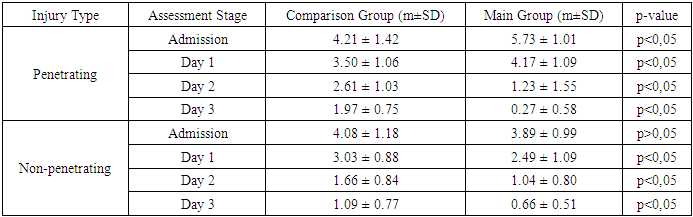
On the first day following inpatient medical care, a statistically significant difference in pain intensity between the groups was observed. In the main group, the mean pain score was 2.79 ± 1.27 points, whereas in the comparison group it was 3.09 ± 0.91 points (p < 0.01). This difference was attributed to the specific effects of the antiseptic gel used, which exhibited not only antimicrobial properties but also a pronounced local analgesic effect due to its active components, thereby contributing to a rapid reduction in pain intensity. This effect was further enhanced when the gel was applied during dressing changes after surgical intervention.By the third day, pain intensity in the main group was significantly lower than in the comparison group (1.07 ± 0.98 points versus 1.79 ± 0.92 points, respectively; p < 0.001). Although pain levels decreased in both groups, the reduction was more pronounced in the main group, likely due to the gel's efficacy in suppressing inflammatory responses and accelerating regenerative processes in the wound. This trend persisted on the fifth day, with pain intensity remaining markedly lower in the main group (0.59 ± 0.54 points) compared to the comparison group (1.20 ± 0.82 points; p < 0.001). The sustained reduction in pain throughout the observation period supports the pronounced and prolonged effectiveness of the new first aid method, which fosters favorable conditions for rapid wound healing and tissue recovery.Analysis of the presence of pain over time using the chi-square (χ²) test revealed no significant changes in patient distribution regarding the presence or absence of pain from day 1 to day 3 (χ² = 1.069; p = 0.301). Despite the continued gradual decrease in average pain intensity, the proportion of patients experiencing any level of pain remained relatively stable. However, in the final observation phase (from day 3 to day 5), significant and statistically meaningful changes were detected (χ² = 140.494; p < 0.001). The proportion of patients who reported complete pain relief increased substantially, confirming the high efficacy of the new method, which is attributable to the prolonged analgesic effect of the gel, facilitated by its multicomponent formulation and potent anti-inflammatory properties.The analysis of pain dynamics according to the Visual Analog Scale (VAS), stratified by injury type—penetrating and non-penetrating—yielded the results presented in Table 1. Table 1. Pain Dynamics According to the Visual Analog Scale (VAS) by Type of Injury
 |
| |
|
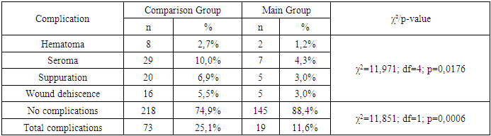
On the first day of observation, no statistically significant differences between the groups were found in relation to the type of injury (p > 0.05). However, by the third day of observation, a significant improvement in the condition of patients in the main group was noted for both penetrating and non-penetrating injuries, with statistically significant differences identified between the main and comparison groups (p < 0.05). By the fifth day, these differences became even more pronounced and statistically significant (p < 0.05) for both injury types. The main group exhibited a marked reduction in pain intensity to nearly minimal levels, indicating the high effectiveness and sustained impact of the proposed method.Thus, the new method of providing first aid for soft tissue injuries demonstrates significant efficacy in pain reduction, particularly from the third day of observation, for both penetrating and non-penetrating wounds.The dynamics of wound edema were then analyzed. At admission, the distribution of patients by the degree of local edema severity was comparable between the main and control groups. In the main group, 15.2% of patients showed no edema, mild edema was observed in 47.6%, and moderate edema in 37.2%. In the comparison group, the corresponding figures were 11.7%, 42.3%, and 46.0%, respectively. No severe edema was observed in either group. There were no statistically significant differences between the groups at this stage (χ² = 3.608; df = 2; p = 0.1646; p > 0.05), indicating an initially comparable clinical status of the patients.Already by the first day of observation, statistically significant differences between the groups were recorded (χ² = 25.093; df = 3; p = 0.000015; p < 0.001). In the main group, 39.0% of patients had no edema, compared to only 19.9% in the comparison group. The proportion of patients with moderate and severe edema was significantly lower in the main group (11.6% and 7.9%, respectively) than in the comparison group (24.7% and 12.0%). On the third day, this trend persisted: 36.0% of patients in the main group had no edema, compared to 25.4% in the comparison group. Moderate and severe edema were observed in 20.7% and 4.9%, respectively, in the main group, and in 10.7% and 9.6% in the comparison group. The differences remained statistically significant (χ² = 19.879; df = 3; p = 0.0002; p < 0.001).By the fifth day, positive edema dynamics in the main group were sustained: complete absence of edema was recorded in 34.1% of patients versus 22.0% in the comparison group. The proportion of severe edema in the main group decreased to 2.4%, compared to 5.2% in the control group (χ² = 10.741; df = 3; p = 0.0132; p < 0.05). By the seventh day of observation, the differences between the groups had leveled out. Both groups showed a significant reduction in edema severity: 81.7% of patients in the main group and 77.7% in the comparison group had no edema. The proportion of severe edema was less than 1% in the main group and 1.7% in the comparison group. At this stage, the differences were not statistically significant (χ² = 1.731; df = 3; p = 0.6301; p > 0.05).In general, the analysis of wound edema dynamics revealed that on the first day after surgical intervention, edema persisted in 80.1% of patients in the comparison group and in only 61.0% of patients in the main group. Meanwhile, 39.0% of patients in the main group exhibited no edema, compared to 19.9% in the comparison group. These differences were statistically significant (χ² = 19.485; df = 1; p = 0.00001; p < 0.001), indicating a strong positive effect of the proposed method in the early reduction of edema. By the third day, the proportion of patients without edema increased to 36.0% in the main group, compared to 25.4% in the comparison group (χ² = 5.639; df = 1; p < 0.05). On the fifth day, edema was absent in 34.1% of patients in the main group and in only 22.0% in the comparison group (χ² = 7.978; df = 1; p = 0.0047; p < 0.01). Thus, the gel’s effect manifested early and persisted throughout the acute phase. By the seventh day, intergroup differences leveled out: 81.7% of patients in the main group and 77.7% in the comparison group showed no signs of edema, with no statistically significant differences observed at this stage (χ² = 1.038; df = 1; p = 0.3082).The next evaluated criterion was the degree of wound hyperemia. At admission, hyperemia status was comparable between groups: it was absent in 87 (53.0%) patients in the main group and 165 (56.7%) in the comparison group. Mild and moderate hyperemia were observed in 33.5% and 13.4% of the main group, versus 31.6% and 11.7% in the control group. No statistically significant differences were identified (χ² = 0.628; df = 2; p = 0.7306; p > 0.05).By postoperative day 1, the main group showed a marked reduction in hyperemia severity: 39 (23.8%) patients had no hyperemia, compared to only 23 (7.9%) in the control group. The number of patients with pronounced hyperemia was only 9 (5.5%) in the main group and 39 (13.4%) in the control group. The differences were statistically significant (χ² = 26.681; df = 3; p < 0.001). On day 3, hyperemia was absent in 86 (52.4%) patients in the main group compared to 71 (24.4%) in the control group, again with significant differences (χ² = 37.208; df = 3; p < 0.001). Moderate and severe hyperemia were less frequent in the main group (12.8% and 3.0%) than in the comparison group (24.4% and 5.2%).On day 5, the positive trend persisted: hyperemia was absent in 114 (69.5%) patients in the main group, compared to 129 (44.3%) in the control group (χ² = 27.563; df = 3; p < 0.001). The main group had fewer cases of mild, moderate, and severe hyperemia. By day 7, the differences between the groups diminished: hyperemia was absent in 138 (84.1%) patients in the main group and 215 (73.9%) in the control group (χ² = 7.212; df = 3; p > 0.05). Severe hyperemia was rare—1 case (0.6%) in the main group and 4 cases (1.4%) in the comparison group.When considering the overall criterion of hyperemia presence or absence, no differences were observed at admission: hyperemia was absent in 87 (53.0%) patients in the main group and 165 (56.7%) in the comparison group. By day 1, the main group demonstrated a statistically significant advantage: 39 (23.8%) patients showed no hyperemia compared to 23 (7.9%) in the comparison group (χ² = 22.464; p < 0.001), indicating an earlier anti-inflammatory effect of the method. On day 3, this trend intensified—86 (52.4%) patients in the main group had no hyperemia compared to 71 (24.4%) in the control group (χ² = 36.492; p < 0.001). By day 5, hyperemia persisted in only 50 (30.5%) patients in the main group compared to 162 (55.7%) in the control group (χ² = 26.73; p < 0.001), while 114 (69.5%) patients in the main group and 129 (44.3%) in the control group exhibited no hyperemia. Even by day 7, the differences remained statistically significant: hyperemia was absent in 138 (84.1%) patients in the main group and 215 (73.9%) in the comparison group (χ² = 6.352; p = 0.0117; p < 0.05).The observed dynamics in pain severity, edema, and wound hyperemia clearly demonstrate the anti-inflammatory and antiseptic potential of the proposed first aid method. The accelerated resolution of the local inflammatory response in the early post-traumatic period creates favorable conditions for subsequent wound healing progression. A logical continuation of this analysis is the assessment of the frequency and structure of wound complications as a final clinical outcome indicator of the method’s effectiveness.Analysis of wound complication rates and structure revealed significant differences between the main and comparison groups, as visually represented in the charts. The overall complication rate in the main group was 19 cases (11.6%) versus 73 cases (25.1%) in the comparison group (χ² = 11.851; p < 0.001), indicating nearly a twofold reduction in risk when the proposed first aid method was employed. This advantage is attributed to the multistage application of the combined antiseptic gel, which was used from the initial point of injury care.The application of the gel on the wound surface and its pressurized introduction into deep wound tracts immediately after injury provided early antiseptic action and reduced bacterial contamination of tissues even before hospital admission. This created a local protective barrier that prevented wound infection during the critical early hours. Additionally, the gel was applied after surgical wound debridement—prior to closure of the subcutaneous tissue and skin—which contributed to the suppression of residual microflora and reduced the risk of secondary infection. The gel was also used during dressing changes in the early postoperative period, enhancing its antimicrobial effect and ensuring prolonged wound protection.The most common complication observed in both groups was seroma: 29 cases (10.0%) in the comparison group versus only 7 cases (4.3%) in the main group. The incidence of suppuration was also lower with gel use—5 cases (3.0%) versus 20 cases (6.9%). Wound dehiscence occurred in 5 patients (3.0%) in the main group and in 16 patients (5.5%) in the comparison group. Hematomas were recorded in 2 patients (1.2%) in the main group and 8 patients (2.7%) in the comparison group (χ² = 11.971; df = 4; p = 0.0176) (Table 2).Table 2. Frequency and Structure of Wound Complications
 |
| |
|
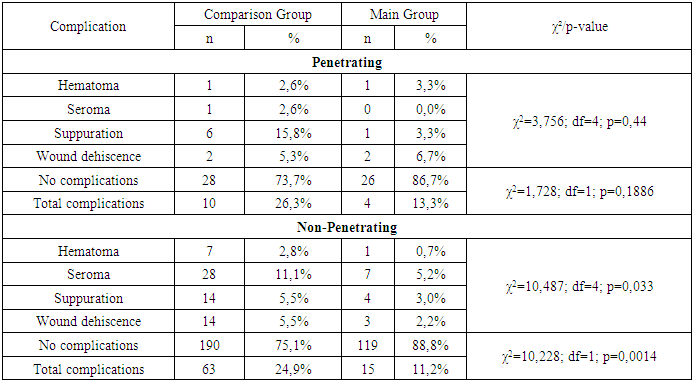
At the same time, the proportion of patients without complications in the main group was 145 (88.4%), significantly higher than 218 (74.9%) in the comparison group. This outcome confirms the high clinical efficacy of the proposed method, achieved through early antiseptic action of the gel, its application both before and after surgical wound debridement, and during dressing changes, thereby ensuring sustained suppression of inflammation and protection against microbial contamination.In cases of penetrating injuries, the overall complication rate in the comparison group was 26.3% (10 out of 38), compared to 13.3% (4 out of 30) in the main group; however, the difference was not statistically significant (χ² = 1.728; df = 1; p = 0.1886). Within the complication structure, isolated cases of hematomas were registered in both groups (2.6% and 3.3% respectively), seromas occurred only in the comparison group (2.6%), suppuration rates were 15.8% versus 3.3%, and wound dehiscence was noted in 5.3% and 6.7% of cases respectively. Despite the visible reduction in complication rates with gel use, statistical significance was not achieved (χ² = 3.756; p = 0.44), likely due to the limited sample size in these subgroups.For non-penetrating injuries, differences were more pronounced: complications were observed in 63 of 253 patients in the comparison group (24.9%) and in 15 of 134 patients in the main group (11.2%), which was statistically significant (χ² = 10.228; df = 1; p = 0.0014). The structure of complications included: seromas (11.1% vs. 5.2%), suppuration (5.5% vs. 3.0%), wound dehiscence (5.5% vs. 2.2%), and hematomas (2.8% vs. 0.7%). The structural differences were also statistically significant (χ² = 10.487; df = 4; p = 0.033), indicating the pronounced prophylactic effectiveness of the proposed method in cases of extensive soft tissue trauma (Table 3).Table 3. Frequency and Structure of Complications in Penetrating Injuries
 |
| |
|
Significant differences were also identified between the groups regarding the need for repeat surgical interventions. In the second group (which used the proposed method), secondary suturing was required in only 8 out of 164 cases (4.9%), whereas in the comparison group such interventions were performed in 34 out of 291 patients (11.7%) (χ² = 5.798; df = 1; p = 0.016).Analysis of hospitalization duration (among hospitalized patients) demonstrated that the use of the proposed first aid method significantly reduced inpatient treatment times. Overall, the average length of hospital stay was 5.9 ± 2.3 days in the comparison group (227 patients) and 4.6 ± 1.9 days in the main group (122 patients) (p < 0.001), reflecting an overall acceleration of the wound healing process under the influence of the proposed method.The most pronounced differences were noted in patients with penetrating wounds, where the average duration of hospitalization decreased from 9.0 ± 3.5 days in the comparison group (38 patients) to 6.8 ± 2.5 days in the main group (30 patients) (p < 0.001). This reduction of more than two days may be attributed to the prevention and early suppression of inflammation through deep antiseptic treatment of tissues and complication prevention due to the use of the gel starting from the pre-hospital phase.In non-penetrating injuries, a significant reduction in hospitalization time was also observed: from 5.3 ± 1.9 days in the comparison group (189 patients) to 3.9 ± 1.3 days in the main group (92 patients) (p < 0.001). This effect is primarily associated with the reduction of edema and hyperemia, faster healing, and decreased need for repeat interventions due to the multi-phase antiseptic approach.In all presented categories, the main group showed significantly better outcomes than the comparison group (p < 0.001 for all parameters). For instance, the number of return visits for dressing changes was significantly lower with the proposed method—2.5 ± 1.0 versus 3.5 ± 1.6—indicating a reduced need for additional wound management and more stable wound conditions. The time to initial healing was 7.3 ± 1.8 days in the main group compared to 8.6 ± 2.3 days in the comparison group, while the time to complete epithelialization was 11.6 ± 2.9 days versus 14.1 ± 3.0 days, respectively.Effectiveness of the Primary Treatment Method for Non-Gunshot Soft Tissue Injuries in Military Personnel. The initial stage of analysis revealed no statistically significant difference in microbial growth frequency between the main and comparison groups at the time of injury. In the comparison group, microbial growth was detected in 17 out of 23 patients (73.9%), and in the main group, in 21 out of 26 patients (80.8%) (χ² = 0.33; p = 0.5659). This can be attributed to the inherently high level of contamination in field-acquired injuries among military personnel in both groups. However, the situation changed markedly at the hospital stage (during surgical intervention). At that point, the proportion of sterile cultures in the main group was 57.7% (15 out of 26 patients), whereas in the comparison group it was only 13.0% (3 out of 23) (χ² = 10.468; p = 0.0012), indicating a statistically significant difference between the groups. Despite the initially comparable microbial status, by the time of surgery, those military personnel who had received treatment involving the antiseptic gel and algorithm-based assistance showed a 2.5 to 3-fold reduction in microbial load.These findings underscore the effectiveness not only of the method itself (the use of an antiseptic gel), but also of the organizational component—the implementation of simulation-based training among junior military medical personnel. In field trauma settings, where the risk of environmental contamination is extremely high, the proposed approach not only compensates for these conditions but significantly improves the wound’s microbiological profile by the time of surgery. This provides a foundation for reducing complications and improving outcomes in military personnel operating in combat or training environments. Analysis of the frequency and structure of postoperative complications in military personnel with non-penetrating soft tissue injuries showed that in the main group—where the proposed antiseptic treatment method and simulation training program were applied—complication rates were significantly lower compared to the comparison group.In the comparison group, complications developed in 11 out of 23 military personnel (47.8%). Among these, seromas accounted for 17.4%, suppuration for 13.0%, wound dehiscence for 13.0%, and hematomas for 4.3%. In the main group, where an enhanced primary care system was implemented, complications were registered in only 4 out of 26 patients (15.4%). These included seromas in 7.7%, with isolated cases of suppuration and wound dehiscence (3.8% each).Although the overall difference in the structure of complications between the groups did not reach statistical significance (χ² = 6.448; df = 4; p = 0.1681), the difference in the overall complication rate was statistically significant (χ² = 6.047; df = 1; p = 0.0139). The proportion of patients without complications in the main group was 84.6% (22 out of 26), compared to only 52.2% (12 out of 23) in the comparison group.These data indicate that the proposed approach—combining the use of an antiseptic gel with the training of junior personnel in the military medical service—not only reduces microbial contamination but also significantly decreases the risk of clinically relevant complications in military personnel under field care conditions.
4. Conclusions
According to data from the Visual Analog Scale (VAS), as early as the first day after care, pain intensity in the main group was significantly lower compared to the comparison group (2.79 ± 1.27 points vs. 3.09 ± 0.91 points; p < 0.01). This difference became more pronounced by day 3 (1.07 ± 0.98 vs. 1.79 ± 0.92; p < 0.001) and persisted through day 5 (0.59 ± 0.54 vs. 1.20 ± 0.82; p < 0.001). By day 7, values in both groups were comparable; however, the application of the gel provided a significant advantage in pain management during the critical initial 3–5 days.The proposed method, involving multistage application of an antiseptic gel (at the point of first aid, post-surgical wound management, and during dressing changes), leads to a marked reduction in the risk of postoperative complications—especially in cases of non-penetrating soft tissue injuries. This confirms its effectiveness in preventing microbial contamination, controlling inflammation, and maintaining favorable healing conditions.Comprehensive use of the gel at all treatment stages—from pre-medical first aid to postoperative care—ensures a substantial reduction in early complications by suppressing local inflammation, preventing infection, stabilizing wound edges, and significantly shortening treatment duration and accelerating healing. This is particularly crucial in environments with limited access to specialized medical assistance. The obtained data affirm that the proposed approach—combining the antiseptic gel with the training of junior military medical personnel—effectively reduces microbial contamination and significantly lowers the risk of clinically significant complications in military personnel under field care conditions. In military personnel with non-penetrating injuries included in the main group, the use of the proposed first aid method in combination with a simulation training program for junior military medical staff resulted in a significantly lower level of wound microbial contamination by the time of surgery (57.7% sterile cultures vs. 13.0% in the comparison group; χ² = 10.468; p = 0.0012), as well as a lower incidence of postoperative complications (15.4% vs. 47.8%; χ² = 6.047; p = 0.0139). These results confirm that the proposed approach ensures comprehensive effectiveness at both the microbiological control level and in clinical outcomes—especially critical in field medicine scenarios with limited time for intervention.
References
| [1] | Babajanov A.Kh., Sadikov R.A., Usmonov U.D. Current state of the problem of gunshot and non-gunshot wounds and the development of wound infections. Biologiya va tibbiyot muammolari. 2024; (6): 307–311. |
| [2] | Volkov P.V., Grin' A.A. Tactics of surgical treatment of patients with gunshot and stab wounds of the spine and spinal cord. Neurosurgery. 2010; (2): 72–79. |
| [3] | Grzhibovsky A.M., Ivanov S.V., Gorbatova M.A. Comparison of quantitative data of two independent samples using Statistica and SPSS software: parametric and nonparametric criteria. Science and Healthcare. 2016; (2): 5–28. |
| [4] | Samokhvalov I.M. and others. Principles of medical triage in a military medical organization during mass admission of the wounded. Anniversary scientific and practical conference. 2018: 122–126. |
| [5] | Tolmosov Yu.V. et al. Trends in the organization of medical support for military units in extreme situations of peacetime and wartime based on the principles of the "golden hour". Modern problems of science and education. 2020; (4): 153. |
| [6] | Shelepov A.M. et al. Definition of indicators of treatment and evacuation characteristics and assessment of their impact on the organization of medical care for the wounded (sick) of a surgical profile in a military field medical organization. Bulletin of the Russian Military Medical Academy. 2015; (1): 173–177. |
| [7] | Yadgarov M.Ya., Kuzovlev A.N., Berikashvili L.B., Baeva A.A., Likhvantsev V.V. The Importance of Assessing the Distribution Law of Data: Theory and Practical Guide. Anesthesiology and Resuscitation. 2021; 2: 136–142. |
| [8] | Bulanov NM, Blyuss OB, Munblit DB, et al. Studies and research design in medicine. Sechenov Med J. 2021; 12(1): 4–17. |
| [9] | Fair KA, Gordon NT, Barbosa RR, et al. Traumatic diaphragmatic injury in the American College of Surgeons National Trauma Data Bank: a new examination of a rare diagnosis. Am J Surg. 2015; 209 (5): 864–869. |
| [10] | Ghasemi A, Zahediasl S. Normality tests for statistical analysis: a guide for non-statisticians. Int J Endocrinol Metab. 2012; 10(2): 486–489. |
| [11] | Kholikov I. Ethical and legal basis for the standards of triage used in the Russian military medical service. In: Resource Scarcity in Austere Environments: An Ethical Examination of Triage and Medical Rules of Eligibility. Cham: Springer International Publishing; 2023: 77–87. |
| [12] | Kirkwood B, Stern J. Essential Medical Statistics. 2nd ed. Blackwell Publishing; 2003. 512 p. ISBN: 978-0-865-42871-3. |
| [13] | Langford E. Quartiles in elementary statistics. J Stat Educ. 2017; 14(3). |
| [14] | Mohd Razali NM, Wah YB. Power comparisons of Shapiro-Wilk, Kolmogorov-Smirnov, Lilliefors and Anderson-Darling tests. J Stat Model Anal. 2011; 2: 21–33. |
| [15] | Motulsky H. Intuitive Biostatistics. 4th ed. Oxford University Press; 2018. 568 p. ISBN-13: 978-0190643560. |
| [16] | Nguyen MP, Reich MS, O’Donnell JA, et al. Infection and complications after low-velocity intra-articular gunshot injuries. J Orthop Trauma. 2017; 31(6): 330–333. |
| [17] | Nuzzo RL. Histograms: a useful data analysis visualization. PMR. 2019; 11(3): 309–312. |
| [18] | Rothman K. Random error and the role of statistics. In: Epidemiology: An Introduction. 2nd ed. Oxford University Press; 2012: 148–163. |
| [19] | Spriestersbach A, Röhrig B, Prel JB, et al. Descriptive statistics: the specification of statistical measures and their presentation in tables and graphs – part 7 of a series on evaluation of scientific publications. Dtsch Arztebl. 2009; 106(36): 578–583. |


 Abstract
 Reference
 Full-Text PDF
 Full-text HTML