F. F. Altiev1, M. A. Gafur Akhunov2, D. A. Nishanov3, G. N. Saidov1
1Bukhara Regional Branch of the Republican Specialized Scientific and Practical Medical Center of Oncology and Radiology, Uzbekistan
2Center for the Development of Professional Qualifications of Medical Workers, Uzbekistan
3Republican Specialized Scientific and Practical Medical Center of Oncology and Radiology, Uzbekistan
Copyright © 2025 The Author(s). Published by Scientific & Academic Publishing.
This work is licensed under the Creative Commons Attribution International License (CC BY).
http://creativecommons.org/licenses/by/4.0/

Abstract
In this study, 60 out of 103 patients who underwent surgery for malignant thyroid tumors and a course of chemotherapy at the Bukhara branch of the Republican Specialized Scientific and Practical Medical Center of Oncology and Radiology in 2015-2023, were examined by immunohistochemical method. Patients were divided into three groups: papillary malignant tumors (20), follicular malignant tumors (20), and undifferentiated cancer (20). In immunohistochemical analyses, the markers Ki67, p53, and CD34 were studied. According to the results, a high positive reaction to Ki67, p53, and CD34 markers was observed in undifferentiated cancer, which indicates its aggressive nature and high metastasis potential. In papillary and follicular malignant tumors, moderate and low levels of positive reactions were observed. The CD34 marker was 100% positive in all cases, and a high level of tumor vascularization was confirmed. This study serves as the basis for a deeper study of the morphological and molecular features of thyroid cancer.
Keywords:
Thyroid gland, Malignant tumors, Immunohistochemistry, Ki67, p53, CD34, Cancer, Metastasis, Oncology
Cite this paper: F. F. Altiev, M. A. Gafur Akhunov, D. A. Nishanov, G. N. Saidov, Immunohistochemical Study of Patients with Thyroid Cancer, American Journal of Medicine and Medical Sciences, Vol. 15 No. 8, 2025, pp. 2437-2443. doi: 10.5923/j.ajmms.20251508.03.
1. Introduction
Thyroid tumors are widespread among oncological diseases worldwide and are one of the most pressing problems in medicine today. The increasing incidence of these diseases in recent years, especially the increasing number of highly aggressive tumors such as undifferentiated (anaplastic) cancer, indicates the need for a deeper study of the complexities of their diagnosis and treatment [1,2].There are various forms of thyroid cancer, and their biological characteristics differ from each other. For example, papillary and follicular malignant tumors are well differentiated, grow relatively slowly, and have a better prognosis. On the other hand, undifferentiated (anaplastic) cancer has rapid growth and metastasis, the effectiveness of treatment is low, and the prognosis is unfavorable [3,4]. Therefore, the early detection of such malignant tumors, the in-depth study of their molecular and biological properties, and the development of effective treatment methods are among the important tasks of modern oncology.Immunohistochemical analysis plays an important role in the study of the biological characteristics of malignant thyroid tumors. Through these analyses, the progression of the tumor, its cell proliferation, genetic changes, and the level of blood supply are assessed. In this study, the expression of important immunohistochemical markers such as Ki67, p53, and CD34 was studied.The Ki67 marker is one of the main indicators reflecting cell proliferation. This sign is expressed in all active phases of the cell cycle and indicates how quickly the tumor grows [5,6]. Studies have shown that undifferentiated cancer cells have high Ki67 expression, which confirms their rapid reproduction and high invasiveness. In papillary and follicular tumors, Ki67 is low or moderate, which indicates their relatively slow growth.p53 is one of the main gene suppressors that ensures cell death (apoptosis) and genetic stability. Mutations in this gene can lead to the formation of malignant tumors and their aggressiveness [7]. An increase in p53 expression in thyroid cancer indicates the aggressive nature of the tumor. It was found that p53 is expressed at a high level in undifferentiated cancer cells, and at a medium or low level in papillary and follicular malignant tumors. This indicates the need for a deeper study of the role of the p53 gene in thyroid cancer.CD34 is an endothelial marker, reflecting the process of angiogenesis in cells. It is used to assess the density of blood vessels and the degree of blood supply to the tumor [8,9]. In the conducted studies, CD34 was 100% expressed in all malignant tumors. Especially in undifferentiated cancer, the number of vessels marked with CD34 is higher than in other types, which indicates rapid tumor growth and a high probability of metastasis.Immunohistochemical assessment of malignant thyroid tumors based on various markers makes it possible to predict the degree of their aggressiveness, the potential for metastasis, and the effectiveness of treatment. Especially, the assessment of the biological characteristics of the tumor through the markers Ki67, p53, and CD34 serves as the basis for the development of new individual treatment tactics in clinical oncology.The conducted studies showed that high expression of these markers in undifferentiated malignant tumors increases the degree of their malignancy, which indicates the need for a more aggressive orientation of the treatment strategy in patients. In papillary and follicular malignant tumors, a lower expression of these markers indicates their relatively slow growth, which increases the likelihood of a better response to treatment.This study will contribute to a deeper study of the clinical and morphological characteristics of malignant thyroid tumors, improving methods of diagnosis and treatment. It can also be considered as an important scientific basis for developing individual treatment strategies based on immunohistochemical markers.
2. Materials and Methods of Research
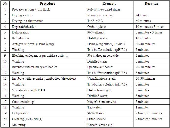
103 patients who underwent surgery and received chemotherapy courses at the Bukhara branch of the Republican Specialized Scientific and Practical Medical Center of the Republic of Uzbekistan for Surgery in 2015-2023 were selected, of which 60 patients were selected for immunohistochemical examination. Of these, 20 patients with papillary malignant tumors of the thyroid gland, 20 patients with follicular malignant tumors, and 20 patients with undifferentiated cancer were selected and studied by the immunohistochemical method. Molecular-genetic markers Ki67, P53, and CD34 were studied by immunohistochemical research (Table 1).Table 1. Stages of immunohistochemical (IHC) examination
 |
| |
|
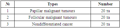
Table 2. The following patients were selected from each group for immunohistochemical examination (n = 60)
 |
| |
|
3. Results
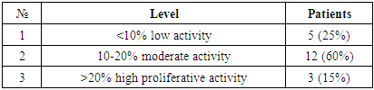
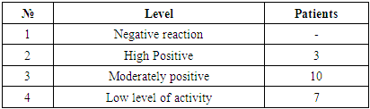
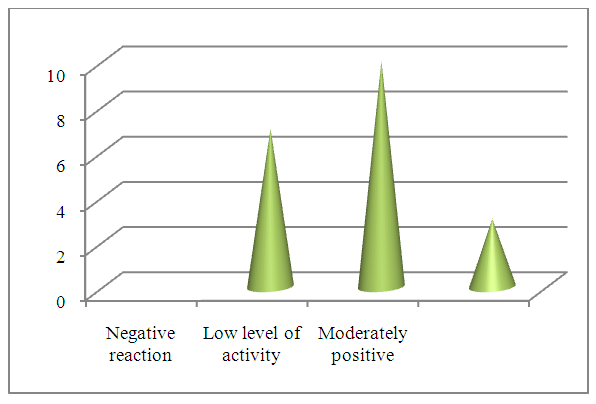
In papillary malignant tumors of the thyroid gland, the staining of wound cells for the purpose of diagnosing the Ki67 marker is described as follows. <10% mild activity, 10-20% moderate activity, >20% high proliferative activity. Of the 20 patients, 5 (25%) had a low degree of positive reaction, 12 (60%) had a moderate degree of positive reaction, and 3 (15%) had a high degree of positive reaction. No negative reaction was observed. (Table 3).Table 3. Degree of proliferative activity of the Ki67 marker in papillary tumors of the thyroid gland
 |
| |
|
According to the immunohistochemical picture, malignant tumor cells with polymorphism in the papillary structure of the thyroid gland are identified, the nuclei of which are stained dark brown, the nuclei are hyperchromatic, and malignant tumor cells with a large number of mitoses are detected.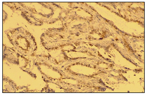 | Figure 1. Moderate positive reaction of the Ki67 marker in papillary tumors of the thyroid gland. IGX - Dab chromogen. Ob10xok40 |
 | Figure 2. Degree of proliferative activity of the Ki67 reagent in papillary malignant tumors of the thyroid gland |
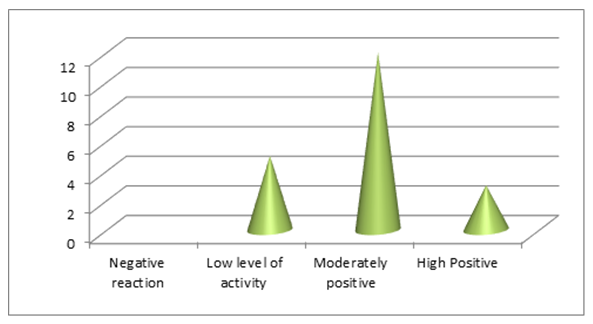
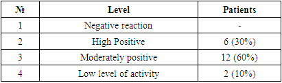
20 patients were selected for the p53 test for papillary malignant tumors of the thyroid gland. The results obtained in all patients were assessed by mild (10-30%), moderate (30-60%), and high (60-100%) positive reactions. Of the 20 patients, 6 (30%) had a low positive reaction, 12 (60%) had a medium positive reaction, and 2 (10%) had a high positive reaction. No negative reaction process was observed. In the papillary structure of the thyroid gland, malignant tumor cells that underwent polymorphism with a dark brown nucleus, a hyperchromatic nucleus, and a large number of mitoses were identified.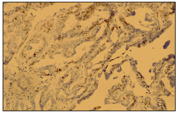 | Figure 3. Moderate positive reaction of the p53 gene suppressor in malignant tumors of the papillary form of the thyroid gland. IGX - Dab chromogen. Ob10xok40 |
Table 4. Results of expression of the p53 gene suppressor in papillary malignant tumors of the thyroid gland
 |
| |
|
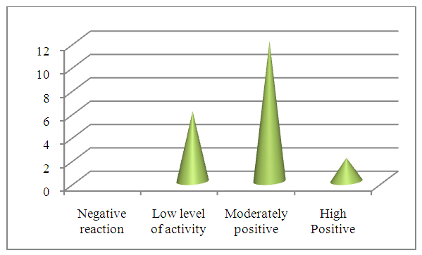 | Figure 4. Level of expression of the p53 gene suppressor in papillary malignant tumors of the thyroid gland |
In papillary tumors of the thyroid gland, the CD34 marker was assessed by studying the vascular richness of the tumor and its dependence on the nature of tumor spread to neighboring organs. The obtained results were evaluated by negative and positive reactions. A 100% positive reaction was observed in all 20 patients (Fig. 5).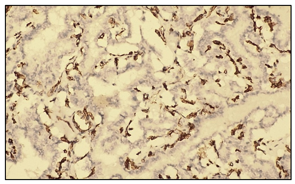 | Figure 5. Positive reaction of the CD34 marker in the papillary form of malignant thyroid tumors. IGX - Dab chromogen. Ob10. Ok40 |
Under a microscope, a positive reaction of vascular endothelium and a density of up to 20-30 vessels of various sizes were determined in one field of view. No cases of negative reaction were observed. | Figure 6. The positive reaction of the reagent CD34 in the papillary form of malignant thyroid tumors is presented in the form of a diagram |
In follicular malignant tumors of the thyroid gland, the proliferative activity of Ki67 tumor cells was assessed as a percentage. The staining of nuclear cells was characterized as follows: <10% light activity, 10-20% moderate activity, >20% high proliferative activity. Based on these results, it is possible to determine the prognostic factor of cancer.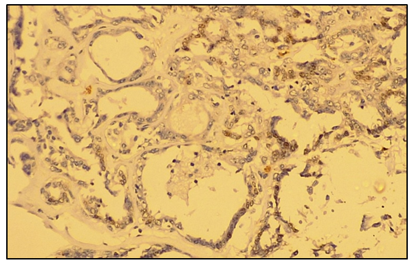 | Figure 7. Low positive Ki67 marker reaction in follicular malignant tumors of the thyroid gland. IGX - Dab chromogen. Ob10xok40 |
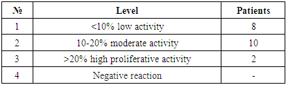
Of the 20 patients, 8 (40%) had a low degree of positive reaction, 10 (50%) had a moderate degree of positive reaction, and 2 (10%) had a high degree of positive reaction. No negative reaction was observed. Microscopically, according to the immunohistochemical picture, in the follicular structure of the thyroid gland, malignant tumor cells with polymorphic nuclei stained dark brown in small quantities, hyperchromatic nuclei, and multiple mitoses were detected. (Fig. 8).Table 5. Degree of proliferative activity of the Ki67 marker in follicular malignant thyroid tumors
 |
| |
|
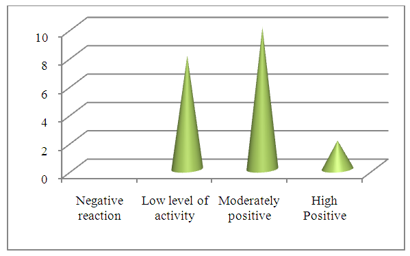 | Figure 8. Proliferative activity of the Ki67 marker of follicular malignant thyroid tumors |
In follicular malignant tumors of the thyroid gland - expression of the p53 suppressor gene, a low positive reaction was observed in 10 (50%) of 20 patients, a moderate positive reaction in 7 (35%), and a high positive reaction in 3 (15%) patients. No negative reaction process was observed. Immunohistochemically, in the follicular-cellular structure of the thyroid gland, malignant tumor cells with polymorphic nuclei stained dark brown, hyperchromatic nuclei, and a large number of mitosis were identified. (Fig. 9).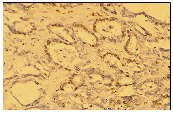 | Figure 9. Low positive reaction of the p53 gene suppressor in follicular malignant tumors of the thyroid gland. IGX - Dab chromogen. Ob10xok40 |
Table 6. Result of p53 gene suppressor expression in follicular malignant thyroid tumors
 |
| |
|
 | Figure 10. Level of expression of the p53 gene suppressor in follicular malignant thyroid tumors |
The results of the study of the CD34 marker in follicular malignant tumors of the thyroid gland were assessed by negative and positive reactions. A 100% positive reaction was observed in all 20 patients. Under a microscope, the density of 20-25 vessels of various sizes and the positive reaction of the vascular endothelium were determined in one field of view. No cases of negative reactions were observed.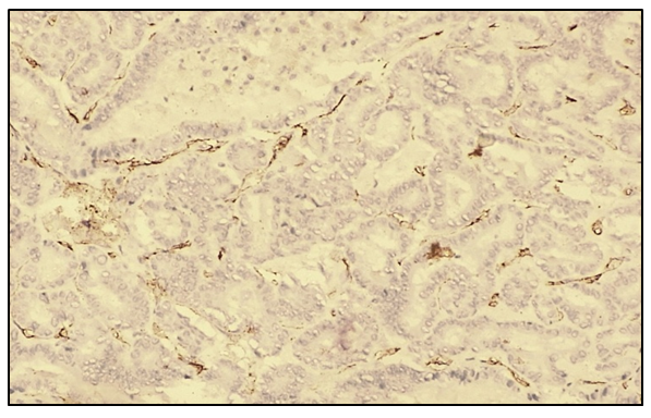 | Figure 11. Positive reaction of the CD34 marker in follicular type in malignant thyroid tumors. The density of 20-25 blood vessels was determined in one field of view. IGX - Dab chromogen. Ob10. Ok40 |
 | Figure 12. Positive reaction of the CD34 marker in malignant follicular tumors of the thyroid gland |
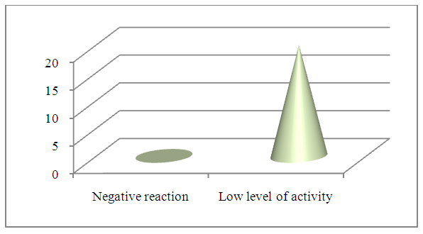
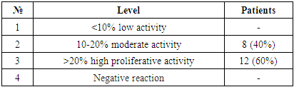
In undifferentiated malignant tumors of the thyroid gland, the proliferative activity of Ki67 tumor cells was assessed as a percentage. Of the 20 patients, 8 (40%) had a moderate positive reaction and 12 (60%) had a high positive reaction. Low and negative reactions were not observed.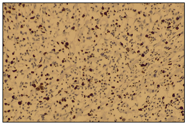 | Figure 13. High positive reaction of the Ki67 marker in undifferentiated malignant thyroid tumors. IGX - Dab chromogen. Ob10xok40 |
Microscopic appearance: tumor cells of the thyroid gland with cell polymorphism, isolated areas of tumor tissue, hyperchromatic nuclei, wide undifferentiated cells, polymorphism of tumor cells, nuclei stained dark brown, nuclei hyperchromatic, and malignant tumor cells with a large number of mitoses were detected.Table 7. Level of proliferative activity of the Ki67 marker in undifferentiated thyroid cancer
 |
| |
|
 | Figure 14. Degree of proliferative activity of the Ki67 marker in undifferentiated thyroid cancer |
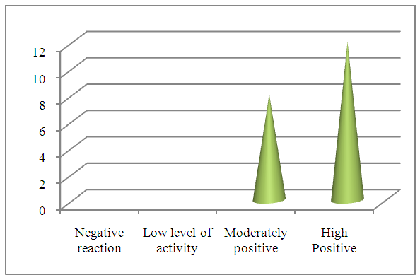
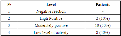
When studying the suppressor of the p53 gene in undifferentiated thyroid cancer, a low positive reaction was observed in 2 (10%) of 20 patients, a medium positive reaction in 10 (50%), and a high positive reaction in 8 (40%). No negative reaction process was observed. Microscopic appearance: tumor cells of the thyroid gland with cell polymorphism, isolated areas of tumor tissue with hyperchromatic nuclei of wide, undifferentiated cells of tumor cells with polymorphism with dark brown nuclei stained, hyperchromatic nuclei and a large number of malignant tumor cells with mitosis (Fig. 15).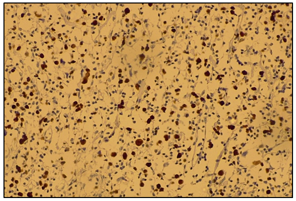 | Figure 15. High positive reaction to the suppressor of the p53 gene in undifferentiated thyroid cancer. IGX - Dab chromogen. Ob10xok40 |
Table 8. Level of expression of the p53 suppressor gene in undifferentiated thyroid cancer
 |
| |
|
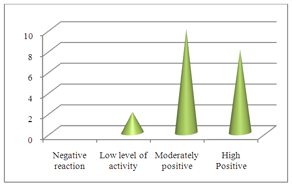 | Figure 16. Level of suppressorinin expression of the p53 gene in undifferentiated thyroid cancer |
20 patients with undifferentiated thyroid cancer were selected. The results obtained in all patients were assessed by studying the vascular richness of the tumor and the dependence of the CD34 marker on the nature of tumor spread to neighboring organs.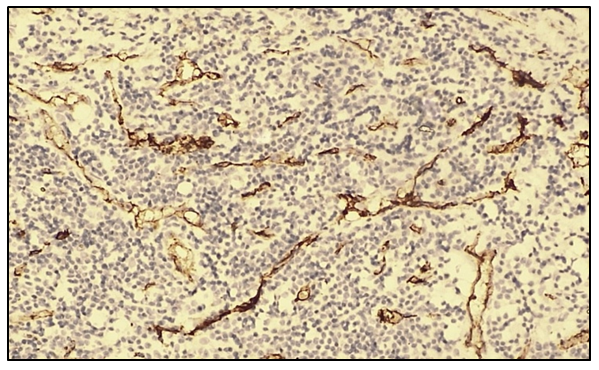 | Figure 17. Positive reaction of the CD34 marker in the type of undifferentiated thyroid cancer. The density of 30-40 blood vessels was determined in one field of view. IGX - Dab chromogen. Ob10. Ok40 |
In all 20 patients, a positive reaction was observed in 100% of patients. Under a microscope, up to 30-40 vessels of varying sizes were observed in one field of view, and a positive reaction of the vascular endothelium was noted. No cases of negative reaction were observed.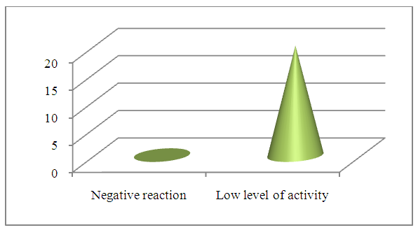 | Figure 18. Positive response of CD34 marker in undifferentiated thyroid cancer |
4. Conclusions
Malignant thyroid tumors are one of the important problems in oncology, and their various morphological and molecular properties directly affect treatment tactics and prognosis. In this study, the expression of immunohistochemical markers such as Ki67, p53, and CD34 in papillary, follicular, and undifferentiated malignant tumors was studied.The results showed that in papillary and follicular malignant tumors, Ki67 and p53 markers were expressed at a low or medium level, which is associated with their relatively slow growth and low invasiveness. The CD34 marker gave a 100% positive result in all patients, indicating good vascularization of the tumor tissue.On the other hand, it was confirmed that undifferentiated (anaplastic) cancer has an extremely aggressive nature. In this species, the expression of Ki67 and p53 is at a high level, which indicates rapid cell proliferation and disruption of genetic stability. Also, vascular density, determined by the CD34 marker, is highest in undifferentiated cancer, which indicates a strong ability of the tumor to rapidly grow and metastasize.These results contribute to a better understanding of the biological characteristics of malignant thyroid tumors. In particular, high expression of Ki67 and p53 markers can be one of the important indicators of tumor aggressiveness and poor prognosis. Assessment of the degree of vascularization through CD34 also provides information about the invasiveness of the tumor and the potential for metastasis.The results of the study show that a deeper study of the immunohistochemical properties of thyroid tumors allows for their early diagnosis and the development of individual treatment tactics. Especially in undifferentiated cancers, the nature of the aggressive course and the high probability of metastasis indicate the need for a comprehensive approach to the treatment process. Therefore, in the future, the development of new biological markers and individual treatment strategies for such patients will have important scientific and practical significance.
References
| [1] | Siegel R.L., Miller K.D., Jemal A. Cancer statistics, 2023. CA Cancer J Clin. 2023; 73(1): 17-48. |
| [2] | Cabanillas M.E., McFadden D.G., Durante C. Thyroid cancer. Lancet. 2016; 388(10061): 2783-2795. |
| [3] | Xing M. Molecular pathogenesis and mechanisms of thyroid cancer. Nat Rev Cancer. 2013; 13(3): 184-199. |
| [4] | Nikiforov Y.E., Seethala R.R. Anaplastic thyroid carcinoma. Surgical Pathology Clinics. 2019; 12(4): 865-878. |
| [5] | Scholzen T., Gerdes J. The Ki-67 protein: from the known and the unknown. J Cell Physiol. 2000; 182(3): 311-322. |
| [6] | Zlobec I., Lugli A. Prognostic and predictive factors in tumor pathology. Histopathology. 2008; 53(1): 1-9. |
| [7] | Levine A.J. p53, the cellular gatekeeper for growth and division. Cell. 1997; 88(3): 323-331. |
| [8] | Ribatti D., Nico B., Crivellato E. The history of the angiogenic switch concept. Leukemia. 2007; 21(1): 44-52. |
| [9] | Weidner N. Tumor angiogenesis: review of current applications in tumor prognostication. Semin Diagn Pathol. 1993; 10(4): 302-313. |

 Abstract
 Reference
 Full-Text PDF
 Full-text HTML