Fozilov Uktam Abdurazzoqovich, Eronov Yoqub Quvatovich
Bukhara State Medical Institute named after Abu Ali Ibn Sino, Bukhara, Uzbekistan
Correspondence to: Eronov Yoqub Quvatovich, Bukhara State Medical Institute named after Abu Ali Ibn Sino, Bukhara, Uzbekistan.
| Email: |  |
Copyright © 2025 The Author(s). Published by Scientific & Academic Publishing.
This work is licensed under the Creative Commons Attribution International License (CC BY).
http://creativecommons.org/licenses/by/4.0/

Abstract
This study focuses on the evaluation of maxillary protrusions in children with disabilities using cephalometric radiography, a modern orthodontic diagnostic method. The application of this method enables the precise assessment of the spatial relationship between the skull and dental arches, as well as the alignment of the teeth in relation to the surrounding bone structures. The findings contribute to improving orthodontic diagnosis and treatment planning in pediatric patients with special needs.
Keywords:
Children with disabilities, Maxillary protrusions, Orthodontic examination methods, Cephalometric radiography methods
Cite this paper: Fozilov Uktam Abdurazzoqovich, Eronov Yoqub Quvatovich, Indications of Orthodontics Examination of Maxillary Protrussions in Disabled Children, American Journal of Medicine and Medical Sciences, Vol. 15 No. 6, 2025, pp. 1663-1666. doi: 10.5923/j.ajmms.20251506.06.
1. Introduction
The increasing trend in the prevalence of maxillary protrusions, dental anomalies and deformations in children with disabilities is characterized by differences in the growth rate of the jaw bones. In patients at the stage of development of the dento-maxillary system during the period of temporary and permanent dentition, temporary disproportions in the size of the jaws are determined, which are associated with the order of eruption of teeth and their sequence.During the period of permanent dentition, the tendency to reduce anomalies and deformations of the dento-maxillofacial system is attributed to the high efficiency of self-regulation processes in the dento-maxillofacial system of the body and early orthodontic treatment, but the period of the appearance of permanent teeth is characterized by the appearance of new deformations and primary anomalies resulting from the early loss of teeth due to caries and its complications. During the period of primary dentition, anomalies of individual teeth in children occur in 16.5% of cases, occlusal anomalies in 13.59% of cases, and dental anomalies in this period occur only in 2.92% of cases [1,3,5,7,9,11,13].Thus, it is one of the effective methods for correcting distal occlusion and jaw pathologies, as well as for normalizing the masticatory height. In addition, this radiograph provides a posterior-anterior projection to analyze any potential asymmetry of the skull. In the practice of orthodontic dentistry, cephalometry is an important tool for diagnosis and treatment planning of facial and jaw deformities.
2. Results and Analysis
The data obtained as a result of cephalometric radiography help the orthodontist to identify pathologies and developmental anomalies of the facial skeleton, to assess the symmetry and balance of the face. In orthodontics, it is not enough to make a correct diagnosis by examining the teeth alone, since in 90% of cases the cause of pathological occlusion is the skeletal component. To determine the true cause of dental problems, the size, position and ratio of the upper and lower jaws are studied. In addition, cephalometry includes: projections of the front teeth;jaws (jaw growth in front, side, rear projections);made it possible to determine the presence of significant irregularities in the structure of the jaws.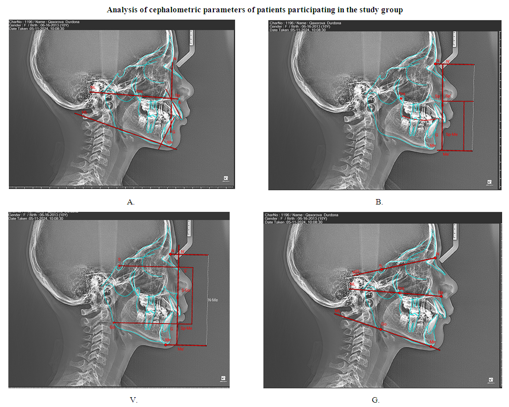 | Figure 1. Cephalometric parameters in patients in the control group (B.A. -12 years old), A – plane of upper and lower jaw, angle of scapular teeth; B – measurement of the middle and lower height of the face, V – measurement of the front and back height of the face, G – the front angle of the base of the skull, the lines and angle of the lower and upper jaw plane |
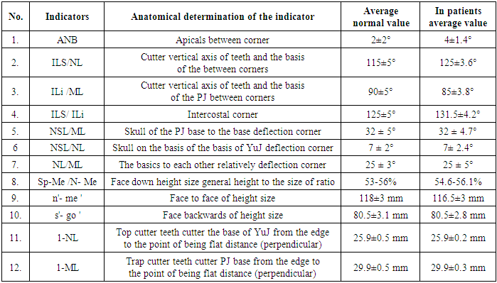
Teleroentgenogram - hereinafter referred to as TRG. A face profile or full face X-ray image that determines the correct proportion of the skull. TRG reflects the contours of the maxillofacial skeleton and soft tissues of the face.The tele- radiographic image, it was possible to determine the characteristics of the growth and development of the facial bones, the localization of their altered growth. By comparing the images before, during and after treatment, changes associated with orthodontic treatment were studied.Teleroentgenogram is a precise image and measurement of the various structures and features of the skull bones (cephalometric analysis). This diagnostic method is also an effective method for determining the size of the upper and lower jaw dentition. Studies of this type have been conducted mainly in patients with the third group. The TRG examination method allows for a highly accurate anatomical examination of the skull from any angle.According to the results of the study We found it appropriate to use this method of teleradiography (TRG) in patients of groups II and III of our study. The method used in our study group patients It is a method designed to obtain X-ray images of the actual dimensions of the brain and maxillofacial bones, and is distinguished by its high medical and economic efficiency over other X-ray methods due to its informativeness, the possibility of conducting high-precision dimensional analyses, and the wide scope and convenience of its application.Also, the possibility of morphometric study of anthropometric points located at different depths and layers using TRG in two-dimensional projection (2D) allows us to accurately perform angular, linear and planimetric dimensional analyses between these points. Taking into account the high informativeness of the TRG image, its volumetric and spatial proportionality, this method is included in the “gold standard” of orthodontic practice, and therefore, from an orthodontic point of view, it is possible to collect more information as a result of using this method than modern visual methods [2,4,6,8,10,12].Images can be taken in five projections (lateral, straight anterior-posterior or posterior-anterior, oblique, maxillary-parietal). The lateral projection is most often used for cephalometric analysis. Among the many indicators used in the calculation of TRG, information on the development of TJT in KTChT is reflected in table 1.Table 1. Comparative analytical parameters of TRH testing in patients in the study group
 |
| |
|
The following results were obtained when analyzing the TRG test results presented in table 1.N (nazion) - nasal and frontal bones are combined the point where the sphere intersects the midline;B (basion) - the point where the horizontal line passing through the highest point of the edge of both eye sockets intersects the contour of the skull;Zg (zygion) - lateral protrusion of the cheekbone;Or (orbital) - eye cup outline the lowest point;Maxilla - the area where the alveolar process transitions to the cheek process;A (ANS) – anterior tip of the nose;O - the point where the medial corners of the upper central incisors meet;D - points in the widest area of the nasal cavity;Ye is the most convex point on the vestibular surface of teeth 17 and 27.In TRG, as in profile TRG, anthropometric points are marked on the skin contours with lowercase Latin letters: the presence of n, b, zn, or points was determined.According to the analysis of the results of the study, two different methods were used in the radiographic analysis of TRG: the first method was aimed at identifying a specific teleradiographic sign of anomaly or deformation, and the second was considered a method that combined all existing analyzes and incorporated into a computer program.The first method is a variety of analyzes proposed by scientists, which are aimed at identifying a specific pathology, while software methods are used to make a conclusive diagnosis of existing anomalies or deformations. One of the advantages of this method is that the child's maxillofacial structure is also taken into account, taking into account the normal TRG indicators for the child's age. The study studied and compared the normal angular, linear, planimetric and objective analyzes.
3. Conclusions
The angle of inclination of the upper canines to the vestibular side in group III patients with maxillary protrusion was studied in relation to the flatness of the Fracfort Horizon. In the dentoalveolar form, the angle of inclination of the canines was shifted to the normal side by 3 - 3.7°. The angle of inclination of the lower incisors was determined in relation to the flatness of the MR. During the treatment, the retrusion and density of the lower incisors to the vestibular side by 2 - 2.1° was achieved. The tooth rows were brought into a parabolic shape.
References
| [1] | Arunachalam, S. Orthodontic appliances and oral hygiene: Are we asking the right questions? / S. Arunachalam, J. Sharan, I. Sivakumar [et al.] // Amer.j. of orthodontics and dentofacial orthopedics. - 2018. - Vol. 154, № 2. - P. 155-156. |
| [2] | Ardani, I. G. Cephalometric Characteristic of Skeletal Class II Malocclusion in Javanese Population at Universitas Airlangga Dental Hospital | I. G. Ardani, M. L. Sanjaya, J. Sjamsudin // Contemp. Clin. Dent. - 2018. - Vol. 9 (Supp l.2). - P. 342-346. |
| [3] | Alobeid, A. Comparison of the force levels among labial and lingual self-ligating and conventional brackets in simulated misaligned teeth / A. Alobeid, T. El-Bialy, S. Khawatmi [et al.] // Eur. J. Orthod. - 2017. - Vol. 39 (4). - P. 419-425. |
| [4] | Bazarova, K. M. Ortodontic problems in the treatment of patients with dental defects / K. M. Bazarova, R. A. Zhartybaev, B. B. Salymbekov, M. K. Iskakova // В сборник: Stience and educationA problems and innovations/ сборник стaтeй VII Meждунapoднoй нaучнo-пpaктичeскoй кoнфepeнции. - neroa, 2021. - C. 117-125. |
| [5] | Cantarella, D. Changes in the midpalatal and pterygopalatine sutures induced by microimplant-supported skeletal expander, analyzed with a novel 3D method based on CBCT imaging / D. Cantarella, R. Dominguez-Mompell, S. M. Mallya, [et al.] // Prog Orthod. - 2017. - P. 18-34. |
| [6] | Vorobeva, M. V. Causes behind distal occlusion / M. V. Vorobeva, S. V. Konnov, N. V. Bulkina, [et al.] // Archiv EuroMedica. - 2019. - Т. 9. № 1. - С. 191-193. |
| [7] | Choi, S. H. Effect of malocclusion severity on oral health-related quality of life and food intake ability in a Korean population / S. H. Choi, J. S. Kim, J. Y. Cha [et al. ] // Am J Orthod Dentofacial Orthop. - 2016. - № 149 (3). - P. 38490. |
| [8] | Felter, M. Comparative study of the usability of two software programs for visualization and analysis of digital orthodontic models / M. Felter, M. M. Lenza, M. G. Lenza [et al.] // J. Dent. Res. Dent. Clin. Dent. Prospects. - 2018. -Vol. 12 (3). - P. 213-220. |
| [9] | Postnikov, M. A. Functional evidence-based dentistry in osteopathic correction of distal occlusion / M. A. Postnikov, O. N. Pavlova, F. G. Klochkov, E. O. Guseva // International Research Journal. - 2021. - № 4-2 (106). - C. 148-152. |
| [10] | Gay, G. Root resorption during orthodontic treatment with Invisalign®: a radiometric study / G. Gay, T. Ravera, F. Castroflorio, [et al.] // Prog. Orthod. -2017. - № 18(1). - P. 12. |
| [11] | Hourfar, J. Influence of interradicular and palatal placement of orthodontic mini-implants on the success (survival) rate / J. Hourfar, D. Bister, G. Kanavakis [et al.] // Head Face Med. - 2017. - Vol.13. - P. 14-18. |
| [12] | Ishchenko, T. The instant diagnostics of obstruction of the upper airway for children with distal occlusion / Ishchenko T., Kharke M., Kharke V.V. // В книге: мaтеpиaлы Х Междутародной таучной конференции. Российский университет дружбы тародов. 2019. С. 83. |
| [13] | Jayaratne, Y. S. Maxillary incisors changes during space closure with conventional and skeletal anchorage methods: a systematic review / Y. S. Jayaratne, F. Uribe, N. Janakiraman // J. Istanb. Univ. Fac. Dent. - 2017. - Vol. 51 (3 Suppl 1). - P. 90-101. |

 Abstract
 Reference
 Full-Text PDF
 Full-text HTML