-
Paper Information
- Next Paper
- Previous Paper
- Paper Submission
-
Journal Information
- About This Journal
- Editorial Board
- Current Issue
- Archive
- Author Guidelines
- Contact Us
American Journal of Medicine and Medical Sciences
p-ISSN: 2165-901X e-ISSN: 2165-9036
2025; 15(5): 1483-1486
doi:10.5923/j.ajmms.20251505.39
Received: Apr. 17, 2025; Accepted: May 15, 2025; Published: May 27, 2025

Immunohistochemical Analysis of Morphological Changes in the Pancreas of Rats under the Influence of Alcohol and Energy Drinks (On the Example of Bcl-2)
 Abstract
 Reference
 Full-Text PDF
 Full-text HTML
Davronova M. S.1, Ilyasov A. S.2, Avezov A. U.3, Aytimova G. Y.3
1Bukhara State Medical Institute, Bukhara, Uzbekistan
2Navoi Innovation University Department, Navoi, Uzbekistan
3Urgench Branch of Tashkent Medical Academy, Urgench, Uzbekistan
Copyright © 2025 The Author(s). Published by Scientific & Academic Publishing.
This work is licensed under the Creative Commons Attribution International License (CC BY).
http://creativecommons.org/licenses/by/4.0/

This study aimed to investigate the histological changes induced by energy drinks in the pancreas of Wistar Albino rats. Comparisons were made between the control group and rats given energy and alcohol drinks. Histopathological examination revealed the following: vascular occlusion, mononuclear cell infiltration, and tissue edema. Here, vascular hyperemia refers to dilation and swelling of blood vessels. Infiltration of mononuclear cells means that tissues are infiltrated primarily by lymphocytes. Tissue edema refers to the accumulation of free fluid in tissue spaces.
Keywords: Energy drinks, Energy drinks, Caffeine, Taurine, Dietary supplements
Cite this paper: Davronova M. S., Ilyasov A. S., Avezov A. U., Aytimova G. Y., Immunohistochemical Analysis of Morphological Changes in the Pancreas of Rats under the Influence of Alcohol and Energy Drinks (On the Example of Bcl-2), American Journal of Medicine and Medical Sciences, Vol. 15 No. 5, 2025, pp. 1483-1486. doi: 10.5923/j.ajmms.20251505.39.
1. Introduction
- It is known that energy drinks are products that stimulate and excite the human central nervous system, causing a feeling of vivacity, increased efficiency and increased motor activity [2,11,14,19,20]. Although these products were created by mankind recently, the ingredients they contain have long been used to stimulate the nervous system. Energy drinks have become a real salvation for young people preparing for exams, for company employees who were unable to submit work on time, for fitness trainers trying to set records in sports, for tired drivers, in a word, for those who are tired but want to feel rested and full of energy [4,8,11,16].Energy drinks first appeared in Europe and Asia in the 1960s as a result of increased consumer demand for energy-giving food supplements [1,9,16,20]. Several studies conducted in Saudi Arabia show that almost half of consumers are young people (13–35 years old), and more than half of them have consumed such drinks, 40% of whom have consumed more than 3 cans per week for more than a year [6,7,9,13,17,18].According to the Centers for Disease Control and Prevention, high school students consume energy drinks at a rate similar to that of soda [5,7,11,14,16]. In fact, energy drink consumption may be much higher than reported in this survey, and such surveys are likely to be more likely to underestimate consumption. Energy drinks have been found to contain taurine, glucuronolactone, caffeine, ginseng, and guarana [8,10,15,18,20]. Energy drinks often contain stimulants, which are not on the U.S. Food and Drug Administration’s list of substances. The amounts of these stimulants vary among energy drinks and often exceed the legal limits [1,12,17,20].Studies have shown that the caffeine content of energy drinks ranges from 50 mg/can to 505 mg/can, which is significantly higher than the caffeine content in a can of cola [4,14,16,18].According to the US Department of Mental Health and Substance Abuse in 2013, the number of emergency room visits related to energy drinks has doubled, from 10,068 in 2011 to 20,000 in 2017 [3,7,12,14,19]. A major obstacle to understanding the associations between energy drink consumption and adverse outcomes is the paucity of data on the toxic effects of the various compounds in the drinks [7,8,14,17,19,20].In Europe, energy drinks are considered biologically active supplements and are allowed to be sold only in pharmacies. In Russia, there are also certain restrictions, such as the content of the drink should not exceed two components with a sedative effect, its consumption should be limited in a can, its sale in schools is prohibited, and it must be indicated, as stated in the Resolution of the Chief State Sanitary Doctor of the Russian Federation Zelepukhina L.P. (2012) “On strengthening control over drinks containing sedative components” [9,15,17,18].Caffeine reduces drowsiness and fatigue, increases heart rate, and helps a person cope with mental stress. On the one hand, all this is a temporary effect, which is replaced by even more severe fatigue. If at this time the body is not given a full rest and another cup of coffee or black tea is drunk, the possible dose of caffeine is increased again, and although it acts for 3 hours, it is slowly excreted from the body [15,16,19,20].The aim of the study was to study the effect of energy and alcoholic drinks on morphological changes in the pancreas.
2. Research Material and Research Methods
- In the experiment, 3-month-old rats were given an energy drink in a dose of 5 ml per day for 3,6,9 months. The calculation of energy drinks for the experiment was carried out based on the indicators of the “Hygienic standards of maximum permissible concentrations of pollutants in the air and atmosphere of populated areas in the territory of the Republic of Uzbekistan”.
3. Research Results
- Immunohistochemical examination was used to assess the morphological substrates of changes in specific cells and to recognize their specific features in the pancreas.
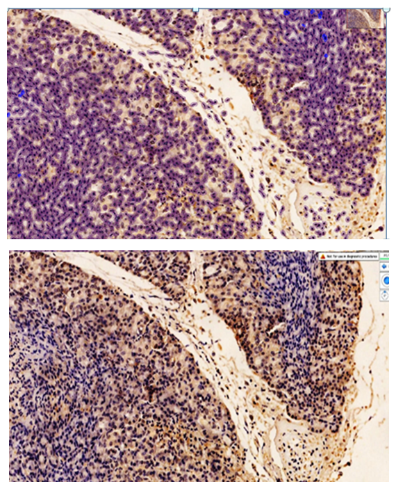 | Figure 1. Moderately positive expression of the Bcl-2 marker in the pancreas of rats that consumed ethyl alcohol and energy drinks for 3 months. Scanned and expression levels were determined using the QuPath-0.4.0.ink. program. Expressed cells are dark brown. Staining is Dab chromogen. Size 10X10 |
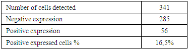
 | Figure 2. Low positive expression of the Bcl-2 marker in the pancreas of rats that consumed ethyl alcohol and energy drinks for 6 months. Scanned and the level of expression was determined using the QuPath-0.4.0.ink. program. Expressed cells are dark brown. Staining is Dab chromogen. Size 10X10 |
 In the next immunohistochemical study, the pancreatic acinar and islet cells of the exocrine and endocrine systems were examined. In particular, it inhibits the death (apoptosis) of pancreatic epithelial cells and most other cells. It regulates programmed cell death mainly by controlling the permeability of the mitochondrial membrane. It inhibits caspases by preventing the release of cytochrome-C from mitochondria or by binding the apoptosis-activating factor APAF1. In experimental conditions, artificial inactivation of the Bcl-2 gene in rats (under the influence of ethyl alcohol and energy drinks) is manifested in the experimentally accelerated programmed cell death, mainly by morphological changes in the pancreas of rats.The results were evaluated as follows: no expression - 0 points, weak expression - 1 point, moderate expression - 2 points, strong expression - 3 points.In our work, rats that consumed ethyl alcohol and energy drinks for 3 months developed changes in the form of effects on the pancreas, increased programmed death.The moderate positive expression of the Bcl-2 marker, in turn, is explained by the induction of cell apoptosis (enhancement of any influencing factors) in rats given ethyl alcohol and energy drinks. The positive expression of the Bcl-2 marker is explained by the increased apoptosis process under the influence of ethyl alcohol and energy drinks and the high number of labile cells in the pancreatic acini (labile cells always have a normal number of mitotic foci and epithelial cells are rapidly self-renewing). The positive expression of the Bcl-2 marker is not localized or localized, but rather uniformly distributed throughout the tissue, and is explained by the fact that ethyl alcohol and energy drinks stimulate the apoptosis process in most tissue cells through the blood vessels.The moderate positive expression of the BcL-2 marker, in turn, is explained by the induction (enhancement of any influencing factors) of cell apoptosis in rats given ethyl alcohol and energy drinks. The positive expression of the BcL-2 marker is explained by the fact that the apoptosis process is enhanced under the influence of ethyl alcohol and energy drinks and by the abundance of labile cells in the pancreatic acini (labile cells always have a normal number of mitotic foci, and epithelial cells quickly self-renew). The positive expression of the BcL-2 marker is not focal or in any one area, but is evenly distributed in all areas of the tissue, and is explained by the fact that ethyl alcohol and energy drinks stimulate the apoptosis process in most tissue cells through the blood vessels.
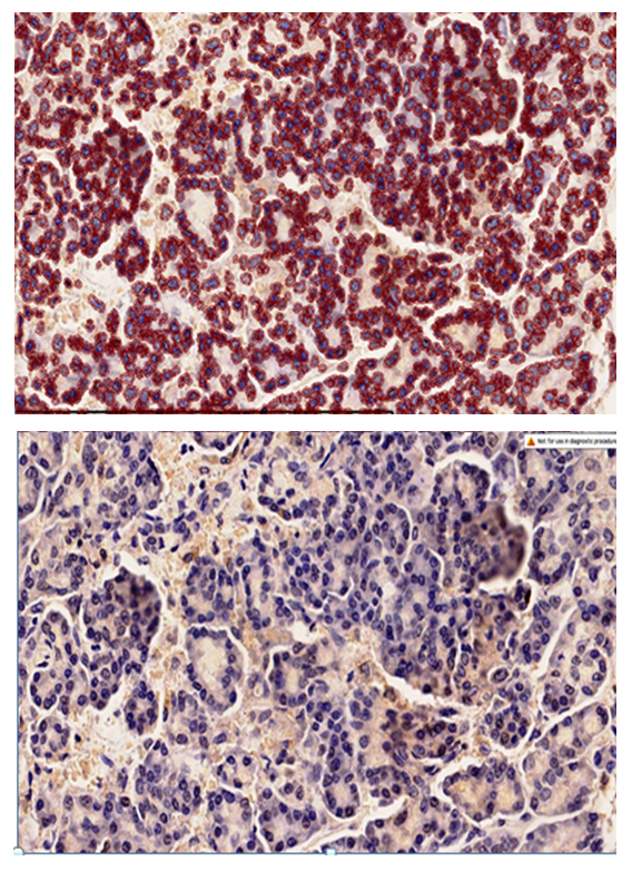
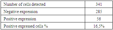
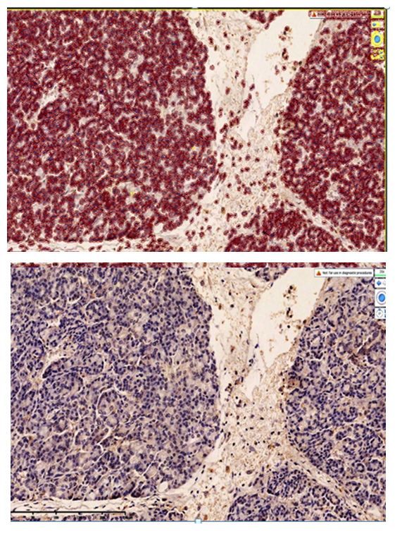 | Figure 3. Low positive expression of the Bcl-2 marker in the pancreas of rats that consumed ethyl alcohol and energy drinks for 9 months. Scanned and expression levels were determined using the QuPath-0.4.0.ink. program. Expressed cells are dark brown. Staining is Dab chromogen. Size 10X10 |
 Chronic 3-month toxic intoxication with ethyl alcohol and energy drinks resulted in strong regenerative changes in the acinar epithelium of the pancreas, manifested by the binding of the apoptosis-blocking factor APAF1 protein and the expression of the Bcl-2 marker.In this case, the premature death of most of the cells undergoing apoptosis depends on the concentration of the influencing factor, and microscopically, the expression of the Bcl-2 marker in monocellular and segmental cells in the glandular epithelium is determined by the results of 2-point Bcl-2 markers, which are dead cells (see figure). There are no changes in the histotopographic structure of the pancreas, the trajectory of the glandular structures is preserved, and the positive expression of the Ki-67 marker in paracellular glandular cells indicates that the process of proliferation and apoptosis occurs simultaneously in parallel to restore the lost glandular cell.It was recently found that in rats exposed to ethanol and energy drinks for a chronic period of 6 months, moderate regenerative changes occurred in the glandular epithelium of the pancreas, resulting in a moderate decrease in the binding of the apoptosis-blocking factor APAF1 protein and the expression of the Bcl-2 marker.In this case, the majority of the premature death of cells undergoing apoptosis at the 6th month, compared to the 3rd month, is explained by the morphological adaptation, the decrease in the size of the glandular epithelium during its life, and depending on the duration and concentration of the influencing factor, microscopically detected in the glandular epithelium as foci of monocellular cell death. Changes in the histotopographic structure of the pancreas were mainly manifested by an increase in the number of cells that had shrunk in size. The trajectory of the glandular structures is preserved, and in the paracellular glandular cells, a small number of glandular epithelium proliferating in place of the lost glandular cells is detected with a low positive expression of the Ki-6 marker. This is explained by the continuity of influencing factors, as mentioned above.Therefore, at 6 months, low positive expression of the Bcl-2 marker is determined by the number of cells that show positive expression by staining in a light golden yellow color in the cytoplasm of the acinar gland epithelium, and low expression of the Bcl-2 marker is determined as 1 point.In the last 9 months, rats given ethyl alcohol and energy drinks had moderate to low-grade regenerative changes in the glandular epithelium, due to which the binding of the intracellular protein APAF1-factor that blocks apoptosis and the expression of the Bcl-2 marker were found to be low. At the 9th month, the decrease in the rate of premature death of cells undergoing apoptosis compared to the 3rd and 6th months was mainly explained by the morphological adaptation, the significant decrease in the glandular epithelium during its life, and the decrease in its morphofunctional aspects. In the case of chronic exposure to the influencing factor for 9 months, focal foci of monocellular cell death were detected microscopically in the glandular epithelium. Changes in the histotopographic structure of the pancreas were mainly manifested by the proliferation of acinar glandular epithelium, which significantly reduced the volume of the cell mass. The trajectory of the glandular structures was preserved, the periacinar blood vessels appeared to be poorly vascularized, the paraacinar glandular cells became smaller and uniform, and a small number of light golden cells with positive expression of the Bcl-2 marker masking in their cytoplasm were detected. It is at this point in time, over a period of 9 months, that the low proliferation was directly correlated with the low positive expression of the Ki-6 marker in the glandular epithelium. This, as mentioned above, is explained by the duration of the influencing factors and the process of adaptation that occurred in the morphological aspect.Thus, rats that consumed ethyl alcohol and energy drinks under experimental conditions for 9 months showed persistent low positive expression of the Bcl-2 marker in the pancreas. Microscopically, the number of cells that showed positive expression was determined by a small number of light golden yellow staining in the cytoplasm of the acinar gland epithelium. The expression of the Bcl-2 marker was determined as weak expression - 1 point.