American Journal of Medicine and Medical Sciences

p-ISSN: 2165-901X    e-ISSN: 2165-9036

2025;  15(5): 1474-1480

doi:10.5923/j.ajmms.20251505.37

Received: Apr. 22, 2025; Accepted: May 21, 2025; Published: May 27, 2025


Immunohistochemical Study of Morphological Changes in Patients with Uterine Cancer

Juraeva Gulbakhor1, Mamadalieva Yashnar2, Amanov Akma1

1Assistant, Termez Branch of the Tashkent Medical Academy, Termez, Uzbekistan

2Professor, Department of Oncology of the Center for the Development of Professional Qualifications of Medical Workers of the Ministry of Health of the Republic of Uzbekistan, DSc, Professor, Tashkent, Uzbekistan

Correspondence to: Juraeva Gulbakhor, Assistant, Termez Branch of the Tashkent Medical Academy, Termez, Uzbekistan.

Email:

Copyright © 2025 The Author(s). Published by Scientific & Academic Publishing.

This work is licensed under the Creative Commons Attribution International License (CC BY).
http://creativecommons.org/licenses/by/4.0/

Abstract

In this study, an immunohistochemical analysis of the expression of Ki-67, p53, and Bcl-2 markers was conducted in 60 patients with uterine body adenocarcinoma, divided into three groups depending on the degree of tumor differentiation (high, moderate, low; 20 patients in each group). The study was conducted using a Bond (Leica, Australia) automated immunohistochemical processor. It has been established that the expression of Ki-67, p53, and Bcl-2 increases as the degree of tumor differentiation decreases. Thus, in highly differentiated adenocarcinoma, a weak positive reaction (up to 30%) prevailed for all three markers, while in low-differentiated tumors, a high expression (more than 60%) was significantly more common. Microscopically, signs of pronounced cellular atypia, endometrial gland hyperplasia, myometrial invasion, and pathological mitoses were identified, especially in the G2 and G3 groups. The obtained data indicate that the indicators of proliferative activity (Ki-67), anti-apoptotic activity (Bcl-2), and mutational instability (p53) can be used as prognostic markers of tumor biological aggressiveness and contribute to determining the treatment tactics of patients with endometrial cancer.

Keywords: Endometrial cancer, Ki-67, p53, Bcl-2, Immunohistochemistry, Degree of differentiation, Morphology, Proliferation

Cite this paper: Juraeva Gulbakhor, Mamadalieva Yashnar, Amanov Akma, Immunohistochemical Study of Morphological Changes in Patients with Uterine Cancer, American Journal of Medicine and Medical Sciences, Vol. 15 No. 5, 2025, pp. 1474-1480. doi: 10.5923/j.ajmms.20251505.37.

1. Introduction

According to WHO estimates, more than one billion people on our planet are overweight or obese. In Western European countries, 20-25% of women are obese. Statistics show that excess weight is detected in 54% of the Russian population. A characteristic feature of obesity is that it is often combined with serious diseases: type 2 diabetes mellitus, arterial hypertension, dyslipidemia, atherosclerosis, coronary heart disease, some types of reproductive diseases [9]. According to preliminary calculations by American Cancer RE, in 2019, 61,880 cases of endometrial cancer were diagnosed in the United States, with a mortality rate of 12,160. [22].
In the period from 40 to 59 years, there is a sharp increase in the incidence of endometrial cancer, increasing approximately 4 times, gradually reaching a peak at 60-64 years, after which it significantly decreases. Thus, by age 65, the risk increases threefold, and by age 75, it increases 4.7 times. [23]. A number of biological factors may be involved in the relationship between obesity and endometrial cancer. Excessive accumulation of adipose tissue in postmenopausal women leads to an increase in estrogen levels, which, in turn, contributes to an increase in the mitotic activity of endometrial cells in the absence of a balancing effect of progesterone. [17].
Bcl2 suppresses apoptosis in multicellular systems, including lymphohematopoietic and neuronal cells. It regulates cell death by controlling the permeability of the mitochondrial membrane.
Bcl 2 suppresses apoptosis, including in lymphohematopoietic and neuronal cells. Depending on the prevalence and intensity of the immunohistochemical reaction, the eyepiece and objective 10-40 objects were examined in the field of view depending on the percentage of stained cells, and the absence of expression or staining of less than 10% of cells was assessed systematically - 0 points, from 10 to 30% - 1 point, from 30 to 60 50% - 2 points, from 60 to 100% - 3 points. The expression of Bcl 2 manifested itself in the form of brown staining of the cytoplasm of epithelial cells of varying intensity.
Ki-67-protein (also known as MKI 67) is a cellular marker for proliferation and can be used in immunohistochemistry. This is closely related to cell proliferation. The proportion of Ki-67-positive tumor cells (Ki-67 marking index) is often associated with the clinical course of cancer. Ki-67-nuclear protein is a marker of the proliferative activity of cells and is evaluated as a percentage. Ki 67 is used for diagnostic purposes to determine the biological potential of malignant tumors in humans. The staining of nuclear cells is described as follows. <10% low activity, 10-20% moderate activity, >20% high proliferative activity. Through these results, it is possible to determine the prognostic factor of cancer.
The antigen for these antibodies is the protein p53, which controls the course of cell cycle processes, as well as the presence of damage in the genome, which can lead to the further development of pathology. p53-dependent apoptosis is a powerful selector, preventing the accumulation of mutations, and if they have already appeared, p53-dependent apoptosis allows the destruction of such potentially dangerous cells for the organism. This gene encodes a transcription factor that controls the entry of cells into the cell cycle. Like all tumor suppressants, p53 controls the normal course of the cell cycle. p53 is a transcription factor that regulates the cell cycle; this reagent acts as a suppressor of the formation of malignant tumors. The p53 gene is an anti-oncogenic agent.

2. Research Material and Method

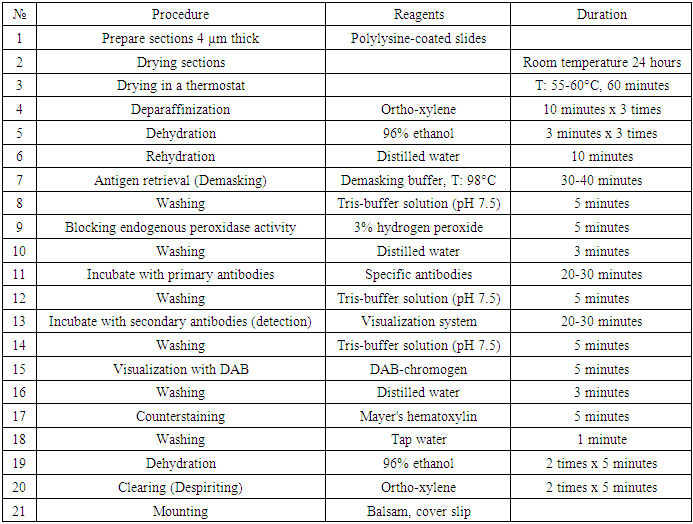
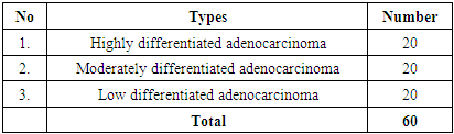
To study pathomorphologically malignant tumors by the immunohistochemical method, a total of 60 patients with uterine cancer were examined by immunohistochemistry for the degree of selective differentiation, 20 each from 3 groups. In addition to the results of morphological studies, immunohistochemical studies are currently recognized worldwide as the gold standard. For immunohistochemical examination, cell expression through monoclonal antibodies Ki67, Bcl 2, and p53 was studied using an immunohistoprocessor from Bond Leica Australia (Australia). In all patients, candle blocks were cut, treated for immunocytochemical examination, slices were taken on a slide and examined using the immunohistochemical examination method for Ki67, Bcl 2, and p53 antigens. The conducted immunohistochemical examination method was technically carried out as follows (Table 1).
Table 1. Stages of immunohistochemical (IHC) examination
     
Table 2. Number of patients selected from each group for immunohistochemical examination n=60)
     
Immunohistochemical research is conducted to analyze various processes. This study is important for identifying molecular structures in cells, studying the location of cells, studying the prevalence or histogenesis of tumor diseases, observing these processes in the development of precancerous processes, determining prognostic complications of diseases, determining the stages of tumors and treatment tactics, dynamic monitoring and control of treatment processes, and identifying risk groups for the development of tumor diseases.

3. Results

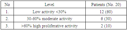
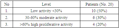
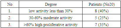
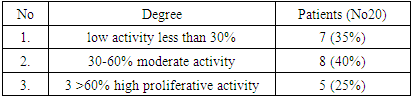
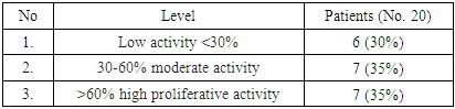
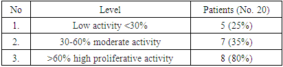
As a result, highly differentiated adenocarcinoma: For this study, 20 patients with highly differentiated cancer were selected. The results obtained in all patients were evaluated as a percentage of the marker of proliferative activity of Ki 67 tumor cells. The obtained results were assessed as mild, moderate, and severe positive reactions. Of the 20 observed patients, 12 (60%) had a mild positive reaction, 6 (30%) had a moderate positive reaction, and 2 (10%) had a high positive reaction. No negative reaction processes were observed (Table 3).
Table 3. Degree of proliferative activity of the reagent Ki 67 in highly differentiated adenocarcinoma of uterine body cancer (No 20)
     
By microscopic appearance: endometrial glands with hyperplasia and polymorphism and foci of pathological mitosis. Loose fibrous connective tissue between polymorphosed tumor cells and with signs of invasion into the myometrium. The tumor cells, undergoing polymorphism, are stained dark brown.
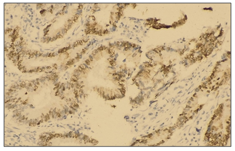
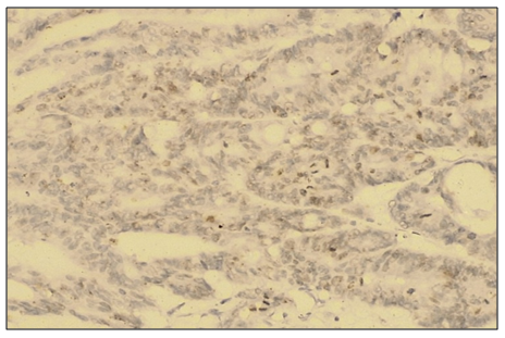
Figure 1. Medium positive Ki 67 reagent reaction in highly differentiated adenocarcinoma in uterine body cancer. IGX - Dab chromogen. Ob10. Ok40
The obtained results show that the lower the degree of cancer differentiation, the lower the indicator of its malignancy or aggressive course.
Expression analysis was performed under a microscope magnification of 40-60 mm. During the study, up to 100 cells were examined under the microscope's field of view. When more than 10% of tumor cell nuclei were stained brown, the reaction was considered positive and assessed in the region of maximum marker expression. The proliferative activity of the tumor was assessed as the percentage of Ki-67 positive cells.
In the study, a positive reaction to the tumor marker p53 was observed in 10 (50%) of 20 patients with a mild positive reaction. A moderate positive reaction was observed in 6 (30%) patients, and a high positive reaction in 4 (20%) patients.
Table 4. Degree of proliferative activity of reagent p53 in highly differentiated adenocarcinoma of uterine body cancer (No. 20)
     
By microscopic appearance: endometrial glands with hyperplasia and polymorphism and foci of pathological mitosis. Loose fibrous connective tissue between polymorphosed tumor cells and with signs of invasion into the myometrium. The tumor cells, undergoing polymorphism, are stained dark brown.
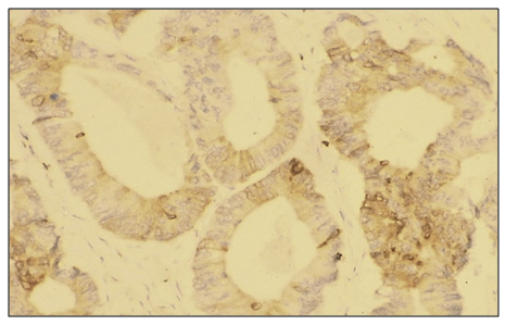
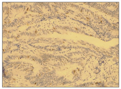
Figure 2. Cancer of the uterine body in highly differentiated adenocarcinoma with a low degree of positive reaction to reagent p53
In the study, a positive reaction to the tumor marker Bcl 2 was observed in 9 (45%) of 20 patients with a mild degree of positive reaction, mainly with the differentiation of uterine corpuscular adenocarcinoma G-1. A moderate positive reaction was observed in 7 patients (35%), and a high positive reaction was observed in 4 (20%). By microscopic appearance: endometrial glands with hyperplasia and polymorphism and foci of pathological mitosis. Loose fibrous connective tissue between polymorphosed tumor cells and with signs of invasion into the myometrium. The membranes of tumor cells undergoing polymorphism are stained dark brown.
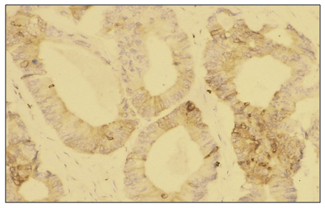
Figure 3. Immunohistochemical micropreparation: Moderate expression of the Bcl-2 gene in a highly differentiated type of uterine body cancer. cytoplasmic reaction, tumor cell membranes stained brown. Ob10 × ok40
Table 5. Degree of proliferative activity of the reagent Bcl-2 in highly differentiated adenocarcinoma of uterine body cancer (No. 20)
     
The obtained results show that the low degree of Bcl2 differentiation of cancer facilitates its treatment possibilities. However, the high proliferative activity of Bcl2 in patients indicates a high frequency of tumor recurrence and metastasis.
Moderately differentiated adenocarcinoma: High, medium, and low differential levels were studied in patients with uterine body cancer using proliferation markers (studying the Ki-67 protein and mitotic index). For this study, 20 patients with moderately differentiated cancer were selected. The results obtained in all patients were evaluated as a percentage of the marker of proliferative activity of Ki 67 tumor cells. Of the 20 observed patients, 8 (40%) had a mild positive reaction, 5 (25%) had a moderate positive reaction, and 7 (35%) had a high positive reaction. No negative reaction processes were observed.
Table 6. Degree of proliferative activity of reagent Ki 67 in moderately differentiated adenocarcinoma of uterine body cancer (No. 20)
     
By microscopic appearance: endometrial glands with hyperplasia and polymorphism and foci of pathological mitosis. Loose fibrous connective tissue between polymorphosed tumor cells and with signs of invasion into the myometrium. The tumor cells, undergoing polymorphism, are stained dark brown.
Expression analysis was performed under a microscope magnification of 40-60 mm. During the study, up to 100 cells were examined under the microscope's field of view. When more than 10% of tumor cell nuclei were stained brown, the reaction was considered positive and assessed in the region of maximum marker expression. The proliferative activity of the tumor was assessed as the percentage of Ki-67 positive cells. In the obtained results, an average positive reaction was observed in 5 (25%) patients, and a high positive reaction in 7 (35%) patients.
In the study, a positive reaction to the tumor marker p53 was observed in 7 (35%) of 20 patients with a mild positive reaction. A moderate positive reaction was observed in 8 patients (40%) and a high positive reaction in 5 patients (25%).
Table 7. Degree of proliferative activity of reagent p53 in moderately differentiated adenocarcinoma of uterine body cancer (No. 20)
     
By microscopic appearance: endometrial glands with hyperplasia and polymorphism and foci of pathological mitosis. Loose fibrous connective tissue between polymorphosed tumor cells and with signs of invasion into the myometrium. The tumor cells, undergoing polymorphism, are stained dark brown.
Figure 4. Low level positive reaction of p53 reagent in moderately differentiated adenocarcinoma in uterine body cancer. IGX - Dab chromogen. Ob10. Ok40
In the study, a positive reaction to the tumor marker Bcl 2 was observed in 6 out of 20 patients (30%) with a mild positive reaction, mainly with the differentiation of uterine corpuscular adenocarcinoma G-2. A moderate positive reaction was observed in 7 patients (35%), and a high positive reaction was observed in 7 (35%). By microscopic appearance: endometrial glands with hyperplasia and polymorphism and foci of pathological mitosis. Loose fibrous connective tissue between polymorphosed tumor cells and with signs of invasion into the myometrium. The membranes of tumor cells undergoing polymorphism are stained dark brown.
Figure 5. Immunohistochemical micropreparation: Moderate expression of the Bcl-2 gene in moderately differentiated uterine cancer cytoplasmic reaction, tumor cell membranes stained brown. Ob10 ×ok40
Table 8. Degree of proliferative activity of the Bcl-2 reagent in moderately differentiated adenocarcinoma of uterine body cancer (No. 20)
     
The obtained results show that the low degree of differentiation of cancer Bcl 2 facilitates its and treatment possibilities. However, the high proliferative activity of Bcl2 in patients indicates a high frequency of tumor recurrence and metastasis.
Low-differentiated adenocarcinoma: High, medium, and low differential levels were studied in patients with uterine carcinoma using proliferation markers (studying the Ki-67 protein and mitotic index).
For the purpose of this study, 20 patients with low-differentiated cancer were selected. The results obtained in all patients Ki 67 - tumor cell proliferative activity was assessed as a percentage. The obtained nresults were evaluated as mild, moderate, and severe positive reactions. Of the 20 observed patients, 4 (20%) had a mild positive reaction, 6 (30%) had a moderate positive reaction, and 10 (50%) had a high positive reaction. No negative reaction processes were observed.
Table 9. In low differentiated adenocarcinoma of uterine body cancer Level of proliferative activity of reagent Ki 67 (No. 20)
     
By microscopic appearance: endometrial glands with hyperplasia and polymorphism and foci of pathological mitosis. Loose fibrous connective tissue between polymorphosed tumor cells and with signs of invasion into myometrium. The tumor cells, undergoing polymorphism, are stained dark brown.
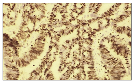
Figure 6. In low-differentiated adenocarcinoma with uterine carcinoma High positive reaction to reagent Ki 67. IGX - Dab chromogen. Ob10. Ok40
The obtained results show that the lower the degree of differentiation of cancer, the higher the risk or aggressive course of the process.
To assess nuclear expression, the percentage of tumor cells with positively colored nuclei from the total number in the zones with the highest Ki-67 content was calculated and used. Expression analysis was performed at a microscope magnification of 40-60 mm. During the study, up to 100 cells were studied in the microscope's field of view. When staining more than 10% of tumor cell nuclei brown, the reaction was considered positive and assessed in the region of maximum marker expression. The proliferative activity of the tumor was assessed as the percentage of Ki-67 positive cells.
In patients with low-differentiated adenocarcinoma with uterine carcinoma, p53 apoptosis using p53 protein markers is a strong selector, preventing the accumulation of mutations. High, medium, and low differential levels of p53 protein were studied.
In the study, a positive reaction to the tumor marker p53 was observed in 5 (25%) of 20 patients with a mild positive reaction. A moderate positive reaction was observed in 6 patients (30%) and a high positive reaction in 9 patients (45%).
Table 10. In low-differentiated adenokarycinoma with uterine body cancer p53 level of proliferative activity of the reagent (No. 20)
     
In the study, a positive reaction to the immunohistochemical tumor Bcl 2 marker was observed, mainly in the G-3 differentiation of uterine corpus adenocarcinoma, in 5 (25%) of 20 patients, a mild positive reaction was noted. A moderate positive reaction was observed in 7 patients (35%), and a high positive reaction was observed in 8 (40%). By microscopic appearance: endometrial glands with hyperplasia and polymorphism and foci of pathological mitosis. Loose fibrous connective tissue between polymorphosed tumor cells and with signs of invasion into myometrium. The membranes of tumor cells undergoing polymorphism are stained dark brown.
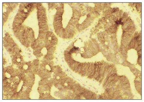
Figure 7. Immunohistochemical micropreparation: Moderate expression of the Bcl-2 gene in low-differentiated uterine body cancer cytoplasmic reaction, tumor cell membranes stained brown. Ob10 ×ok40
Table 11. Uterine body cancer with highly differentiated adenocarcinoma Bcl -2 level of proliferative activity of the reagent (No. 20)
     

4. Conclusions

The conducted study showed that immunohistochemical study of the expression of proliferative (Ki-67), apoptotic (Bcl-2), and suppressor (p53) markers in patients with endometrial cancer allows for an objective assessment of the biological characteristics of tumors depending on the degree of their differentiation.
A significant correlation was found between the expression level of the studied markers and the degree of tumor differentiation. Thus, in highly differentiated uterine body adenocarcinoma, low expression of Ki-67, Bcl-2, and p53 prevailed, indicating less aggressive biological potential. At the same time, in moderate and especially low-differentiated forms of adenocarcinoma, high proliferative activity was observed, increased expression of the p53 and Bcl-2 marker, which indicates a greater tendency of the tumor to invasive growth, metastasis, and recurrence.
The obtained data confirm the diagnostic and prognostic significance of immunohistochemical prophylaxis in endometrial adenocarcinoma. Elevated expression of Ki-67 and p53 in G2-G3 tumors can be considered a potential unfavorable prognostic factor. Increased Bcl-2 expression, despite its antiapoptotic nature, is also associated with an unfavorable clinical course of the disease in certain cases.
Thus, determining the expression of Ki-67, p53, and Bcl-2 markers can serve as a reliable tool for risk stratification, choosing treatment tactics, and assessing the prognosis in patients with uterine body adenocarcinoma.

References

[1]  O.S. Danilova, S.A. Velichko, L.A. Kolomiets, I.G. Frolova, N.G. Truchacheva. "The Role of Radiation Research Methods in the Accurate Diagnosis of Endometrial Cancer in Combination with Metabolic Syndrome," Siberian Oncological Journal, 2012, No. 3 (51).
[2]  L.A. Ashrafyan, I.B. Antonova, S.V. Ivashina, N.A. Babayeva, O.A. Alyoshikova, I.I. Baranov. "Optimizing Diagnostic Tactics in Patients with Abnormal Uterine Hemorrhage in Peri- and Postmenopausal Periods," Obstetrics and Gynecology: News, Opinions, Training, 2019, Vol. 7, No. 1, pp. 24-30. doi: 10.24411/2303-9698-2019-11003.
[3]  L.F. Gulyayeva, S.E. Krasilnikov. "Molecular mechanisms of endometrial carcinogenesis," Bulletin of the Higher Attestation Commission of the Russian Academy of Medical Sciences, 2012, No. 3 (85), part 1.
[4]  N.A. Demakova, O.B. Altukhova, S.P. Pakhomov, V.S. Orlova. "Molecular-genetic mechanisms of endometrial hyperplastic processes development," Medical Series. Pharmacy, 2014, No. 4 (175), issue. 25.
[5]  A.B. Muntyan. "Uterine fibroids as a risk factor for endometrial hyperplastic processes and cancer," Siberian Oncological Journal, 2009, appendix. No. 1, p. 140.
[6]  A.T. Amiraslanov, S.I. Safarova. "Risk factors and prognostic indicators of atypical endometrial hyperplasia," Bulletin of Modern Clinical Medicine, 2019, vol. 12, issue. 2, pp. 8-9.
[7]  R.G. Guseynova, I.M. Ordiants, A.A. Yamurzina, N.L. Sargsyan, S.Ya. Ismailzade. "Malignant Transformation of Hyperplastic Endometrium: Epidemiology and Prognosis," Obstetrics and Gynecology: News, Opinions, Training, 2020, Vol. 8, No. 3, appendix, pp. 100-105. doi: 10.24411/2303-9698-2020-13916.
[8]  A.T. Amiraslanov, S.I. Safarova. "Risk factors and prognostic indicators of atypical endometrial hyperplasia," Bulletin of Modern Clinical Medicine, 2019, vol. 12, issue. 2, pp. 8-9.
[9]  O.R. Grigoryan, E.N. Andreeva. "Pathogenetic aspects of endometrial hyperplastic processes in women with carbohydrate metabolism disorders in perimenopause," Dr.ru, 2010, No. 7 (58), pp. 39-43.
[10]  Yu.S. Sidorenko, E.A. Shurygina, O.G. Shishkina, V.V. Gorobtsova. "Informativeness of some indicators of endocrine homeostasis in the formation of risk groups for the development of endometrial cancer," FSBU RNIOI MZsotsro, Rostov-on-Don, 2012, pp. 83-87.
[11]  L.M. Bershtein, A.G. Ievleva, M.S. Mukhina, D.A. Vasiliev, T.E. Poroshina. "Connection of hormone-associated properties and plasticity of omental fat with clinical and morphological features of endometrial cancer," Oncology Questions, 2016, Vol. 62, No. 1, pp. 79-84.
[12]  L. Dossus, S. Rinaldi, S. Becker, A. Lukanova, et al. "Obesity, inflammatory markers, and endometrial cancer risk: a prospective case-control study," Endocr Relat Cancer, 2010, vol. 17, no. 4, pp. 1007-1019.
[13]  A.L. Chernishova, L.A. Kolomiets, N.V. Yunusova, I.V. Kondakova, A.A. Bulanova, E.V. Shanshashvili. "Pathogenetic justification of the need for correction of metabolic syndrome in patients with endometrial hyperplastic processes and cancer," Russian biotherapeutic journal, 2013, Vol. 12, No. 1, pp. 3-10.
[14]  O.N. Asadchikova. "Endometrial cancer in patients with metabolic syndrome: features of the insulin-like growth factor system," Abstract.
[15]  A.I. Zhirnyakov. "Metabolic syndrome as a risk factor for the development of endometrial cancer and cardiovascular pathology," Bulletin of Medical Internet Conferences, 2015, Vol. 5, No. 11, pp. 1383-1384.
[16]  A.Yu. Kishkina, N.V. Yunusova, L.A. Kolomiets, E.S. Kolegova, I.V. Kondakova. "The Significance of Clinical and Hormonal-Metabolic Parameters in Predicting the Risk of Lymphogenous Metastasis in Patients with Endometrial Cancer in Clinical Stage I," Oncology Questions, 2021, Vol. 67, No. 1, pp. 105-110.
[17]  L. Dossus, S. Rinaldi, S. Becker, A. Lukanova, et al. "Obesity, inflammatory markers, and endometrial cancer risk: a prospective case-control study," Endocr Relat Cancer, 2010, vol. 17, no. 4, pp. 1007-1019.
[18]  A.Yu. Kishkina, L.A. Kolomiets, N.V. Yunusova. "Clinical variants of metabolic syndrome in patients with endometrial cancer," Siberian Oncological Journal, 2019, Vol. 18, No. 5, pp. 38-44.
[19]  K.M. Jurabekova, M.B. Sayfutdinova. "The influence of age-related aspects and metabolic syndrome on the clinical course of endometrial cancer," Payomi Academy of Sciences and Medicine of Tajikistan, 2019, Vol. IX, No. 2, pp. 133-138.
[20]  S.B. Radynova, A.G. Kenyaikina, M.A. Turayeva, M.S. Lodyreva. "The Effect of Metabolic Syndrome on Women's Reproductive Function," Medical Sciences / Colloquium-journal, No. 4 (28), 2019, pp. 13-16.
[21]  A.Yu. Kishkina, N.V. Yunusova, L.A. Kolomiets, E.S. Kolegova, I.V. Kondakova. "Significance of clinical and hormonal-metabolic parameters in the preoperative prediction of lymphogenous metastasis risk," Oncology Questions, 2021, Vol. 67, No. 1, pp. 105-110.
[22]  A.Yu. Kishkina, L.A. Kolomiets, N.V. Yunusova. "Clinical variants of metabolic syndrome in patients with endometrial cancer," Siberian Oncological Journal, 2019, Vol. 18, No. 5, pp. 38-44.
[23]  K.M. Jurabekova, M.B. Sayfutdinova. "The influence of age-related aspects and metabolic syndrome on the clinical course of endometrial cancer," Payomi Academy of Sciences and Medicine of Tajikistan, 2019, Vol. IX, No. 2, pp. 133-138.