-
Paper Information
- Next Paper
- Previous Paper
- Paper Submission
-
Journal Information
- About This Journal
- Editorial Board
- Current Issue
- Archive
- Author Guidelines
- Contact Us
American Journal of Medicine and Medical Sciences
p-ISSN: 2165-901X e-ISSN: 2165-9036
2025; 15(5): 1468-1470
doi:10.5923/j.ajmms.20251505.35
Received: Apr. 25, 2025; Accepted: May 17, 2025; Published: May 24, 2025

Pathomorphology Changes in the Associated Tissue of Monochorial Twins Related to the Degree of Premature Birth
 Abstract
 Reference
 Full-Text PDF
 Full-text HTML
Jumanov Ziyadulla Eshmamatovich1, Abdullaev Sheroz Shavkatovich2
1Professor, Doctor of Medical Sciences, Department of Pathological Anatomy, Samarkand State Medical University, Uzbekistan
2Independent Researcher of the Department of Pathological Anatomy of Samarkand State Medical University, Uzbekistan
Correspondence to: Jumanov Ziyadulla Eshmamatovich, Professor, Doctor of Medical Sciences, Department of Pathological Anatomy, Samarkand State Medical University, Uzbekistan.
| Email: |  |
Copyright © 2025 The Author(s). Published by Scientific & Academic Publishing.
This work is licensed under the Creative Commons Attribution International License (CC BY).
http://creativecommons.org/licenses/by/4.0/

Pathomorphological examination of tissue samples taken from the placenta of 44 monochorionic twins with varying degrees of prematurity was performed. It was found that pathomorphological changes in the placental tissue of monochorionic twins differed depending on the duration and degree of prematurity. This indicates the degree to which placental tissue affects fetal development.
Keywords: Twin, Prematurity, Degree, Monochorionic, Placenta, Inflammation, Fibrinoid necrosis
Cite this paper: Jumanov Ziyadulla Eshmamatovich, Abdullaev Sheroz Shavkatovich, Pathomorphology Changes in the Associated Tissue of Monochorial Twins Related to the Degree of Premature Birth, American Journal of Medicine and Medical Sciences, Vol. 15 No. 5, 2025, pp. 1468-1470. doi: 10.5923/j.ajmms.20251505.35.
1. Introduction
- Multiple pregnancy, according to various authors, occurs in 0.5-4% of cases [1]. The increase in the average age of childbirth and the frequent use of assisted reproductive technologies has led to an increase in the number of twins worldwide. Thus, the use of successful in vitro fertilization methods has led to an increase in the rate of pregnancy by 24%, compared to only 3% for natural conception [3]. The issue of multiple pregnancy remains an urgent problem today due to the high incidence of developmental complications, the peculiarities of the course of pregnancy, the high incidence of complications after childbirth, and the high incidence of diseases and lethality even in the surviving second twin. Even with the current level of development of perinatal medicine, the mortality rate of newborns is 5 times higher than in multiple singleton pregnancies [4]. The rate of stillbirth is 2-10 times higher than in patients with singleton pregnancies. Perinatal morbidity and mortality are primarily associated with preterm birth [5]. More than half (56%) of multiple births occur before 37 weeks of gestation, and 6% of singleton births [4]. Angiogenesis plays a central role in the development and normal function of placental tissues. The placenta is the organ responsible for providing all nutrients for normal fetal growth and development [2].The purpose of the study: To determine the pathomorphology of changes in placental tissue in monochorionic twins with varying degrees of prematurity.
2. Materials and Methods
- The placental tissue of 38 twins born prematurely with monochorionicity in Samarkand city maternity hospitals was studied at the Department of Pathological Anatomy of the Multidisciplinary Clinic of Samarakand State Medical University. Anamnestic, macroscopic, microscopic, morphometric and statistical research methods were used to assess the dependence of morphological and morphometric changes in the structures of placental tissue on the degree of prematurity. The causes of premature birth of 38 twins included in our study are as follows: 11 twins were born prematurely due to excessive contraction of uterine muscles during pregnancy, 14 twins were born due to inflammatory diseases of the placenta, 4 twins were born prematurely due to placental blood circulation disorders, and 9 twins were born prematurely due to maternal pathologies. Based on the purpose of the study, preterm monochorionic twins were divided into the following groups: Group I: monochorionic twins of 22-27 weeks (body weight 500-999 g); Group II: monochorionic twins of 28-31 weeks (body weight 1000-1499 g); Group III: monochorionic twins of 32-27 weeks (body weight 1500-1999 g). Histological sections were stained with hematoxylin-eosin. Microphotographic techniques were conducted. Histological preparations were studied and photographed using a LeicaGME microscope (Leica, India) coupled with a LeicaEC3 digital camera (Leica, Singapore) and a Pentium IV computer. Photo processing was carried out using Windows Professional applications.
3. Results and Discussion
- The results of the study conducted on monochorionic twins born to varying degrees of prematurity show that out of a total of 38 twins included in our study, 28 (74%) were male and 10 (26%) were female. Monochorionic twins aged 22-27 weeks (body weight 500-999 g) were 12, of which 9 (75%) were male twins, and 3 (25%) were female twins; 11 monochorionic twins aged 28-31 weeks (body weight 1000-1499 g), of which 8 (67%) were male twins, and 4 (33%) were female twins; There were 15 monochorionic twins aged 32-27 weeks (body weight 1500-2000 g), of which 11 were male twins (73%) and 4 were female twins (27%). The placental tissue of the first group of monochorionic twins revealed ischemia of the placental villi, fibrinoid necrosis, and hypertrophy of the terminal villi. Intervillous fibrin deposition, fibrinoid necrosis, pronounced fibrosis of the villi, and thrombosis of small blood vessels were noted (Figure 1).
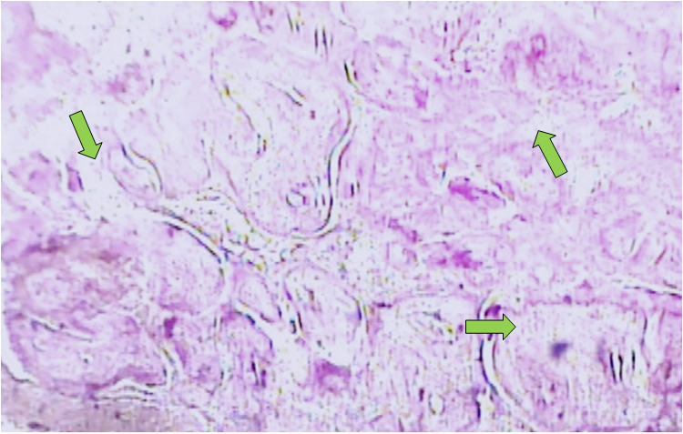 | Figure 1. Dystrophic and necrotic changes in the chorionic tissue of monochorionic twins. Stained with hematoxylin-eosin. Ob.40, ok.10 |
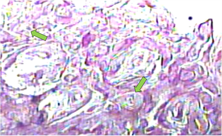 | Figure 2. Dystrophic and necrotic changes in the chorionic tissue of monochorionic twins. Stained with hematoxylin-eosin. Ob.40, ok.10 |
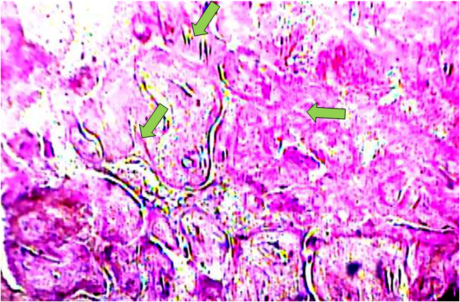 | Figure 3. Fibrous tissue growth and necrotic changes in the placenta of monochorionic twins. Hematoxylin-eosin stained. Ob.40, ok.10 |
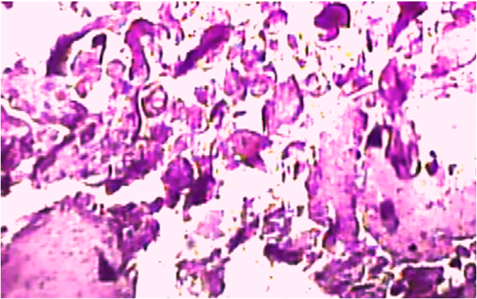 | Figure 4. Fibrous tissue growth and necrotic changes in the placenta of monochorionic twins. Hematoxylin-eosin stained. Ob.40, ok.10 |
4. Conclusions
- Thus, the pathomorphological changes in the placental tissue of monochorionic twins differ depending on the duration and degree of prematurity. In the placental tissue of monochorionic twins with varying degrees of prematurity, ischemia of the placental villi, fibrinoid necrosis, and growth of the terminal villi are detected. Pathomorphological changes such as intervillous fibrin deposition, fibrinoid necrosis, pronounced fibrosis of the villi, and thrombosis of small blood vessels are detected. As the gestation period increases, the intensity of the formation of fibrous tissue increases. This indicates the degree to which the placental tissue affects the development of the fetus.