-
Paper Information
- Next Paper
- Previous Paper
- Paper Submission
-
Journal Information
- About This Journal
- Editorial Board
- Current Issue
- Archive
- Author Guidelines
- Contact Us
American Journal of Medicine and Medical Sciences
p-ISSN: 2165-901X e-ISSN: 2165-9036
2025; 15(5): 1465-1467
doi:10.5923/j.ajmms.20251505.34
Received: Apr. 19, 2025; Accepted: May 16, 2025; Published: May 24, 2025

Pathomorphology of Endometrial Hyperplasia Detected by Ultrasound in Women Due to Abnormal Bleeding
 Abstract
 Reference
 Full-Text PDF
 Full-text HTML
Khamidov Obid Abdurakhmanovich1, Jumanov Ziyadulla Eshmamatovich2, Usarov Mukhriddin Shukhratovich3
1Associate Professor Department of Medical Radiology, Faculty of Postgraduate Education, Samarkand State Medical University, Doctor of Medical Sciences (DSc), Associate Professor, Uzbekistan
2Professor of the Department of Pathological Anatomy of Samarkand State Medical University, Doctor of Medical Sciences (DSc), Uzbekistan
31st year Basic Doctoral Student of Medical Radiology of the Faculty of Postgraduate Education of Samarkand State Medical University, Uzbekistan
Correspondence to: Khamidov Obid Abdurakhmanovich, Associate Professor Department of Medical Radiology, Faculty of Postgraduate Education, Samarkand State Medical University, Doctor of Medical Sciences (DSc), Associate Professor, Uzbekistan.
| Email: |  |
Copyright © 2025 The Author(s). Published by Scientific & Academic Publishing.
This work is licensed under the Creative Commons Attribution International License (CC BY).
http://creativecommons.org/licenses/by/4.0/

In order to investigate the hyperplasia of the endometrium layer detected by ultrasound examination due to abnormal bleeding in women, uterine scrapings from 51 patients were examined. It was found that the hyperplasia of the uterine endometrium layer detected by ultrasound examination is of various forms, most of which are simple hyperplasia. It is noted that the incidence of simple atypical hyperplasia of the endometrium has increased recently. It is noted that complex atypical hyperplasia is rare among the patients studied, and it is noted that the detection of squamous cell metaplasia in most of them does not mean that it has transformed into adenocarcinoma.
Keywords: Abnormal bleeding, Ultrasound examination, Uterus, Endometrium, Hyperplasia
Cite this paper: Khamidov Obid Abdurakhmanovich, Jumanov Ziyadulla Eshmamatovich, Usarov Mukhriddin Shukhratovich, Pathomorphology of Endometrial Hyperplasia Detected by Ultrasound in Women Due to Abnormal Bleeding, American Journal of Medicine and Medical Sciences, Vol. 15 No. 5, 2025, pp. 1465-1467. doi: 10.5923/j.ajmms.20251505.34.
1. Introduction
- The number of patients with various pathologies of the endometrium of the uterus in women around the world is increasing day by day [1]. Also, the most frequently detected pathological condition among endometrial pathologies is endometrial hyperplasia [3]. A number of metabolic processes play a role in the development of endometrial hyperplasia: hyperestrogenism, endometrial cells, endometrial receptor retinal cells and dysfunction [4,6,7]. The general features of endometrial hyperplasia are transformed cells and hypertrophic sclerosis [2]. At the same time, the problem of endometrial hyperplasia remains relevant due to the diseases associated with the functions of the female reproductive system, as well as a decrease in the quality of life and the need for hospitalization for interventions such as removal of the endometrial layer and cleaning of the mucous membrane [5].The purpose of the study: Conducting a pathomorphological analysis of endometrial hyperplasia detected by ultrasound examination in women with abnormal bleeding.
2. Materials and Methods
- Endometrial scrapings from 51 patients with endometrial hyperplasia detected by ultrasound were examined at the Department of Gynecology of the Multidisciplinary Clinic of Samarakand State Medical University. Histological sections were stained with hematoxylin-eosin and subjected to pathomorphological analysis.Hematoxylin and eosin staining procedure:It is the most widely used method for staining histological sections.Sections are deparaffinized in chloroform, washed in distilled water, then a solution of hematoxylin is dripped onto the surface of the section and fixed for 3 minutes. It is washed in running water for 10 minutes and stained with eosin for 0.2 to 3 minutes. Dehydrated in 70° and 96° alcohol, transferred to carbol-xylene and xylene and covered with balsam.Result: cell nuclei are stained blue-black, cytoplasm - dark purple.Histological preparations were studied and photographed using a LeicaGME microscope (Leica, India) coupled with a LeicaEC3 digital camera (Leica, Singapore) and a Pentium IV computer. Photo processing was carried out using Windows Professional applications.
3. Results and Discussion
- The results of the study show that microscopic examination of preparations prepared from uterine scrapings reveals thickening of the endometrial layer, an increase in the number of glands and stromal tissue elements. Enlargement of the glands of various shapes and sizes is noted. In particular, cystic enlargement of the glands is observed (Figure 1).
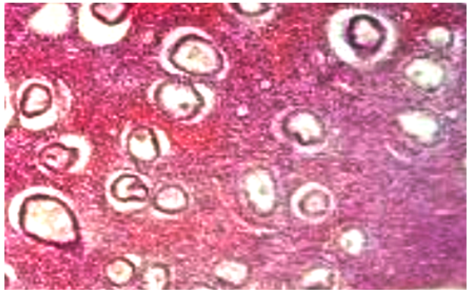 | Figure 1. Cystic enlargement (hyperplasia) of the glands of the uterine endometrium. Stained with hematoxylin-eosin. Ob.40, ok.10 |
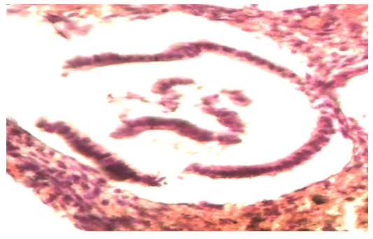 | Figure 2. Cystic dilated glands of the uterine endometrium contain cells of the same size and shape. Stained with hematoxylin-eosin. Ob.40, ok.10 |
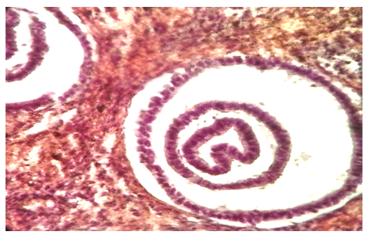 | Figure 3. Formation of glands within the glands located in the endometrial layer of the uterus (endometrial hyperplasia). Stained with hematoxylin-eosin. Ob.40, ok.10 |
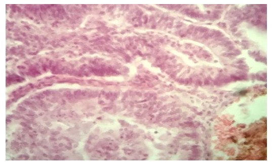 | Figure 4. Hyperplasia of the uterine endometrium. Malignant state of the glands and signs of adenocarcinoma. Stained with hematoxylin-eosin. Ob.40, ok.10 |
 | Figure 5. Hyperplasia of the uterine endometrial layer. Signs of adenocarcinoma of the glands. Stained with hematoxylin-eosin. Ob.40, ok.10 |
4. Conclusions
- Thus, the various forms of hyperplasia of the uterine endometrium detected by ultrasound are diverse, the majority of which is simple hyperplasia. In addition, many glands are formed inside the glands, which are even located in three layers. A recent increase in simple atypical hyperplasia of the endometrium is noted. Complex atypical hyperplasia is rare among the patients studied, and in most of them the detection of squamous cell metaplasia does not mean that it has transformed into adenocarcinoma.