-
Paper Information
- Next Paper
- Previous Paper
- Paper Submission
-
Journal Information
- About This Journal
- Editorial Board
- Current Issue
- Archive
- Author Guidelines
- Contact Us
American Journal of Medicine and Medical Sciences
p-ISSN: 2165-901X e-ISSN: 2165-9036
2025; 15(5): 1463-1464
doi:10.5923/j.ajmms.20251505.33
Received: Apr. 12, 2025; Accepted: May 9, 2025; Published: May 24, 2025

Characteristics of Pathomorphology Changes in Knee Joint Structures in Secondary Form of Gonartirosa
 Abstract
 Reference
 Full-Text PDF
 Full-text HTML
Ochilov Jamshid Temur Ugli1, Mamadiyarova Dilfuza Umirzakovna2
1Independent Researcher of the Department of Pathological Anatomy of Samarkand State Medical University, Uzbekistan
2Associate Professor, Department of Pedagogy and Psychology, Samarkand State Medical University, Uzbekistan
Correspondence to: Ochilov Jamshid Temur Ugli, Independent Researcher of the Department of Pathological Anatomy of Samarkand State Medical University, Uzbekistan.
| Email: |  |
Copyright © 2025 The Author(s). Published by Scientific & Academic Publishing.
This work is licensed under the Creative Commons Attribution International License (CC BY).
http://creativecommons.org/licenses/by/4.0/

A total of 35 patients who underwent surgical procedures for the development of secondary gonarthrosis underwent pathomorphological examination of surgical materials from the knee joint in order to determine the specificity of morphological and morphometric pathomorphological changes in the structures of the knee joint. It was found that in gonarthrosis, in parallel with the development of dystrophic changes in the hyaline cartilage tissue of the knee joint surface, dystrophic changes in the subchondral bone tissue are actively developing. In blood vessels, edema of the intimal layer is noted, obliteration process.
Keywords: Gonarthrosis, Knee joint, Synovial membrane, Dystrophy, Sclerosis
Cite this paper: Ochilov Jamshid Temur Ugli, Mamadiyarova Dilfuza Umirzakovna, Characteristics of Pathomorphology Changes in Knee Joint Structures in Secondary Form of Gonartirosa, American Journal of Medicine and Medical Sciences, Vol. 15 No. 5, 2025, pp. 1463-1464. doi: 10.5923/j.ajmms.20251505.33.
1. Introduction
- Gonarthrosis is a disease based on damage to the joint (its cartilage, synovial membrane, capsule) and the surrounding tissues (ligaments and muscles), accompanied by impaired mobility and support function. Osteoarthritis is a health problem that is becoming increasingly serious due to the prevalence of obesity and the increase in the number of elderly people in the population. Osteoarthritis is found everywhere [1]. In the United States, 21 million people (approximately 7% of the population) suffer from it. According to epidemiological studies, more than 14% of the world's population suffers from knee osteoarthritis, and in recent years, disability due to it has increased 3-5 times [3]. Women are almost twice as likely to develop osteoarthritis as men. They suffer almost twice as much. Two-thirds of patients are people of working age, aged 40-60 years [2,4].The purpose of the study: To identify the specific features of pathomorphological changes in the structures of the knee joint in the secondary form of gonarthrosis.
2. Materials and Methods
- Surgical materials from the knee joints of a total of 28 patients who underwent surgical procedures for the secondary form of gonarthrosis at the Samarkand branch of the Republican Specialized Traumatology and Orthopedics Scientific-Practical Medical Center were studied pathomorphologically at the Department of Pathological Anatomy of the Multidisciplinary Clinic of Samarakand State Medical University. Microscopic methods were used to assess the morphological and morphometric changes in the structures of the knee joint in the secondary form of gonarthrosis. The obtained tissue fragments were fixed in 10% neutral formalin, passed through an alcohol battery, and paraffin blocks were prepared. The prepared histological sections were stained with hematoxylin and eosin, according to Van-Gieson. Microphotographic techniques were conducted. Histological preparations were studied and photographed using a LeicaGME microscope (Leica, India) coupled with a LeicaEC3 digital camera (Leica, Singapore) and a Pentium IV computer. Photo processing was carried out using Windows Professional applications.
3. Results and Discussion
- Patients with secondary gonarthrosis are athletes who have suffered injuries to the knee joint, people who are constantly in motion, and patients with obesity, most of whom are female patients. When the chondrocyte surface of the knee joint removed due to surgery was subjected to pathological examination, it was found that the amount of hyaline cartilage decreased, that is, its thickness became 2.82-3.21 mm, and due to the development of degenerative and dystrophic changes in chondrocytes, their size averaged 21.3±2.1 μm (Figure 1).
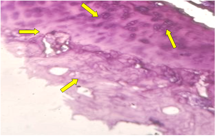 | Figure 1. Dystrophic and necrobiotic changes in hyaline cartilage and subchondral bone tissue. Hematoxylin-eosin stained. Ob.40, ok.10 |
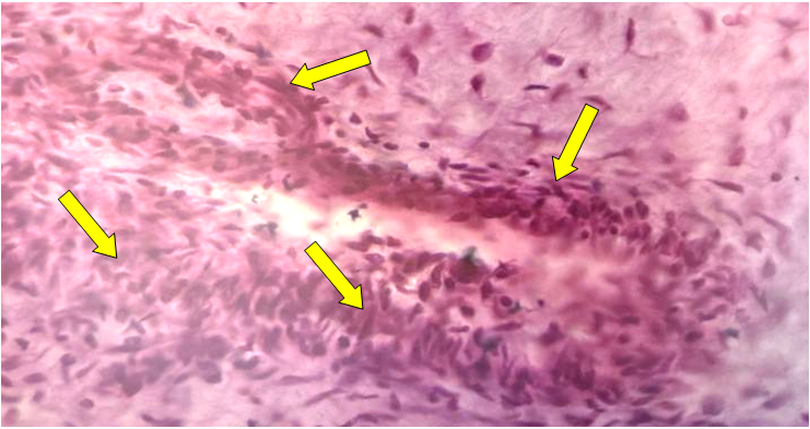 | Figure 2. Swelling of blood vessels and collagen fibers in the periarticular tissues. Stained with hematoxylin-eosin. Ob.40, ok.10 |
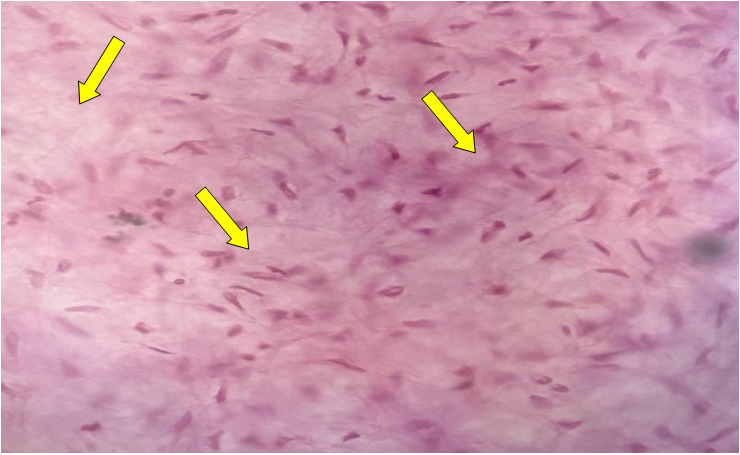 | Figure 3. Changes in periarticular tissues. In the secondary form of gonarthrosis, there is a sharp proliferation of fibrocytes in the synovial tissue of the knee joint. Stained with hematoxylin-eosin. Ob.40, ok.10 |
4. Conclusions
- Thus, in secondary gonarthrosis, in parallel with the development of dystrophic changes in the hyaline cartilage of the knee joint surface, dystrophic changes in the subchondral bone tissue are actively developing. In the blood vessels, the process of obliteration of the intimal layer is noted. In the tissues around the joint, the process of proliferation of cells, tissue fibrosis, and edema of the interstitial tissue are noted. An increased number of adipocytes is detected in the interstitial spaces of the connective tissue. In the blood vessels, edema of the intima and hydropic dystrophy and edema of the nuclei of endothelial cells are observed. In the blood vessels, edema of the collagen and elastic fibers in the walls of the blood vessels, narrowing (obliteration) of their lumens is detected.