-
Paper Information
- Next Paper
- Previous Paper
- Paper Submission
-
Journal Information
- About This Journal
- Editorial Board
- Current Issue
- Archive
- Author Guidelines
- Contact Us
American Journal of Medicine and Medical Sciences
p-ISSN: 2165-901X e-ISSN: 2165-9036
2025; 15(5): 1457-1459
doi:10.5923/j.ajmms.20251505.31
Received: Apr. 17, 2025; Accepted: May 16, 2025; Published: May 24, 2025

Pathomorphology of Structural Changes in Brain Neurons of Patients Who Died from Chronic Ischaemic Heart Disease
 Abstract
 Reference
 Full-Text PDF
 Full-text HTML
Jumanov Ziyadulla Eshmamatovich1, Khasanov Farrukhjon Sherali Ugli2
1Professor, Doctor of Medical Sciences, Department of Pathological Anatomy, Samarkand State Medical University, Uzbekistan
2Independent Researcher of the Department of Pathological Anatomy of Samarkand State Medical University, Uzbekistan
Correspondence to: Jumanov Ziyadulla Eshmamatovich, Professor, Doctor of Medical Sciences, Department of Pathological Anatomy, Samarkand State Medical University, Uzbekistan.
| Email: |  |
Copyright © 2025 The Author(s). Published by Scientific & Academic Publishing.
This work is licensed under the Creative Commons Attribution International License (CC BY).
http://creativecommons.org/licenses/by/4.0/

A total of 42 brain tissue sections from patients who died of chronic ischemic heart disease were subjected to pathomorphological examination. It was found that in chronic ischemic heart disease, ischemic and dystrophic changes develop in neurons in the cortex of the cerebral hemispheres. Also, varying degrees of expansion of the pericellular space and lightening of the neuropil are noted. It is emphasized that such changes should be taken into account in the prevention and treatment of chronic ischemic heart disease.
Keywords: Chronic ischemic heart disease, Brain, Neuron, Pericellular space, Neuropil, Dystrophy
Cite this paper: Jumanov Ziyadulla Eshmamatovich, Khasanov Farrukhjon Sherali Ugli, Pathomorphology of Structural Changes in Brain Neurons of Patients Who Died from Chronic Ischaemic Heart Disease, American Journal of Medicine and Medical Sciences, Vol. 15 No. 5, 2025, pp. 1457-1459. doi: 10.5923/j.ajmms.20251505.31.
1. Introduction
- On a global scale, ischemic heart disease is still one of the most pressing problems facing medicine today. Despite modern advances in the prevention and treatment of cardiovascular diseases, ischemic heart disease still occupies one of the leading positions [2,5,7]. In developed countries, mortality from this disease accounts for 1/3 of all deaths. More than 1 million people die from this disease every year. Changes in brain structures in chronic ischemic heart disease remain one of the pressing problems facing medicine, which has not been fully elucidated to date and is awaiting its solution [1,3,4,6]. Chronic ischemic heart disease has been a leading cause of death in economically developed countries for many years worldwide [8]. In 2018, it amounted to 17.3 million people and, according to a number of forecasts, will only grow and will amount to 23.6 million people by 2030 [9,10]. Based on 2018 statistics, acute heart attack is a frequent manifestation of cardiovascular diseases, and angina pectoris, the first sign of pathology, occurs in approximately 50% of patients [11].The purpose of the study: To determine the pathomorphology of structural changes in brain neurons in patients who died from chronic ischemic heart disease.
2. Materials and Methods
- Tissue sections taken from the brains of 42 patients who died of chronic ischemic heart disease in the departments of therapy and intensive care of the multidisciplinary clinic of the Samarkand State Medical University were studied in the Department of Pathological Anatomy. Anamnestic, macroscopic, microscopic, morphometric and statistical research methods were used to assess the relationship between morphological and morphometric changes in the neuronal structures of the brain and chronic ischemic heart disease. Sections were taken from the cortex and subcortical areas of the cerebral hemispheres. The size of the sections was 1x1 cm, the thickness did not exceed 0.5 cm, they were fixed in 10% neutral formalin, passed through an alcohol battery, and paraffin blocks were prepared. Histological sections 7-10 microns thick were stained with hematoxylin and eosin, Van Gieson and Nissl (nervous tissue) methods. Microphotographic techniques were conducted.Histological preparations were studied and photographed using a LeicaGME microscope (Leica, India) coupled with a LeicaEC3 digital camera (Leica, Singapore) and a Pentium IV computer. Photo processing was carried out using Windows Professional applications.
3. Results and Discussion
- The results of the study show that ischemic-type changes are observed in neurons in the superficial layers of the cerebral cortex of patients who died of chronic ischemic heart disease (Figure 1).
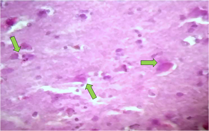 | Figure 1. Development of ischemic-type changes in brain neurons. Stained with hematoxylin-eosin. Ob.40, ok.10 |
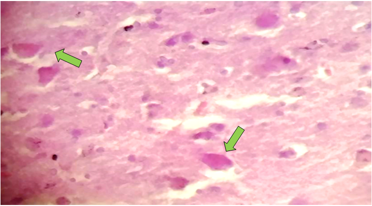 | Figure 2. Necrobiotic and necrotic changes in brain neurons. Stained with hematoxylin-eosin. Ob.40, ok.10 |
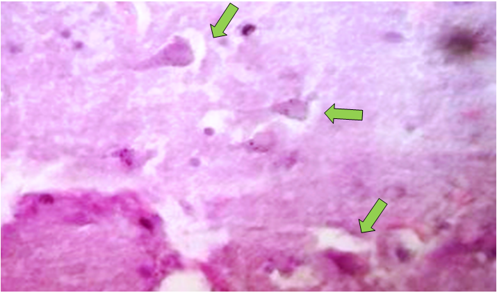 | Figure 3. The state of the space around the neurons of the brain (expansion of the pericellular space). Stained with hematoxylin-eosin. Ob.40, ok.10 |
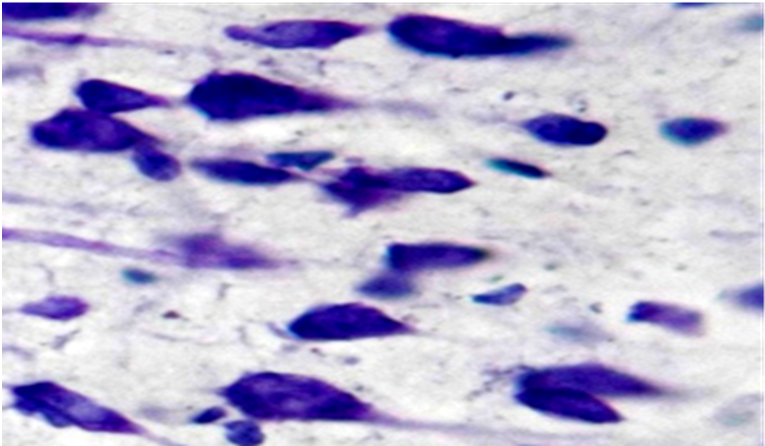 | Figure 4. Perinuclear chromolysis and necrotic changes in brain neurons. Stained by Nissl method. Ob.40, ok.10 |
4. Conclusions
- Thus, in chronic ischemic heart disease, ischemic and dystrophic changes develop in neurons in the cortex of the cerebral hemispheres. Also, varying degrees of expansion of the pericellular space and lightening of the neuropil are noted. Dystrophic changes in neurons are detected, and a decrease in the number of gliocytes is noted. Increased lightening of the neuropil is observed. Necrotic changes in a number of neurons and in the form of karyolysis are observed. Such changes should be taken into account in the prevention and treatment of chronic ischemic heart disease.