-
Paper Information
- Next Paper
- Previous Paper
- Paper Submission
-
Journal Information
- About This Journal
- Editorial Board
- Current Issue
- Archive
- Author Guidelines
- Contact Us
American Journal of Medicine and Medical Sciences
p-ISSN: 2165-901X e-ISSN: 2165-9036
2025; 15(5): 1454-1456
doi:10.5923/j.ajmms.20251505.30
Received: Apr. 12, 2025; Accepted: May 11, 2025; Published: May 24, 2025

Specific Aspects of the Dependence of Morphological Changes of Skin Structures in Psoriasis on Hot Climatic Conditions
 Abstract
 Reference
 Full-Text PDF
 Full-text HTML
Jumanov Ziyadulla Eshmamatovich1, Nazarov Jamshid Abdurashidovich2
1Professor, Department of Pathological Anatomy, Samarkand State Medical University, Doctor of Medical Sciences (DSc), Uzbekistan
2Independent Researcher of the Department of Pathological Anatomy of Samarkand State Medical University, Uzbekistan
Correspondence to: Jumanov Ziyadulla Eshmamatovich, Professor, Department of Pathological Anatomy, Samarkand State Medical University, Doctor of Medical Sciences (DSc), Uzbekistan.
| Email: |  |
Copyright © 2025 The Author(s). Published by Scientific & Academic Publishing.
This work is licensed under the Creative Commons Attribution International License (CC BY).
http://creativecommons.org/licenses/by/4.0/

In order to determine the specific aspects of the morphological and morphometric changes of skin structures in psoriasis, which are associated with hot climate conditions, a pathomorphological examination of biopsies taken from the skin of 66 patients in the summer season was conducted. It was found that in the autumn season, spongiosis is observed in the spinous layer of the skin structures of patients with psoriasis. Perivascular edema also develops strongly in the dermis. It is emphasized that this should be taken into account when treating the disease.
Keywords: Psoriasis, Keratinocytes, Dystrophy, Epidermis, Dermis, Edema
Cite this paper: Jumanov Ziyadulla Eshmamatovich, Nazarov Jamshid Abdurashidovich, Specific Aspects of the Dependence of Morphological Changes of Skin Structures in Psoriasis on Hot Climatic Conditions, American Journal of Medicine and Medical Sciences, Vol. 15 No. 5, 2025, pp. 1454-1456. doi: 10.5923/j.ajmms.20251505.30.
1. Introduction
- Psoriasis is one of the most common skin diseases worldwide. Most studies today report a prevalence of 1–5%, with significant variations depending on climate and geographical region [1,2,3]. For example, in Scandinavian countries and among indigenous populations in the far north of Russia, the incidence is high (up to 4%), while in Kuwait it is 0.11%. These are low rates compared to global rates [4,5,6].The purpose of the study: To determine the specific aspects of the dependence of morphological changes in skin structures in psoriasis on hot climatic conditions.
2. Materials and Methods
- Biopsies taken from the skin of 66 patients registered in the Samarkand regional branch of the Republican Scientific Center for Dermatovenerology and Cosmetology and undergoing regular treatment during the summer season were studied at the Department of Pathological Anatomy of the Multidisciplinary Clinic of Samarakand State Medical University. Macroscopic, microscopic, methods were used to assess the dependence of morphological changes in skin structures in psoriasis on climatic conditions. Histological sections prepared from the slices were stained with hematoxylin-eosin.Hematoxylin-eosin staining procedure:It is the most widely used method for staining histological sections.Sections are deparaffinized in chloroform, washed in distilled water, then a solution of hematoxylin is dripped onto the surface of the section and fixed for 3 minutes. It is washed in running water for 10 minutes and stained with eosin for 0.2 to 3 minutes. Dehydrated in 70° and 96° alcohol, transferred to carbol-xylene and xylene and covered with balsam.Result: cell nuclei are stained blue-black, cytoplasm - dark purple.Histological preparations were studied and photographed using a LeicaGME microscope (Leica, India) coupled with a LeicaEC3 digital camera (Leica, Singapore) and a Pentium IV computer. Photo processing was carried out using Windows Professional applications.Gallbladder adenoma was morphologically evaluated in prepared micropreparations.
3. Results and Discussion
- The results of the study conducted in the summer season show that in the summer months, when examined under a microscope, vacuolar dystrophy of the cells of the basal layer, a decrease in their number, necrosis of basal keratinocytes and the development of leuko-lymphocytic infiltration in their atrophy are detected. Mitotic division is noted in the remaining cells of the basal layer. In particular, the basal layer cells are prismatic in shape, the nucleus is located in the center, and mitotic division is detected in most cells. Rows of spinous layer cells are in a state of division. Perinuclear swelling, caripyknosis, karyrexis, and karyolysis in the nuclei of most cells are detected in the cells. The number of granular layer cells has sharply increased, their granularity has increased, and mitotic divisions are clearly visible. The stratum corneum is thin, in most places it is not detected. The stratum corneum is thickened, divided into layers, its cells are separated from each other, and hyperkeratosis is detected (Figure 1).
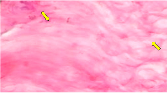 | Figure 1. Hyperkeratosis in psoriasis in warm climates. Stained with hematoxylin-eosin. Ob.40, ok.10. Ob.40, ok.10 |
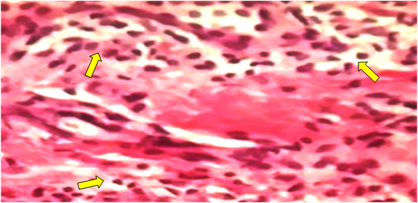 | Figure 2. In the superficial layer of the dermis, there is an inflammatory infiltrate of lymphocytes and histiocytes with an admixture of neutrophilic granulocytes. Stained with hematoxylin-eosin. Ob.40, ok.10 |
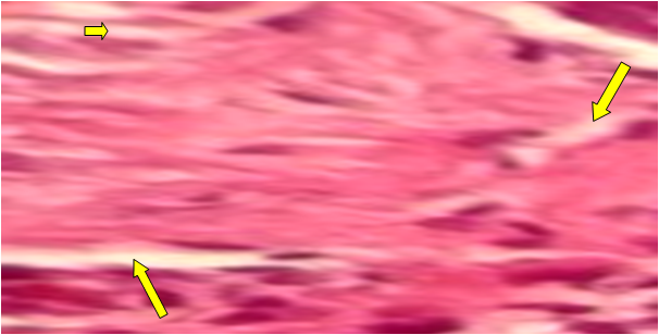 | Figure 3. In psoriasis, the reticular layer is a poorly developed tumor with well-developed collagen and elastic fibers.. Stained with hematoxylin-eosin. Ob.40, ok.10 |
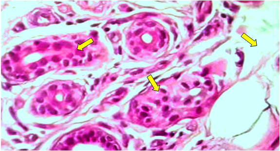 | Figure 4. Dystrophic changes in sweat and sebaceous glands in psoriasis during the summer season. Stained with hematoxylin-eosin. Ob.40, ok.10 |
4. Conclusions
- Thus, in the skin structures of patients with psoriasis, spongiosis is observed in the spinous layer in the summer season. The sebaceous layer of the dermis is well developed, collagen and reticulin fibers develop slightly. Destruction of elastic fibers and a decrease in their number are detected. Collagen and elastic fibers are well developed in the reticular layer. Blood vessels are dilated. Up to 6-8 hair follicles, 5-6 sweat and sebaceous glands are detected in one field of view. The sebaceous layer is well developed. These changes should be taken into account when treating psoriasis in the summer season.