-
Paper Information
- Next Paper
- Previous Paper
- Paper Submission
-
Journal Information
- About This Journal
- Editorial Board
- Current Issue
- Archive
- Author Guidelines
- Contact Us
American Journal of Medicine and Medical Sciences
p-ISSN: 2165-901X e-ISSN: 2165-9036
2025; 15(5): 1451-1453
doi:10.5923/j.ajmms.20251505.29
Received: Apr. 15, 2025; Accepted: May 12, 2025; Published: May 24, 2025

Pathomorphological Analysis of Tissue Sections Taken from the Renal Pelvis in Nephrolithiasis
 Abstract
 Reference
 Full-Text PDF
 Full-text HTML
Jumanov Ziyadulla Eshmamatovich1, Davronov Oybek Otabek Ugli2
1Professor, Doctor of Medical Sciences, Department of Pathological Anatomy, Samarkand State Medical University, Uzbekistan
2Independent Researcher of the Department of Pathological Anatomy of Samarkand State Medical University, Uzbekistan
Correspondence to: Jumanov Ziyadulla Eshmamatovich, Professor, Doctor of Medical Sciences, Department of Pathological Anatomy, Samarkand State Medical University, Uzbekistan.
| Email: |  |
Copyright © 2025 The Author(s). Published by Scientific & Academic Publishing.
This work is licensed under the Creative Commons Attribution International License (CC BY).
http://creativecommons.org/licenses/by/4.0/

Pathomorphological examination of kidney tissue samples removed from 48 patients with kidney stones was performed. It was found that in kidney stones, swelling of the mucous membrane of the renal pelvis, dystrophic, necrobiotic and necrotic changes in epithelial cells are observed. Swelling and fibrosis are noted in the muscle layer. Sclerotic processes have developed in the adventitia layer. It is emphasized that when developing measures for the treatment of kidney stones, it is necessary to take into account the increased activity of changes in the renal pelvis.
Keywords: Kidney stones, Pelvis, Dystrophy, Swelling, Karyopyknosis, Karyorrhexis and karyolysis
Cite this paper: Jumanov Ziyadulla Eshmamatovich, Davronov Oybek Otabek Ugli, Pathomorphological Analysis of Tissue Sections Taken from the Renal Pelvis in Nephrolithiasis, American Journal of Medicine and Medical Sciences, Vol. 15 No. 5, 2025, pp. 1451-1453. doi: 10.5923/j.ajmms.20251505.29.
1. Introduction
- The causes and mechanisms of lithogenesis in urolithiasis remain relevant, as they are not fully understood [1,5]. Existing theories explain only individual links in pathogenesis. However, it is not clearly established whether the factors leading to nephrolithiasis act alone or together, in various combinations [2,7]. The initial stages of crystallization processes, the formation of urinary stones and the accompanying morphofunctional and ultrastructural changes in the tissue elements of the kidney are currently insufficiently studied [3,6]. This is due to the fact that in humans these changes often occur asymptomatically [4,8].The purpose of the study: Identification of pathomorphological changes in pelvis structures in nephrolithiasis.
2. Materials and Methods
- In the Department of Urology of the Multidisciplinary Clinic of Samarkand State Medical University, sections of kidney tissue removed from 48 patients due to kidney stone disease were studied in the Department of Pathological Anatomy. Microscopic methods were used to assess morphological changes in the structures of the renal pelvis in kidney stone disease. 1x1x0.5 cm tissue sections were obtained from the kidney tissue removed due to kidney stone disease. The obtained tissue sections were fixed in 10% neutral formalin, passed through an alcohol accumulator, and paraffin blocks were prepared. The prepared histological sections were stained with hematoxylin and eosin according to Van Gieson. Microphotographic techniques were performed. Histological preparations were studied and photographed using a LeicaGME microscope (Leica, India) coupled with a LeicaEC3 digital camera (Leica, Singapore) and a Pentium IV computer. Photo processing was carried out using Windows Professional applications.
3. Results and Discussion
- Microscopic examination of tissue sections from the renal pelvis of 48 patients who underwent nephroectomy with a diagnosis of nephrolithiasis was performed. The results of the pathomorphological study show that when examining the renal pelvis of patients with advanced nephrolithiasis under a microscope, swelling of the mucous membrane and its thinning are observed. Hydropic dystrophy, swelling of the nucleus and dim staining with hematoxylin-eosin are noted in the cytoplasm of multilayered transitional epithelial cells. Karyopyknosis, karyorrhexis and karyolysis are detected in the nuclei of many epithelial cells. The presence of cells with invisible nuclei is noted in their interstices. Cytorrhexis and cytolysis are detected in a group of cells. In most areas, foci of desquamation are detected. In the sparse connective tissue of the mucosal lamina propria, edema of the interstitial tissue, vascular congestion, and lymphocytic infiltration around the vessels are detected. In the sparse connective tissue fibers of the submucosal base, edema and sparseness of their fibers are noted (Figure 1).
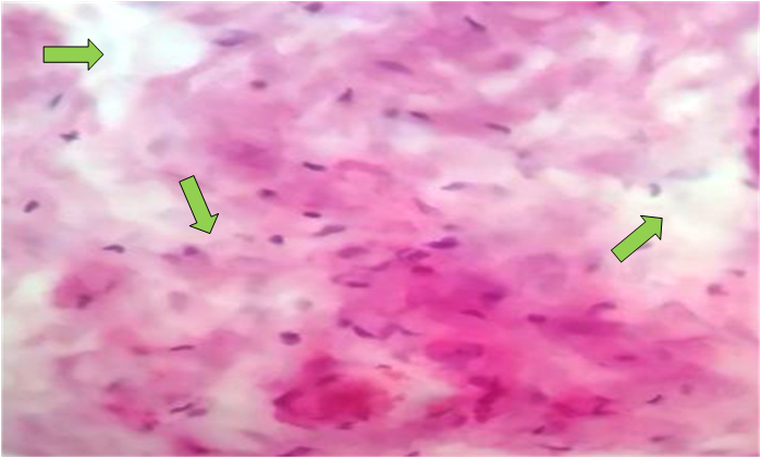 | Figure 1. Swelling of the connective tissue fibers of the renal pelvis. Stained with hematoxylin-eosin. Ob.40, ok.10 |
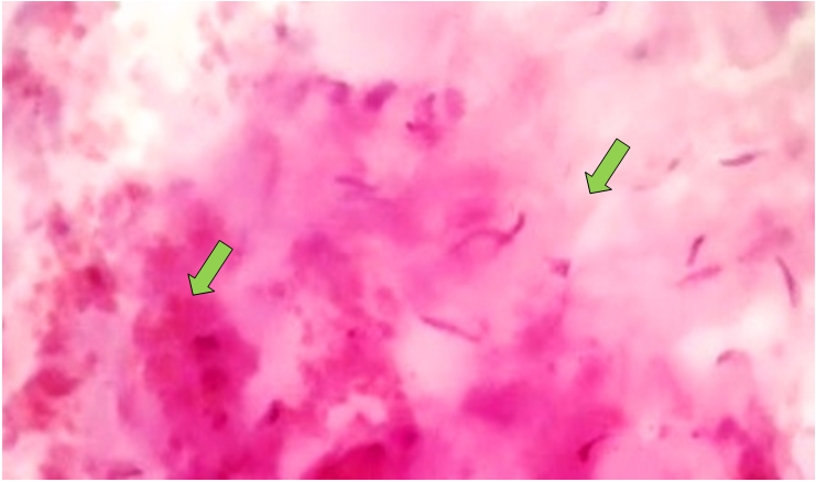
 | Figure 2. Sclerotic changes in the walls of the renal pelvis blood vessels. Stained with hematoxylin-eosin. Ob.40, ok.10 |
 | Figure 3. Swelling and diapedetic hemorrhages in the adventitia layer of the renal pelvis. Stained with hematoxylin-eosin. Ob.40, ok.10 |
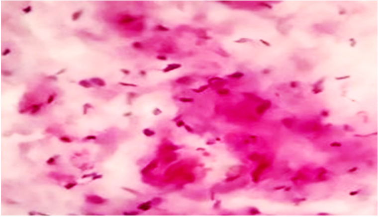 | Figure 4. Swelling in the interstitial and pericellular areas of the renal pelvis. Stained with hematoxylin-eosin. Ob.40, ok.10 |
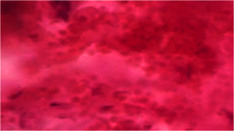 | Figure 5. Diapedetic hemorrhages in the walls of the renal pelvis. Stained with hematoxylin-eosin. Ob.40, ok.10 |
4. Conclusions
- Thus, in kidney stone disease, swelling of the mucous membrane of the renal pelvis, the development of dystrophic, necrobiotic and necrotic changes in epithelial cells are observed. Swelling of the interstitial tissue of the internal longitudinal smooth muscles of the muscular layer and diffuse and fibrous myocytes are observed. Fibrosis and interstitial swelling of the internal longitudinal muscles and external circular muscles are noted. Dilatation and uneven filling of the blood vessels located between them are determined. The development of sclerotic processes is determined in the adventitia layer. When developing measures for the treatment of kidney stone disease, the increased activity of changes in the renal pelvis should be taken into account.