-
Paper Information
- Next Paper
- Paper Submission
-
Journal Information
- About This Journal
- Editorial Board
- Current Issue
- Archive
- Author Guidelines
- Contact Us
American Journal of Medicine and Medical Sciences
p-ISSN: 2165-901X e-ISSN: 2165-9036
2025; 15(5): 1442-1444
doi:10.5923/j.ajmms.20251505.26
Received: Apr. 17, 2025; Accepted: May 19, 2025; Published: May 24, 2025

Patomorphological Aspects of Changes in Skin Structures in Patients with Psoriasis of the Palm of the Hand
 Abstract
 Reference
 Full-Text PDF
 Full-text HTML
Abdullaev Sulton Davlat Ugli1, Mamadiyarova Dilfuza Umirzakovna2
1Samarkand Branch of the Republican Scientific Center of Dermatovenerology and Cosmetology, Doctor of Dermatovenerology, Cosmetologist, Pathologist, Doctor of Philosophy in Medical Sciences (PhD), Uzbekistan
2Associate Professor, Department of Pedagogy and Psychology, Samarkand State Medical University, Uzbekistan
Correspondence to: Abdullaev Sulton Davlat Ugli, Samarkand Branch of the Republican Scientific Center of Dermatovenerology and Cosmetology, Doctor of Dermatovenerology, Cosmetologist, Pathologist, Doctor of Philosophy in Medical Sciences (PhD), Uzbekistan.
| Email: |  |
Copyright © 2025 The Author(s). Published by Scientific & Academic Publishing.
This work is licensed under the Creative Commons Attribution International License (CC BY).
http://creativecommons.org/licenses/by/4.0/

Pathomorphological examination of biopsy specimens from the palm skin of 35 patients was performed. It was found that patients with palm psoriasis have a strong development of spongiosis in the skin structures - the ticanaximon layer. A strong development of perivascular edema is also observed in the dermis layer. This is an important process in the implementation of therapeutic procedures.
Keywords: Psoriasis, Kerotinosit, Dystrophy, Epidermis, Derma, Edema
Cite this paper: Abdullaev Sulton Davlat Ugli, Mamadiyarova Dilfuza Umirzakovna, Patomorphological Aspects of Changes in Skin Structures in Patients with Psoriasis of the Palm of the Hand, American Journal of Medicine and Medical Sciences, Vol. 15 No. 5, 2025, pp. 1442-1444. doi: 10.5923/j.ajmms.20251505.26.
1. Introduction
- Psoriasis is one of the most common chronic dermatoses. The prevalence of this disease in the general population is on average 1-3% in mid-latitudes, and the total number of patients exceeds 200 million people [1,4,5,7]. However, the true incidence of psoriasis has not yet been determined, and the current prevalence and prevalence data are uncertain in different regions of the world. According to data compiled by various researchers, the proportion of patients with psoriasis among dermatological patients is 2-4% [2,3,6].The purpose of the study: to determine the patomorphological aspects of changes in skin structures in patients with psoriasis of the palm of the hand.
2. Materials and Methods
- A total of 35 patients registered at the Samarkand branch of the Republican Scientific Center for Dermatovenerology and Cosmetology and undergoing regular treatment were studied at the Department of Pathological Anatomy of the Multidisciplinary Clinic of Samarakand State Medical University, using biopsies taken from the skin of the palms. Anamnetic, macroscopic, microscopic, morphometric and statistical research methods were used to assess the dependence of morphological and morphometric changes in skin structures in psoriasis on age and climatic conditions. Histological sections prepared from the slices were stained with hematoxylin and eosin.Hematoxylin and eosin staining procedure:It is the most widely used method for staining histological sections.Sections are deparaffinized in chloroform, washed in distilled water, then a solution of hematoxylin is dripped onto the surface of the section and fixed for 3 minutes. It is washed in running water for 10 minutes and stained with eosin for 0.2 to 3 minutes. Dehydrated in 70° and 96° alcohol, transferred to carbol-xylene and xylene and covered with balsam.Result: cell nuclei are stained blue-black, cytoplasm - dark purple.Histological preparations were studied and photographed using a LeicaGME microscope (Leica, India) coupled with a LeicaEC3 digital camera (Leica, Singapore) and a Pentium IV computer. Photo processing was carried out using Windows Professional applications.Gallbladder adenoma was morphologically evaluated in prepared micropreparations.
3. Results and Discussion
- The results of the study show that when examined under a microscope in the summer months, vacuolar dystrophy of the cells of the basal layer, a decrease in their number, and the development of necrosis of basal keratinocytes are detected. Mitotic division is noted in the remaining cells of the basal layer. The rows of the spinous layer are slightly reduced, perinuclear swelling, caripycosis, caryrrhexis, and karyolysis in the nuclei of most cells are detected in the cells of the spinous layer. Keratinocytes in a state of mitotic division are detected in cells where necrobiotic changes are not noted in the spinous layer. The stratum corneum is thickened, and in most places it is thinned. The stratum corneum is thickened, divided into layers, its cells are separated from each other, and hyperkeratosis is detected (Figure 1).
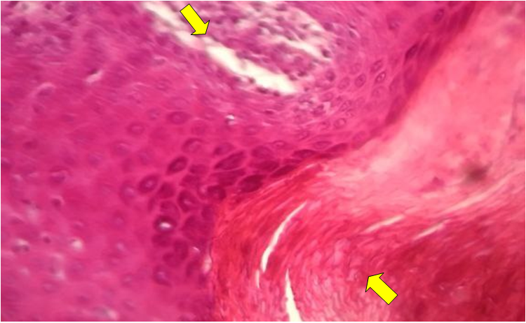 | Figure 1. Palm psoriasis (thickening of the stratum corneum). Stained in hematoxylin-eosin. Ob.40, ok.10 |
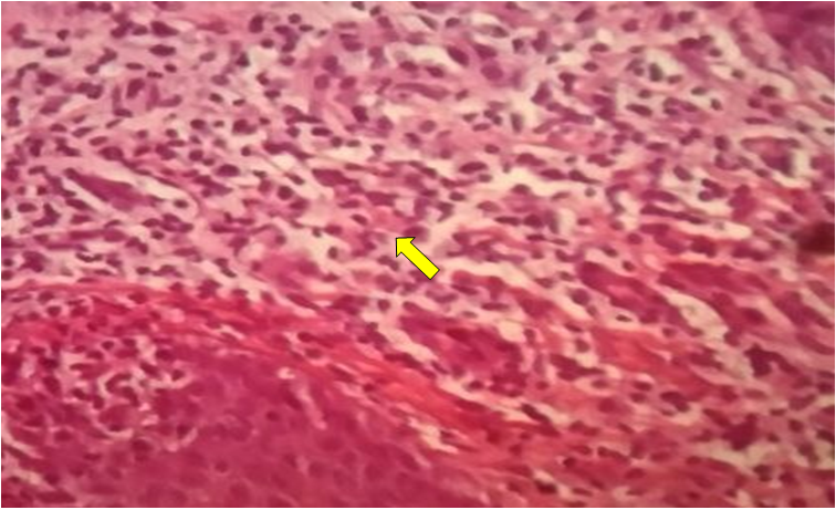 | Figure 2. Palm psoriasis (inflammatory infiltrate of lymphocytes and histiocytes with an admixture of neutrophilic granulocytes). Hematoxylin-eosin stained. Ob.40, ok.10 |
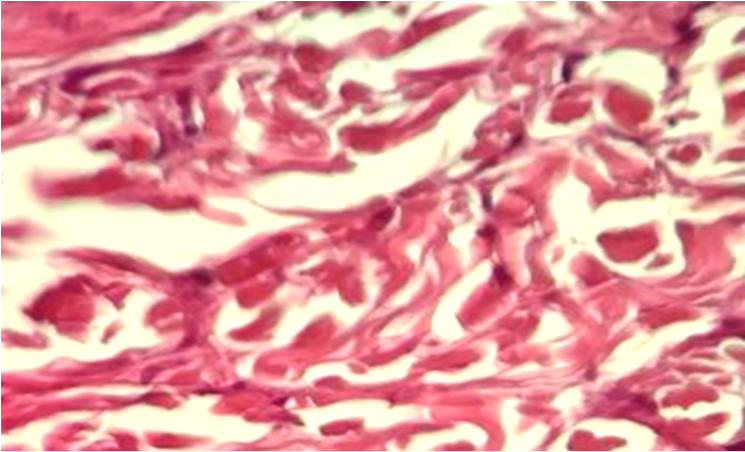 | Figure 3. Palm psoriasis (Destruction of elastic fibers and their reduction in quantity). Hematoxylin-eosin stained. Ob.40, ok.10 |
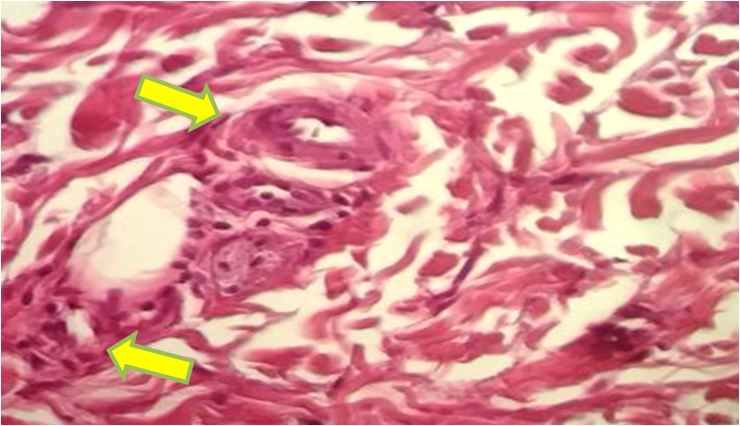 | Figure 4. Palm psoriasis (Sclerotic changes in the vascular wall and narrowing of the blood vessel lumen). Hematoxylin-eosin stained. Ob.40, ok.10 |
4. Conclusions
- Thus, in patients with psoriasis of the palms of the hands, the skin structures - the stratum corneum is thickened, divided into layers, cells are separated from each other, hyperkeratosis is detected. In the spinous layer, a strong spongiosis state is noted. The stratum corneum is thickened, and in most places it is thinned. In the sebaceous layer of the dermis, there is a fullness of blood vessels, dilation of capillaries, swelling and an inflammatory infiltrate of lymphocytes and histiocytes with an admixture of neutrophilic granulocytes. Also, there is a strong development of perivascular edema and thickening of the walls of the blood vessels due to sclerotic changes. This is an important process in carrying out treatment procedures.