American Journal of Medicine and Medical Sciences

p-ISSN: 2165-901X    e-ISSN: 2165-9036

2025;  15(4): 1285-1290

doi:10.5923/j.ajmms.20251504.92

Received: Apr. 6, 2025; Accepted: Apr. 22, 2025; Published: Apr. 30, 2025


Inflammatory Biomarkers in Rheumatoid Arthritis: A Comprehensive Evaluation of Their Prognostic Value for Disease Severity and Outcomes

M. Sh. Karimov , Sh. Х. Mirzayeva , A. A. Eshmurzaeva , A. G. Isroilov

Tashkent Medical Academy, Tashkent, Uzbekistan

Copyright © 2025 Scientific & Academic Publishing. All Rights Reserved.

This work is licensed under the Creative Commons Attribution International License (CC BY).
http://creativecommons.org/licenses/by/4.0/

Abstract

Rheumatoid arthritis (RA) is a chronic autoimmune disease marked by inflamed joints, systemic complications, and progressive disability. The early diagnosis of the disease followed by targeted treatment is essential to avert joint damage and to improve treatment outcomes. C-reactive protein (CRP), erythrocyte sedimentation rate (ESR), rheumatoid factor (RF), anti-citrullinated protein antibodies (ACPAs), and cytokines like TNF-α and IL-6 are so far known to be important inflammatory markers that indicate the diagnosis of RA, severity of the disease, and monitoring of treatment response. The role of toll-like receptors (TLRs), especially TLR-2 and TLR-4, is emerging in inducing synovial inflammation and causes for joint destruction through the enhancement of pro-inflammatory pathways. In this regard, other emerging biomarkers include calprotectin and miRNAs, which may well hold promise in predicting disease clicks and efficacy of treatment. The present review highlights the relevance of both the old and new biomarkers in progressing RA diagnosis, personalized therapy, and patient management by reiterating that these should be combined to achieve better clinical outcomes.

Keywords: Rheumatoid arthritis, Inflammatory biomarkers, Autoimmune disease, TLR-2, TLR-4, CRP, ESR, ACPA, Cytokines, TNF-α, IL-6, IL-1, Autoantibodies, Personalized therapy, Prognostic markers

Cite this paper: M. Sh. Karimov , Sh. Х. Mirzayeva , A. A. Eshmurzaeva , A. G. Isroilov , Inflammatory Biomarkers in Rheumatoid Arthritis: A Comprehensive Evaluation of Their Prognostic Value for Disease Severity and Outcomes, American Journal of Medicine and Medical Sciences, Vol. 15 No. 4, 2025, pp. 1285-1290. doi: 10.5923/j.ajmms.20251504.92.

1. Introduction

Rheumatoid arthritis is a chronic autoimmune disease that has adverse effects on people and healthcare systems worldwide. In the U.S., there are about 1.3 million adults with rheumatoid arthritis [12,47], whereas globally, it is between 0.5% and 1% of adults with rheumatoid arthritis. Women are 2-3 times more likely than men to have rheumatoid arthritis, with the common age range of onset being 30-60 years [12,17]. According to statistics about 35% of the patients become disabled within 10 years, and life expectancy is reduced further, up to five to ten years, that arises from complications of cardiovascular diseases [33,8].
The Pathogenesis of Rheumatoid Arthritis. Rheumatoid arthritis is characterized by synovial inflammation and hyperplasia ("swelling"), production of autoantibodies, including rheumatoid factor and anti-citrullinated protein antibodies, and progressive destruction of cartilage and bone leading to joint deformities. Systemic manifestations of RA include effects on the cardiovascular, pulmonary, psychiatric, and musculoskeletal systems [31].
Rheumatoid arthritis is an autoimmune disease involving genetic factors such as HLA-DRB1 in association with environmental risk factors: smoking and infections. These factors lead to citrullination of proteins, production of autoantibodies such as rheumatoid factors and anti-citrullinated protein antibodies. The formation of immune complexes caused by these autoantibodies activates the immune system, resulting in much chronic inflammation. The respectively inflamed joint synovial tissues cause a pannus, an invasive structure that erodes cartilage and bone. This activity is provoked by proinflammatory cytokines such as TNF-, IL-1, IL-6, and IL-17, as well as by enzymes such as matrix metalloproteinases and osteoclasts activated by RANKL. Chronic inflammation relates to its impact on concurrent cardiovascular disease and osteoporosis as systemic complications of directional immuno-dysregulation [14,46].
The role of Inflammation Biomarkers in Rheumatoid Arthritis. Early diagnosis of rheumatoid arthritis is of utmost importance to hinder the damage to joints and disease progression. Identification of these biomarkers will facilitate earlier diagnosis of RA, including circulating markers such as anti-CCP antibodies, RF, anti-MCV antibodies, and 14–3-3η proteins [43]. Biomarkers of inflammation are important in the diagnosis, target progress, and management of rheumatoid arthritis (RA). Non-specific markers, including C-reactive protein (CRP) and erythrocyte sedimentation rate (ESR), are routinely used to assess inflammation and treatment efficacy. Pro-inflammatory cytokines, such as TNF-α, IL-6, and IL-1, are responsible for joint destruction and placebo markers for RA therapy. Autoantibodies, including rheumatoid factor (RF) and anti-cyclic citrullinated peptide (anti-CCP), are important in the early diagnosis of RA and identifying the potential for aggression. The 14-3-3η protein has also become an important predictor of disease progression. Together, these biomarkers have propelled personalized therapeutic strategies which have led to improved clinical outcomes in RA [1,32,30].
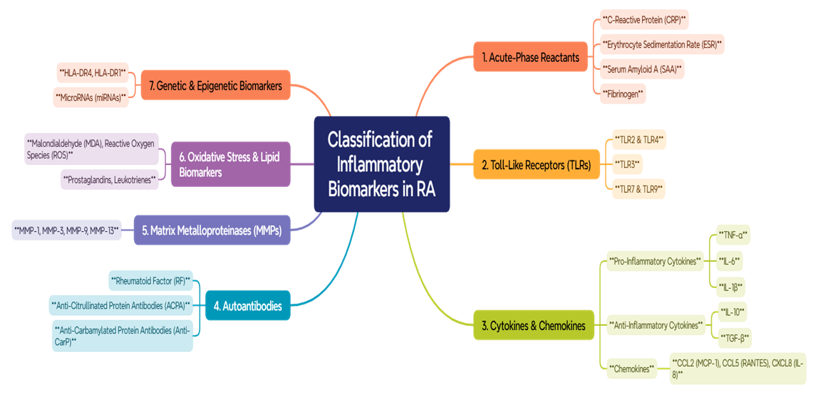
Figure 1. Classification of Inflammatory Biomarkers in RA
Toll-like receptors (TLRs). Toll-like receptors (TLRs) are important pattern recognition receptors that are important for innate immunity since they recognize pathogen-associated molecular patterns (PAMPs) and damage-associated molecular patterns (DAMPs). In rheumatoid arthritis (RA), TLR2 and TLR4 have been recognized to be activated by endogenous ligands such as heat-shock proteins and extracellular matrix damaging patterns which induces pro-inflammatory cytokines (TNF-α, IL-1, IL-6) to be released and promote inflammation in the synovium and destruction of joints [37,22]. These receptors are abundantly expressed in synovial fibroblasts, leading to degradation of cartilage and bone by releasing matrix metalloproteinases (MMPs) and inflammatory cytokines [28]. TLR7 and TLR9 also contribute to the development of RA by inducing production of auto-antibodies (e.g., rheumatoid factor (RF) and anti-citrullinated protein antibodies (ACPAs) that are pertinent to RA pathogenesis [29].
Toll-like receptor 2 (TLR-2) and Toll-like receptor 4 (TLR-4) recognize DAMPs in synovial tissue and lead to the secretion of pro-inflammatory cytokines including TNF-α, IL-1, and IL-6, promoting inflammation and joint damage [25]. There is high expression of TLR-2 and TLR-4 in synovial fibroblasts and macrophages, with the activation promoting the degradation of cartilage and bone by various MMPs [11]. TLR-4 directly promotes the production of various autoantibodies including RF and ACPAs, two biomarkers important in RA [42]. Cross-sectioning analysis among the genetic polymorphisms in TLR-4 suggests an increase in susceptibility and severity to RA, pointing toward their future as biomarkers for diagnosis and prognosis of the disease [44]. This fact strongly suggests that a pathway targeting either TLR-2 or TLR-4 and their downstream implementing molecules like MyD88 and NF-κB will be the most promising in limiting inflammation and disease progression [37].
Acute-phase reactants. C-reactive protein (CRP) is an important acute-phase reactant in Rheumatoid arthritis (RA). It is mainly produced by the liver in response to IL-6 and reflects systemic inflammation and disease activity. CRP furthers RA pathogenesis by promoting complement activation, enhancing phagocytosis, and regulating cytokine production, particularly of IL-6 and TNF-α [39,18]. Higher levels of CRP are associated with joint inflammation, cartilage degradation, and bone erosion and thus serve as useful biomarkers for monitoring disease progression and treatment response [3].
Autoantibodies. Anti-CCP (anti-cyclic citrullinated peptide) antibodies are crystallized highly specific biomarkers for rheumato-nid arthritis-their role is paramount in the early diagnosis and prognosis. Anti-CCP antibodies are believed to target immuno-logically undergone citrullinated protein during the process of inflammation, and they are associated with severe disease such as joint erosion or radiological progression [35,54]. They may be present before clinical symptoms emerge, proving valuable in identifying individuals at risk of developing RA [41]. Their presence correlates well with disease activity and poor treatment response; reaffirming their importance in personalized management of RA [2].
Cytokines and Chemokines. Cytokines are involved in the pathogenesis of rheumatoid arthritis (RA), contextually controlling synovial inflammation, cartilage degradation, and bone erosion, and further serve as important biomarkers and therapeutic targets for disease management.
Interleukin-1 (IL-1) is a cytokine with a molecular weight of 17 kilodaltons secreted principally by monocytes and macrophages. There are, however, two isoforms (a and p) which share -26 percent amino acid sequence homology. IL-1p is the predominant form, mainly synthesized by human monocytes. It binds to the same receptor on target cells as IL-1a and shows-like IL-1a-comparable bioactivities. Its systemic effects include induction of metabolic changes in the central nervous system (CNS), bone marrow, and vascular walls [7].
Interleukin-6 (IL-6) is a multifunctional cytokine that plays a prominent role within the complexity of the pathophysiology of rheumatoid arthritis (RA). Produced by synovial fibroblasts, macrophages, and T cells, IL-6 ultimately participates in local joint destruction and systemic manifestation as a resultant of such stimuli. It stimulates synovial inflammation through the production of acute-phase reactant cytokines like C-reactive protein (CRP) and fibrinogen as well as drives Th17 cell differentiation and autoimmune responses. In addition, IL-6 also activates osteoclasts and causes bone resorption and joint destruction [26,51].
Tumor necrosis factor-alpha (TNF-α) is one of the pro-inflammatory cytokines in rheumatoid arthritis (RA), facilitating synovial inflammation, cartilage destruction, and bone erosion. It activates endothelial cells, fibroblasts, and immune cells, which release such inflammatory mediators as interleukin 1 (IL-1), interleukin 6 (IL-6), and matrix metalloproteinases (MMPs) that will be responsible for joint destruction. TNF-α also promotes the formation of pannus, an invasive tissue attacking joint structures and causing systemic manifestations such as fatigue, anemia, and cardiovascular disease. Because of its critical role in RA pathogenesis, TNF-α is one of the prime targets for some biologic therapies; indeed, TNF inhibitors have a significant effect in controlling inflammation and slowing the disease progression [4,36].
Interleukin-8 (IL-8 or CXCL8), a chemokine that is central to RA because it recruits neutrophils and other immune cells to inflamed joints, is secreted by synovial fibroblasts and macrophages. IL-8 promotes angiogenesis, synovial hyperplasia, and cartilage degradation, contributing to the destruction of the joints [27,49]. Its increased levels in synovial fluid and serum are correlated to disease activity and radiographic progression, thus predicting its usefulness for monitoring RA severity [21,10].
Enzymes & Proteins. Calprotectin, a protein complex released from activated neutrophils and monocytes, is an indicator of inflammation in rheumatoid arthritis (RA). This indicates synovial inflammation with some correlation to disease activity, joint damage, and radiographic damage progression [6,23]. Rheumatoid arthritis is characterized by its elevation in patients in conjunction with heightened production of pro-inflammatory cytokines such as TNF-α and IL-6, thereby serving as an assay in evaluating disease activity and response to treatment [20,13]. The fact that it can predict treatment outcome, particularly for biologic therapy, gives it further clinical relevance [6].
Genetic & Epigenetic Biomarkers. miRNAs are small non-coding RNAs involved in regulating gene expression with high impact on RA. They modulate immune responses, synovial inflammation, and joint destruction by targeting key molecules such as TNF-α, IL-6, and MMPs [9,38,48]. In RA patients, dysregulated miRNAs are correlated with disease activity, joint damage, and response to therapy, suggesting their use as biomarkers and drug targets [34,16].
The contribution of inflammatory biomarkers with respect to the early diagnosis and management of rheumatoid arthritis (RA) has become the subject of continuous research interest. These biomarkers-including CRP, ESR, RF, ACPAs, and novel cytokines-are deemed competitive in assessing disease progression and severity, as well as the treatment response. Intuitively synthesizing these have made the diagnosis of RA easy and treatment more personalized. A brief description of important studies, researchers, and findings is provided below that provide insight into the work done on RA biomarkers:
• Xinping Tian and colleagues have studied some newer biomarkers like IL-34 and IL-33. Their study identifies IL-34 as a potential biomarker to predict joint damage and treatment resistance in patients with RA. These findings imply that newer cytokines could probably provide additional accuracy for RA diagnosis and prognosis [52].
• Kevin D. Deane has advanced the concept of "preventive rheumatology" by studying pre-RA phases. By examining ACPA, RF, and other biomarkers, his research shows that prediction of which high-risk subjects might go on to develop RA is enhanced, thus allowing for consequent preventive interventions [15].
• Tsutomu Takeuchi explores cytokine patterns and their use in predicting treatment responses to biologic therapy. His study identified distinct cytokine patterns in patients with better responses to TNF inhibitors and IL-6 blockers and allows for a personalized treatment approach [50].
• Peter K. Gregersen has searched for genetic and molecular bases of rheumatoid arthritis, also for the role of the interplay between genetic markers and inflammatory cytokines. Such work offered knowledge on how genetic and biomarker information can be jointly applied for disease severity and treatment response prediction [19].
• Dirkjan van Schaardenburg has studied the predictive value of autoantibodies such as ACPAs and RF in pre-RA stages. His work indicates that in individuals with arthralgia (joint pain), ACPA positivity is a strong predictor for progression into clinical RA, thus allowing early intervention [53].

2. Conclusions

Biomarkers of inflammation such as CRP, ESR, RF, ACPAs, TNF-α, and IL-6 are an essential part of the diagnostic, prognostic, and therapeutic management of rheumatoid arthritis. Toll-like receptors primarily TLR-2 and TLR-4 have furthered the understanding of mechanisms responsible for the inflammation of the synovium and subsequent joint destruction. Newer biomarkers namely calprotectin and miRNAs show promise in predicting disease progression and response to treatment. The combined efforts of these above parameters provide an enabling framework for early diagnosis, targeted therapy, and treatment approval enhanced by improved patient outcomes, thus highlighting their critical roles in providing better care for RA. Further studies of new biomarkers and their roles in clinical practice are expected to inform better management for RA.

References

[1]  Aletaha, D., & Smolen, J. S. (2018). Diagnosis and management of rheumatoid arthritis: A review. JAMA, 320(13), 1360–1372. https://doi.org/10.1001/jama.2018.13103.
[2]  Aletaha, D., et al. (2010). 2010 Rheumatoid arthritis classification criteria: An American College of Rheumatology/European League Against Rheumatism collaborative initiative. Arthritis & Rheumatism, 62(9), 2569–2581. https://doi.org/10.1002/art.27584.
[3]  Alluno, A., et al. (2021). CRP and cytokines in rheumatoid arthritis: A comprehensive review. Frontiers in Immunology, 12, 691766. https://doi.org/10.3389/fimmu.2021.691766.
[4]  Alunno, A., et al. (2021). TNF-α in rheumatoid arthritis: Pathogenic mechanisms and therapeutic implications. Frontiers in Immunology, 12, 691766. https://doi.org/10.3389/fimmu.2021.691766.
[5]  Andrés Cerezo, L., et al. (2012). Calprotectin in rheumatoid arthritis: Association with disease activity and treatment response. Journal of Rheumatology, 39(6), 1120–1126. https://doi.org/10.3899/jrheum.111382.
[6]  Andrés Cerezo, L., et al. (2018). Calprotectin as a biomarker for predicting treatment response in rheumatoid arthritis. Arthritis Research & Therapy, 20(1), 1–9. https://doi.org/10.1186/s13075-018-1643-7.
[7]  Arend, W. P., & Dayer, J. (1990). Cytokines and cytokine inhibitors or antagonists in rheumatoid arthritis. Arthritis & Rheumatism, 33(3), 305–315. https://doi.org/10.1002/art.1780330302.
[8]  Avina-Zubieta, J. A., Choi, H. K., Sadatsafavi, M., Etminan, M., Esdaile, J. M., & Lacaille, D. (2008). Risk of cardiovascular mortality in patients with rheumatoid arthritis: A meta-analysis of observational studies. Arthritis Care & Research, 59(12), 1690–1697. https://doi.org/10.1002/art.24092.
[9]  Blüml, S., Bonelli, M., Niederreiter, B., Puchner, A., Mayr, G., Hayer, S., Koenders, M. I., Van Den Berg, W. B., Smolen, J., & Redlich, K. (2011). Essential role of microRNA-155 in the pathogenesis of autoimmune arthritis in mice. Arthritis & Rheumatism, 63(5), 1281–1288. https://doi.org/10.1002/art.30281.
[10]  Brennan, F. M., et al. (1997). Evidence that cytokines play a role in rheumatoid arthritis. Journal of Clinical Investigation, 100(12), 2948–2953. https://doi.org/10.1172/JCI119848.
[11]  Brentano, F., Schorr, O., Gay, R. E., Gay, S., & Kyburz, D. (2005). TLR-2 and TLR-4 expression in synovial fibroblasts and macrophages: Role in cartilage and bone degradation in rheumatoid arthritis. Arthritis Research & Therapy, 7(4), R704-R714. https://doi.org/10.1186/ar1724.
[12]  Centers for Disease Control and Prevention. (2020). Rheumatoid arthritis. Retrieved from https://www.cdc.gov.
[13]  Choi, I. Y., et al. (2017). Calprotectin as a biomarker for rheumatoid arthritis: A systematic review. Journal of Clinical Medicine, 6(7), 69. https://doi.org/10.3390/jcm6070069.
[14]  Choy, E. H., et al. (2020). Translating insights from pathogenesis into therapeutic strategies for rheumatoid arthritis. Nature Reviews Rheumatology, 16(1), 45–56. https://doi.org/10.1038/s41584-019-0339-y.
[15]  Deane, K. D., et al. (2020). Predicting rheumatoid arthritis development in high-risk populations using biomarkers. Annals of the Rheumatic Diseases, 79(3), 345-352. https://doi.org/10.1136/annrheumdis-2019-216123.
[16]  Filková, M., et al. (2014). MicroRNAs in rheumatoid arthritis: Potential role in diagnosis and therapy. BioDrugs, 28(4), 363–377. https://doi.org/10.1007/s40259-014-0095-0.
[17]  Firestein, G. S., Budd, R. C., Gabriel, S. E., McInnes, I. B., & O’Dell, J. R. (2021). Kelley and Firestein’s textbook of rheumatology (11th ed.). Elsevier.
[18]  Gabay, C., & Kushner, I. (1999). Acute-phase proteins and other systemic responses to inflammation. New England Journal of Medicine, 340(6), 448–454. https://doi.org/10.1056/NEJM199902113400607.
[19]  Gregersen, P. K., et al. (2020). Genetic and inflammatory biomarkers in rheumatoid arthritis: Predicting outcomes and treatment response. Nature Reviews Rheumatology, 16(5), 301-312. https://doi.org/10.1038/s41584-020-0408-2.
[20]  Hammer, H. B., et al. (2007). Calprotectin (a major leucocyte protein) is associated with the levels of anti-CCP and rheumatoid factor in a longitudinal study of patients with very early rheumatoid arthritis. Scandinavian Journal of Rheumatology, 36(4), 260–264. https://doi.org/10.1080/03009740701286805.
[21]  Harada, A., et al. (1994). Essential involvement of interleukin-8 (IL-8) in acute inflammation. Journal of Leukocyte Biology, 56(5), 559–564. https://doi.org/10.1002/jlb.56.5.559.
[22]  Huang, Q., & Pope, R. M. (2009). The role of Toll-like receptors in rheumatoid arthritis. Current Rheumatology Reports, 11(5), 357–364. https://doi.org/10.1007/s11926-009-0051-z.
[23]  Inciarte-Mundo, J., et al. (2015). Calprotectin as a biomarker of disease activity in rheumatoid arthritis: A systematic review. Rheumatology, 54(6), 1005–1014. https://doi.org/10.1093/rheumatology/keu462.
[24]  Jochems, C., Islander, U., Erlandsson, M., Verdrengh, M., Ohlsson, C., & Carlsten, H. (2005). Osteoporosis in experimental postmenopausal polyarthritis: the relative contributions of estrogen deficiency and inflammation. Arthritis Research & Therapy, 7(4). https://doi.org/10.1186/ar1753.
[25]  Kim, S. J., et al. (2018). Journal of Immunology, 201(8), 2446–2454. https://doi.org/10.4049/jimmunol.1800116.
[26]  Kishimoto, T. (2010). IL-6: From its discovery to clinical applications. International Immunology, 22(5), 347–352. https://doi.org/10.1093/intimm/dxq030.
[27]  Koch, A. E., et al. (1992). Interleukin-8 as a macrophage-derived mediator of angiogenesis. Science, 258(5089), 1798–1801. https://doi.org/10.1126/science.1281554.
[28]  Kyburz, D., & Rethage, J. (2007). Toll-like receptors in rheumatoid arthritis. Arthritis Research & Therapy, 9(6), 1–7. https://doi.org/10.1186/ar2302.
[29]  Lau, C. M., Broughton, C., Tabor, A. S., Akira, S., Flavell, R. A., Mamula, M. J., Christensen, S. R., Shlomchik, M. J., Viglianti, G. A., Rifkin, I. R., & Marshak-Rothstein, A. (2005). RNA-associated autoantigens activate B cells by combined B cell antigen receptor/Toll-like receptor 7 engagement. The Journal of Experimental Medicine, 202(9), 1171–1177. https://doi.org/10.1084/jem.20050630.
[30]  Maksymowych, W. P., et al. (2014). 14–3-3η protein as a novel biomarker for rheumatoid arthritis. Arthritis Research & Therapy, 16(2), R99. https://doi.org/10.1186/ar4547.
[31]  McInnes, I. B., & Schett, G. (2011). The pathogenesis of rheumatoid arthritis. New England Journal of Medicine, 365(23), 2205–2219. https://doi.org/10.1056/nejmra1004965.
[32]  McInnes, I. B., & Schett, G. (2017). Pathogenetic insights from the treatment of rheumatoid arthritis. The Lancet, 389(10086), 2328–2337. https://doi.org/10.1016/S0140-6736(17)31472-1.
[33]  Myasoedova, E., Davis, J. M., Crowson, C. S., & Gabriel, S. E. (2010). Is the incidence of rheumatoid arthritis rising? Arthritis & Rheumatism, 62(6), 1576–1582. https://doi.org/10.1002/art.27425.
[34]  Nakasa, T., et al. (2011). The inhibitory effect of microRNA-146a expression on bone destruction in collagen-induced arthritis. Arthritis & Rheumatism, 63(6), 1582–1590. https://doi.org/10.1002/art.30281.
[35]  Nielen, M. M., et al. (2004). Specific autoantibodies precede the symptoms of rheumatoid arthritis: A study of serial measurements in blood donors. Arthritis & Rheumatism, 50(2), 380–386. https://doi.org/10.1002/art.20018.
[36]  Nygaard, G., & Firestein, G. S. (2020). Restoring synovial homeostasis in rheumatoid arthritis by targeting fibroblast-like synoviocytes. Nature Reviews Rheumatology, 16(6), 316–333. https://doi.org/10.1038/s41584-020-0413-5.
[37]  Ospelt, C., Brentano, F., Rengel, Y., Stanczyk, J., Kolling, C., Tak, P. P., Gay, R. E., Gay, S., & Kyburz, D. (2008). Overexpression of toll-like receptors 3 and 4 in synovial tissue from patients with early rheumatoid arthritis: Toll-like receptor expression in early and longstanding arthritis. Arthritis & Rheumatism, 58(12), 3684–3692. https://doi.org/10.1002/art.24140.
[38]  Pauley, K. M., et al. (2008). MicroRNA in autoimmunity and autoimmune diseases. Journal of Autoimmunity, 32(3-4), 189–194. https://doi.org/10.1016/j.jaut.2008.12.005.
[39]  Pepys, M. B., & Hirschfield, G. M. (2003). C-reactive protein: A critical update. Journal of Clinical Investigation, 111(12), 1805–1812. https://doi.org/10.1172/JCI200318921.
[40]  Pincus, T., Vogel, S., Burton, A. K., Santos, R., & Field, A. P. (2006). Fear avoidance and prognosis in back pain: A systematic review and synthesis of current evidence. Arthritis & Rheumatism, 54(12), 3999–4010. https://doi.org/10.1002/art.22273.
[41]  Rantapää‐Dahlqvist, S., De Jong, B. a. W., Berglin, E., Hallmans, G., Wadell, G., Stenlund, H., Sundin, U., & Van Venrooij, W. J. (2003). Antibodies against cyclic citrullinated peptide and IgA rheumatoid factor predict the development of rheumatoid arthritis. Arthritis & Rheumatism, 48(10), 2741–2749. https://doi.org/10.1002/art.11223.
[42]  Roelofs, M. F., Joosten, L. A. B., Abdollahi-Roodsaz, S., van Lieshout, A. W. T., Sprong, T., van den Hoogen, F. H. J., van den Berg, W. B., & Radstake, T. R. D. J. (2006). The expression of toll-like receptors 3 and 7 in rheumatoid arthritis synovium is increased and costimulation of toll-like receptors 3, 4, and 7/8 results in synergistic cytokine production by dendritic cells. Arthritis & Rheumatism, 52(8), 2313-2322. https://doi.org/10.1002/art.21278.
[43]  Saffar, M., Alipanah, H., & Ataollahi, M. R. (2019). The role of biomarkers in diagnosis, prognosis, treatment, determining disease activity in rheumatoid arthritis. Majallah-i Dānishgāh-i ̒Ulūm-i Pizishkī-i Fasā, 9(4), 1682–1692. http://jabs.fums.ac.ir/article-1-2141-en.html.
[44]  Sanchez, E., Orozco, G., Lopez-Nevot, M. A., Jimenez-Alonso, J., & Martin, J. (2010). Polymorphisms of toll-like receptor 2 and 4 genes in rheumatoid arthritis and systemic lupus erythematosus. Tissue Antigens, 63(1), 54-57. https://doi.org/10.1111/j.1399-0039.2004.00153.x.
[45]  Sánchez, E., Orozco, G., López-Nevot, M., Jiménez-Alonso, J., & Martín, J. (2003). Polymorphisms of toll-like receptor 2 and 4 genes in rheumatoid arthritis and systemic lupus erythematosus. Tissue Antigens, 63(1), 54–57. https://doi.org/10.1111/j.1399-0039.2004.00162.x.
[46]  Scherer, H. U., Häupl, T., & Burmester, G. R. (2020). The etiology of rheumatoid arthritis. Journal of Autoimmunity, 110, 102400. https://doi.org/10.1016/j.jaut.2019.102400.
[47]  Smolen, J. S., Aletaha, D., & McInnes, I. B. (2016). Rheumatoid arthritis. The Lancet, 388(10055), 2023–2038. https://doi.org/10.1016/S0140-6736(16)30173-8.
[48]  Stanczyk, J., et al. (2008). Altered expression of microRNA in synovial fibroblasts and synovial tissue in rheumatoid arthritis. Arthritis & Rheumatism, 58(4), 1001–1009. https://doi.org/10.1002/art.23386.
[49]  Szekanecz, Z., et al. (2000). Chemokines and angiogenesis in rheumatoid arthritis. Frontiers in Bioscience, 5(1), D23–D29. https://doi.org/10.2741/szekanecz.
[50]  Takeuchi, T., et al. (2021). Cytokine profiles and treatment response in rheumatoid arthritis: A biomarker-driven approach. Rheumatology, 60(4), 789-797. https://doi.org/10.1093/rheumatology/keaa567.
[51]  Tanaka, T., Narazaki, M., & Kishimoto, T. (2014). IL-6 in inflammation, immunity, and disease. Cold Spring Harbor Perspectives in Biology, 6(10), a016295. https://doi.org/10.1101/cshperspect.a016295.
[52]  Tian, X., et al. (2022). IL-34 as a novel biomarker for predicting joint damage in rheumatoid arthritis. Journal of Rheumatology, 49(2), 210-218. https://doi.org/10.3899/jrheum.210123.
[53]  van Schaardenburg, D., et al. (2021). ACPA positivity in arthralgia: Predicting progression to rheumatoid arthritis. Rheumatology Advances in Practice, 5(2), rkab034. https://doi.org/10.1093/rap/rkab034.
[54]  van Venrooij, W. J., van Beers, J. J., & Pruijn, G. J. (2011). Anti-CCP antibodies: The past, the present, and the future. Nature Reviews Rheumatology, 7(7), 391–398. https://doi.org/10.1038/nrrheum.2011.76.