American Journal of Medicine and Medical Sciences

p-ISSN: 2165-901X    e-ISSN: 2165-9036

2025;  15(4): 1023-1030

doi:10.5923/j.ajmms.20251504.35

Received: Mar. 9, 2025; Accepted: Mar. 29, 2025; Published: Apr. 10, 2025


Retrospective Study, Diagnosis, and Clinical Characteristics of Atopic Dermatitis in Children of the Samarkand Region

Khamidova Farida Muinovna, Tolibov Mansur Mahmudovich, Ismoilov Jasur Mardonovich

Samarkand State Medical University, Samarkand, Uzbekistan

Copyright © 2025 The Author(s). Published by Scientific & Academic Publishing.

This work is licensed under the Creative Commons Attribution International License (CC BY).
http://creativecommons.org/licenses/by/4.0/

Abstract

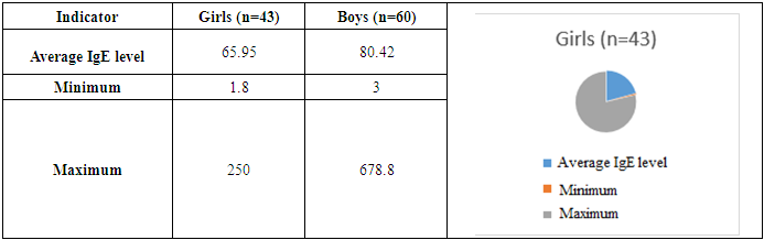
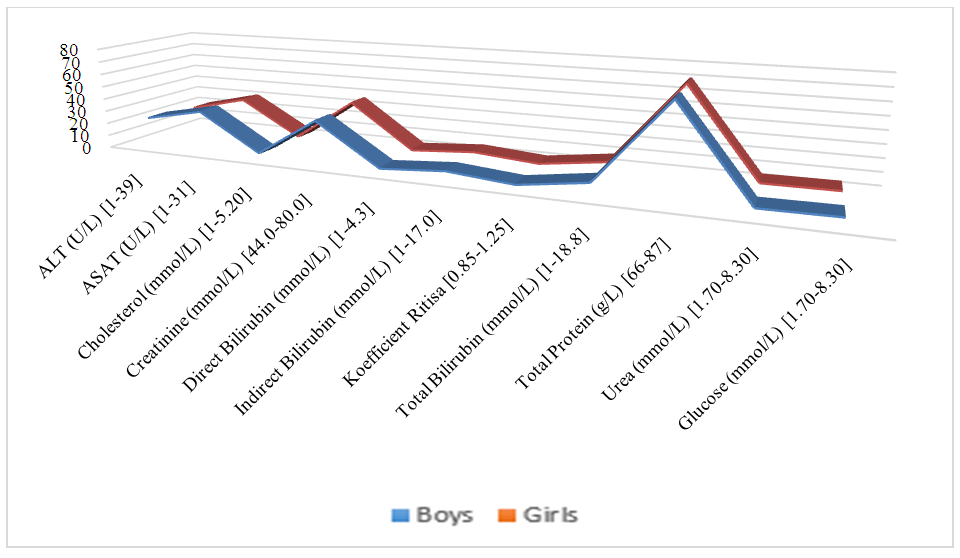
The article presents a comprehensive analysis of a retrospective, diagnostic study of atopic dermatitis in children of the Samarkand region for 2024. The inclusion and exclusion criteria of patients are thoroughly examined, confirming the validity of the selected study groups. Particular attention is paid to the comparative laboratory analysis of young children with atopic dermatitis. Atopic dermatitis is most common in the Taylak, Urgut, and Samarkand districts. Boys are more frequently affected, especially under the age of one. The incidence is seasonal, peaking in the spring and summer months. Blood test data in children with atopic dermatitis show similar results in both genders, but girls have higher levels of platelets and segmented neutrophils, while boys show higher levels of leukocytes and lymphocytes. The average IgE level in girls is 65.95, while in boys, it is 80.42, indicating a slightly higher level in boys. Boys also have higher liver enzyme and bilirubin levels, possibly due to a more active metabolism. Girls show elevated creatinine and Ritis ratio, which may reflect differences in metabolism. The remaining indicators do not show significant differences.

Keywords: Atopic dermatitis, Chronic obstructive bronchitis, Morphometry

Cite this paper: Khamidova Farida Muinovna, Tolibov Mansur Mahmudovich, Ismoilov Jasur Mardonovich, Retrospective Study, Diagnosis, and Clinical Characteristics of Atopic Dermatitis in Children of the Samarkand Region, American Journal of Medicine and Medical Sciences, Vol. 15 No. 4, 2025, pp. 1023-1030. doi: 10.5923/j.ajmms.20251504.35.

1. Introduction

The issue of atopic dermatitis remains one of the most pressing in dermatology due to its widespread prevalence, increasing incidence, severe course, and the insufficient effectiveness of existing treatment methods [2,17,34,37]. The increase in morbidity over the past decade, the chronic nature with frequent relapses, and the insufficient effectiveness of current treatment and prevention methods place atopic dermatitis among the most significant problems in modern medicine [12,14,15,21,36,38]. Atopic dermatitis typically develops in early childhood in individuals with a hereditary predisposition to atopic diseases and has age-related features in the localization and morphology of inflammation foci.
Atopic dermatitis (AD) is one of the most common inflammatory dermatoses and allergic diseases. The incidence among the population of different countries is usually not less than 5-10%, in industrialized countries around 20%, and in children, AD has long been the leading pathology [1,3,12,26,40].
Currently, atopic dermatitis (AD) is understood as a chronic allergic disease characterized by exudative and lichenified rashes, elevated serum IgE levels, and hypersensitivity to specific irritants. However, as shown in several studies by domestic and foreign researchers, only some patients exhibit increased levels of total and allergen-specific IgE, which allows only these patients to be classified in the IgE-mediated type of the disease. In some patients, IgE-mediated hypersensitivity in the pathogenesis cannot be identified, but delayed-type hypersensitivity is present [4,7,11,16,17]. Besides these two groups, there are patients with a mixed type of allergic reactions in the pathogenesis. Finally, in some patients, the pathogenesis of AD is due to other, non-immune mechanisms [5,7,8,10,29].
The concept of "atopy" was introduced into medical practice in 1923 by Coca and Cooke. The term initially encompassed the clinical manifestations of such externally dissimilar diseases as asthma, urticaria, hay fever, food allergies, and eczema, that is, everything that is now considered as variants of type I (immediate) hypersensitivity. In addition to immune mechanisms, all forms of atopy share the fact that they occur significantly more often in people whose parents have similar ailments. This pattern is not strict, however, some authors, who have a weakness for simple explanations of their failures, willingly interpret the term "atopy" precisely as a genetic predisposition to corresponding allergic reactions, which is equivalent to recognizing the incurability of all forms of atopy, including, of course, atopic dermatitis. Such a substitution of concepts, as will be shown below, is absolutely unfounded and brings nothing but a sense of hopelessness. The term "atopic dermatitis" is now widely accepted to refer to this disease [6,8,13,14,18,19,20,32,35].
The prevalence of AD among developed countries is 10-20%. Manifestation of AD symptoms in children is observed at the age of 6 months in 60% of cases, up to 1 year in 75%, and up to 7 years in 80-90%. Over the past decades, there has been a significant increase in the incidence of AD, its course has become more complicated, and its outcome has become more severe. AD is often associated with other allergic diseases — with bronchial asthma in 34%, allergic rhinitis in 25%, and pollinosis in 8% [9,21,22,24,26,39,40].
Atopic dermatitis is the main problem of pediatric dermatology, as the incidence of atopic dermatitis is much higher than other common dermatitis. The terms "atopic eczema" and "neurodermatitis" are used as synonyms for this disease. In everyday life, people simply use the term "eczema." We consider the terms "eczema" and "dermatitis" equivalent and will use exclusively the term "dermatitis" to avoid confusion [19,20,21,23,25,27,28,31].
Recently, studies have begun to explore the deterioration in the quality of life of both the patient and their family — in this disease, it is quite significant. Most patients are children. The incidence of atopic dermatitis is 8-16%, making it the most common skin disease of infancy and childhood [33,34,36,38,40,41,42]. Approximately 60% of patients show the first signs of the disease in the first year of life. Males and females are affected with equal frequency.
Research Aim. To conduct a retrospective, clinical, and diagnostic study of children with atopic dermatitis in the Samarkand region.

2. Materials and Methods of Research

The study used retrospective, clinical, and morphological methods for treating patients with atopic dermatitis. The material consisted of outpatient records, clinical studies, and laboratory studies of children with atopic dermatitis.

3. Research Results

Results of the retrospective study. Patients with atopic dermatitis were examined through retrospective observation of children. In 2024, at the Samarkand branch of the Specialized Scientific and Practical Medical Center of Dermatovenereology and Cosmetology, 42,917 thousand people consulted for dermatovenereological diseases. Of these, a clinical diagnosis of atopic dermatitis was made in 1,330 patients of different ages, which accounted for 3.1% of the total number of patients.
When we conducted an analysis by district within the Samarkand region, the following was revealed:
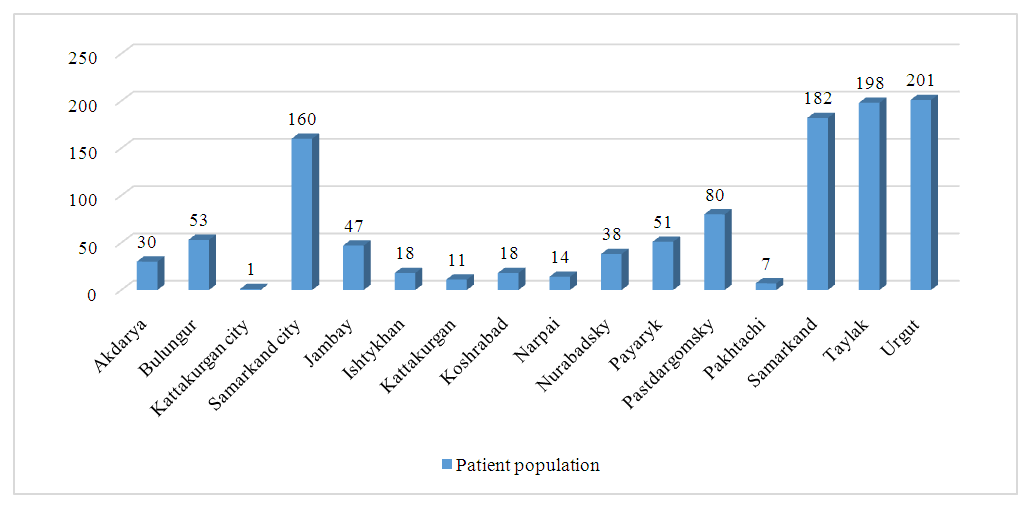
Figure 1. Analysis of the incidence of atopic dermatitis by districts of the Samarkand region
Based on the available data, an analysis was conducted on the incidence among children born in 2024 in each district and region compared to the total number of 148 cases.
Atopic dermatitis was most frequently recorded in the Taylak, Urgut, and Samarkand districts. The lowest number of cases was registered in the Jambay, Kattakurgan, Kushrabot, Nurobod, Narpa districts, and in the city of Tashkent, where only one case was recorded.
The highest incidence rates were observed in the following districts: Taylak: 36 cases (24.3%), Urgut: 28 cases (18.9%), Samarkand: 26 cases (17.6%).
These districts combined account for 60.8% of all registered cases, indicating a higher prevalence of atopic dermatitis in these regions. Possible factors influencing this situation may include environmental conditions, climatic features, and access to medical services.
The average incidence level was noted in: Samarkand city: 13 cases (8.8%), Pastdargom district: 8 cases (5.4%), Bulungur district: 9 cases (6.1%), Payaryk district: 6 cases (4.1%).
In these regions, the incidence is lower than in districts with the highest number of cases, but it still remains significant.
Less frequent cases were recorded in the following districts and areas: Ishtikhan: 3 cases, Nurobod: 3 cases. Among patients diagnosed with "atopic dermatitis" in 2024, 88.3% were born between 2007 and 2024. Girls accounted for 38.4% of all cases and boys for 49.9%.
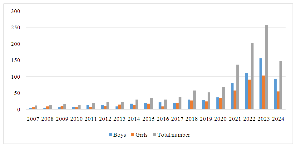
Figure 2. Analysis of children with atopic dermatitis by gender
Among children under the age of one with atopic dermatitis, 63.5% were boys and 36.5% were girls.
The highest incidence was observed in June 2024 (13.5%) and March-April 2024 (12.2%). The lowest cases were recorded in December 2024 (0.7%) and October 2024 (3.4%).
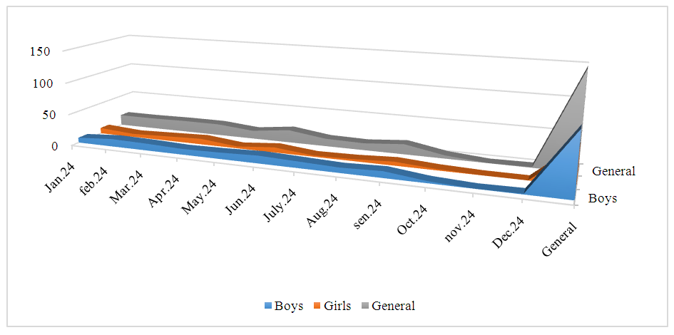
Figure 3. Atopic dermatitis in children born in 2024
In our study, standard clinical and laboratory methods were used to diagnose patients with atopic dermatitis (AD) in accordance with established regulations (Appendix 6 to the Order of the Ministry of Health of the Republic of Uzbekistan No. 273 dated November 30, 2021). At each stage of the work, inclusion and exclusion criteria were determined according to the set objectives.
The clinical group included patients with a confirmed diagnosis of "Atopic Dermatitis" (L20 according to ICD-10).
Inclusion and exclusion criteria for diagnosing atopic dermatitis (AD). To study atopic dermatitis, patients were selected based on the following diagnostic criteria (Hanifin, Rajka. ActaDerm. 92/44, 1980): Skin itching (pruritus), even with minimal rash manifestations; Elevated blood IgE levels; Onset of the disease before 2 years of age; Hyperlinearity of the skin on the palms and between the fingers; Pityriasis alba (discolored spots on the face and shoulders); Follicular hyperkeratosis; Peeling, xerosis, ichthyosis; Nonspecific dermatitis of the hands and feet; Frequent skin infections (staphylococcal, fungal, herpetic); White dermographism; Itching with sweating; Neck folds; Skin irritation after water procedures (in children under 2 years old) and other signs.
When localizing skin rashes, their appearance on the face, neck, armpits, elbow bends, pelvic area, scalp, and behind the ears was considered. Additionally, the presence of an individual or family history of atopy in children, as well as the chronic relapsing course of the disease, was identified.
Exclusion criteria included the absence of such dermatological diseases as seborrheic dermatitis, dermatomycosis, scabies, childhood scabies, dry streptoderma, eczema, and other skin pathologies.
Although the study data allow for effective management of the patient's condition, it should be noted that there is no absolute "gold standard" for diagnosing atopic dermatitis (AD). This is confirmed by medical literature, where authors analyze the frequency of the disease, its prevalence, and its association with other pathologies [39].
Clinical material characteristics. The study was conducted in 2024 at the Samarkand Regional Branch of the Republican Specialized Scientific and Practical Medical Center of Dermatovenereology and Cosmetology. A total of 122 patients participated: 82 in the clinical group and 40 in the control group.
Patients with diagnosed atopic dermatitis (AD) according to the criteria of J. Hanifin and J. Rajka (2000) were included in the study. The participants were children aged 0 to 17 years, with the voluntary consent of their parents.
The severity of the disease was assessed using the SCORAD system [37,40,41], which is an international standard for objective clinical monitoring of atopic dermatitis [36,42].
The SCORAD index was calculated using the following formula:
SCORAD = A / 5 + 7B / 2 + C,
where:
A – Degree of prevalence (affected skin surface area);
B – Intensity of atopic dermatitis symptoms (B1 – Erythema; B2 – Edema/Papules; B3 – Oozing/Crusting; B4 – Excoriations; B5 – Lichenification; B6 – Skin dryness);
C – Subjective symptoms (C1 – Degree of itching; C2 – Sleep disturbance).
The SCORAD index was determined upon admission to the clinic, 10 days after the start of treatment, and after completion of therapy:
Up to 40 points: Mild severity,
From 40 to 60 points: Moderate severity,
Over 60 points: Severe form of the disease [36,37].
Clinical symptoms dynamics. During treatment, the dynamics of clinical manifestations of atopic dermatitis were analyzed in each patient group. The analysis considered the time of full or partial disappearance of objective and subjective symptoms of the disease, as well as the reduction of the SCORAD index.
Clinical group characteristics. The study included patients with a confirmed diagnosis of atopic dermatitis (L20 according to ICD-10).Average age: 7.3 ± 4.2 years. Gender distribution: Boys – 43 (52.4%), Girls – 39 (47.6%).
Distribution by forms of atopic dermatitis: Erythematous-squamous form: 42 patients (51.2%). Eczematous form: 19 patients (23.2%). Lichenoid form: 12 patients (14.6%). Pruriginous form: 9 patients (11.0%).
Patients were divided into groups depending on the stage of the disease (exacerbation or remission within 2–3 months) and treatment methods. Control Group: Received traditional therapy (topical glucocorticosteroids, antihistamines, sedatives, and emollients). Main Group: Additionally received antibiotic therapy, Piracin, and immunomodulators.
Family and allergological history analysis. The study analyzed the family and allergological history of the patients, identified exacerbation factors, peculiarities of clinical course, and comorbidities. Clinical, laboratory, and instrumental diagnostic methods were used before and after treatment. The observation period was 3 months.
Patient examination. All patients were examined by an allergist, therapist or pediatrician, endocrinologist, parasitologist, and neurologist before and after treatment.
Standard laboratory studies conducted during the exacerbation phase:
1. Complete and biochemical blood analysis.
2. Assessment of humoral immunity.
3. Analysis of cytokine status and immunoregulatory protein indicators.
4. Determination of total IgE using the IHLA method.
5. Microscopy to detect fungal diseases.
6. Skin dermatoscopy.
7. Comprehensive diagnosis of helminthiases (ascaris, giardia).
8. ELISA to determine the concentration of C-reactive oxide.
9. Daily measurement of electrolyte content (potassium, sodium, calcium, chlorine, pH).
10. Examination of lesions under Wood’s lamp (in the clinic).
11. Ultrasound examination of internal organs.
12. Studyofskincorneometry.
13. Histological studies (biopsies taken with parental or patient consent).
In addition, 22% of patients were found to have comorbid conditions.
When performing a complete blood count for children with atopic dermatitis, only minor changes were identified (Table 1).
Table 1. Blood test indicators of children with atopic dermatitis
     
The data from the complete blood count of children with atopic dermatitis indicate some similar indicators in boys and girls. However, girls show a slightly higher level of platelets and segmented neutrophils, while boys exhibit a trend towards higher levels of leukocytes and lymphocytes.
A comparative analysis of total IgE levels in girls and boys using the IHLA method showed that the average IgE level in girls was 65.95, while in boys it was 80.42. This indicates a slightly higher IgE level in boys.
Table 2. Comparison of key statistical parameters of ige levels in boys and girls
     
The minimum IgE level in girls is 1.8, while in boys it is 3. The maximum values are 250 in girls and 678.8 in boys, which also indicates a wider range in boys.
Thus, the average total IgE level in boys is higher than in girls, which may indicate individual immunological characteristics or differences in susceptibility to allergens.
Biochemical data analysis in boys (M, n=60) and girls (F, n=43) shows some differences in key indicators (Figure 4). Average ALT Level: Boys: 22.83 U/L, Girls: 19.98 U/L. ASAT Level: Boys: 32.27 U/L, Girls: 30.98 U/L.). These findings may indicate higher metabolic activity of the liver in boys. Average Cholesterol Level: Boys: 3.47 mmol/L, Girls: 3.59 mmol/L. Creatinine Level: Higher in girls (36.48 mmol/L) compared to boys (33.82 mmol/L). Direct Bilirubin: Higher in boys (2.1 mmol/L) compared to girls (1.96 mmol/L). Indirect Bilirubin: Boys: 5.81 mmol/L, Girls: 5.62 mmol/L. Total Bilirubin: Boys: 9.06 mmol/L, Girls: 8.54 mmol/L. These changes may be associated with differences in liver enzyme activity or hemoglobin metabolism.
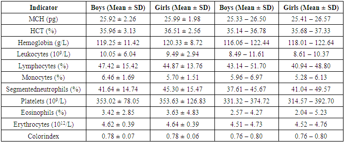
Figure 4. Biochemical analysis indicators of children with atopic dermatitis
Ritis ratio: Higher in girls (1.73) than in boys (1.59), indicating physiological features of liver function. Total protein (g/L), Urea (mmol/L), and Glucose (mmol/L) levels are nearly identical in both sexes. Overall, boys show slightly higher liver enzyme and total bilirubin levels, possibly due to more active metabolism. Girls have higher creatinine and Ritis ratio levels, which may indicate differences in muscle and liver metabolism.
The remaining indicators do not show significant differences. Microscopy was conducted to identify fungal diseases, as part of a broader assessment of treatment effectiveness.
Treatment effectiveness evaluation.
The main therapy to eliminate allergens included: Hypoallergenic diet, Sedative medications, Antihistamines, Membrane-stabilizing medications (Ketotifen), Antibiotics (for infected patients). Additionally, the absence of special treatment for 3 months prior to the study was considered. Piracin-RG (REMEDYGROUP, Uzbekistan) — a combination of zinc and pyridoxine (vitamin B6), was administered intramuscularly at 1.0 ml once daily for 10 days. Pyridoxine (vitamin B6) acts as a coenzyme necessary for metabolism and normal functioning of the nervous system. To enhance treatment efficacy, Immunomodulin (Uzfarmsanoat Gazi) was used for patients with immunological deviations and somatic load.

4. Conclusions

Atopic dermatitis is most common in the Taylak, Urgut, and Samarkand districts. Boys are more frequently affected, especially under the age of one. The incidence has a seasonal nature, peaking in the spring-summer period.
Blood test data in children with atopic dermatitis show similar indicators in both sexes, although girls have higher platelet and segmented neutrophil levels, while boys show higher levels of leukocytes and lymphocytes. The average IgE level in girls is 65.95, while in boys, it is 80.42, indicating a slightly higher level in boys. Boys also have higher liver enzyme and bilirubin levels, likely due to more active metabolism. Girls show elevated creatinine and Ritis ratio, which may reflect differences in metabolism. The remaining indicators do not show significant differences.

References

[1]  Atopic dermatitis: a guide for doctors / edited by Yu. V. Sergeev - M.: Medicine for all, 2002. - 183 p.
[2]  Balabolkin, I. I. Food allergy in children / I. I. Balabolkin; edited by I. I. Balabolkin, V. A. Revyakina. - M.: Dynasty Publishing House, 2010. - 190 p.
[3]  Balabolkin, I. I. Food allergy in children: modern aspects of pathogenesis and approaches to therapy and prevention / I. I. Balabolkin // Immunopathology, allergology, infectology. - 2013. - No. 3. - P. 36-46.
[4]  Bedin P. G. et al. Clinical and morphological characteristics of the skin in children with atopic dermatitis // Journal of the Grodno State Medical University. - 2018. - Vol. 16. - No. 4. - P. 428-434.
[5]  Bragina, E. E. Filaggrin and keratins in the formation of a protective epidermal barrier: a review / E. E. Bragina // Plastic surgery and cosmetology. - 2011. - No. 4. - P. 651-660.
[6]  Varlamov E. E. et al. Clinical significance of FLG gene mutations in children with atopic dermatitis // Russian Bulletin of Perinatology and Pediatrics. - 2015. - Vol. 60. - No. 2. – P. 87-91.
[7]  Varlamov E. E., Pampura A. N. The Importance of Filaggrin in the Development of Atopic Dermatitis // Russian Allergological Journal. – 2011. – No. 5. – P. 26-30.
[8]  Vedler A. A. MORPHOFUNCTIONAL CHARACTERISTICS OF CHILDREN'S SKIN // Scientist. – 2020. – No. 3 (13). – P. 3.
[9]  Davydova E. Yu., Morozova I. V. SKIN: TYPING, CARE, AGE-RELATED CHANGES, DISEASES, IMPROVEMENT OF CONDITION // Biological Sciences through the Eyes of Young Researchers. – 2023. – P. 18-23.
[10]  Illek, Ya. Yu. Immunogenetic parameters in children with different clinical forms of atopic dermatitis / Ya. Yu. Illek, GA Zaitseva, AV Galanina, EV Suslova // Bulletin of the Samara Scientific Center of the Russian Academy of Sciences. - 2014. - Vol. 16, No. 5-2. - P. 715-717.
[11]  Kovaleva Yu. S. et al. Morphofunctional characteristics of the skin in children, methods of protection and restoration // Russian Medical Journal. Medical Review. - 2018. - Vol. 2. - No. 10. - P. 35-39.
[12]  Kovaleva Yu.S., Vedler AA, Kozlova MA, Subbotin EA. Morphofunctional characteristics of the skin in children, methods of protection and restoration // RMJ. Medical review. 2018. No. 10. P. 35–39.
[13]  Kudryavtseva AV, Pakalne R., Ryzhiy E., Makarova IV, Zatulovski M. Atopic dermatitis and food allergy: features of patient management in Russia and other countries (Europe, USA and Japan), the school of atopic dermatitis as the basis for successful treatment of children. Pediatrics. 2018; 97 (4): 173–180.Pediatrics. 2018; 97 (4): 173–180.
[14]  Malyuk EA, Tseluiko SS, Krasavina NP Morphofunctional characteristics of the epidermis in norm and under the influence of extreme factors // Far Eastern Medical Journal. – 2016. – No. 1. - P. 113-117.
[15]  Mishina E. S. et al. Morphofunctional formation of the skin as an organ during ontogenesis // Bulletin of the Volgograd State Medical University. - 2020. - No. 4 (76). - P. 131-135.
[16]  Molokova Galina Vladimirovna, Bukina Ekaterina Viktorovna Diaper dermatitis in children with sensitive skin // Pediatrics. Consilium Medicum. 2019. No. 1. URL: https://cyberleninka.ru/article/n/pelenochnyy-dermatit-u-detey-s-chuvstvitelnoy-kozhey (date of access: 03.03.2025).
[17]  Tamrazova O. B., Kasyanova A. N., Zaplatnikov A. L. Features of skin care for dermatoses in young children // RMJ. Medical review. 2018. No. 1(II). P. 80–84.
[18]  Tsibulevsky A. Yu., Dubovaya T. K. Skin: morphology, histochemistry, histophysiology // Crimean journal of experimental and clinical medicine. - 2021. - Vol. 11. - No. 1. - P. 37-42.
[19]  Yaglova N. V. et al. Organogenesis of the skin and dysmorphogenetic bases of the pathogenesis of its diseases (review) // Modern technologies in medicine. - 2018. - Vol. 10. - No. 4. - P. 195-202.
[20]  Acar N. V. et al. Rare occurrence of common filaggrin mutations in Turkish children with food allergyand atopic dermatitis // Turkish Journal of Medical Sciences. – 2020. – Т. 50. – №. 8. – С. 1865-1871.
[21]  Agrawal R., Woodfolk J. A. Skin barrier defects in atopic dermatitis // Current allergy and asthma reports. – 2014. – Т. 14. – P. 1-11.
[22]  Antonova SB, Ufimtseva MA, Makeev OG, Desyatova MA, Mylnikova ES, Nikolaeva KI, Efimova MS. DNA methylation profile and expression of the phyllagrin gene in patients with atopic dermatitis: case-control study. Vestnik Dermatologiii Venerologii. 2024; 100(2):31–41.
[23]  Berg AK, Nørgaard K, Thyssen JP, Zachariae C, Hommel E, Rytter K, Svensson J. Skin Problems Associated with Insulin Pumps and Sensors in Adults with Type 1 Diabetes: A Cross-Sectional Study. Diabetes TechnolTher. 2018 Jul; 20(7): 475-482.
[24]  Cabanillas B., Novak N. Atopic dermatitis and filaggrin // Current opinion in immunology. – 2016. – Т. 42. – С. 1-8.
[25]  Chen X, Wu Y, Jia R, Fang Y, Cao K, Yang X, Qu X, Xia H. Antioxidant Activity and the Therapeutic Effect of Sinomenine Hydrochloride-Loaded Liposomes-in-Hydrogel on Atopic Dermatitis. Int J Mol Sci. 2024 Jul 12; 25(14): 7676.
[26]  Condrò G, Guerini M, Castello M, Perugini P. Acne Vulgaris, Atopic Dermatitis and Rosacea: The Role of the Skin Microbiota-A Review. Biomedicines. 2022 Oct 9; 10(10): 2523.
[27]  Cramer, C.Elder siblings enhancement tҳe effect of filaggrin mutations on childhood eszema: results from tҳe 2 birtҳcoort studios LISAplus and GINIplus / S. Cramer, E. Link, M. Horster, S. Koletzko, S. P. Bauer, D Berdel, A. von Berg, I. Lehmann, O. Herbart, M. Borte, B. Schaaf, H. Behrendt, S. M. Chen, S. Sauzenthaler, T. Illig,Ҳ. E. Wichmann, J. Heinrich, U. Krämer // Journal of Allergy and Clinical Immunology. – 2010. – Vol. 125, No. 6. –P. 1254–1260.
[28]  Drislane C., Irvine A. D. The role of filaggrin in atopic dermatitis and allergic disease // Annals of Allergy, Asthma & Immunology. – 2020. – Т. 124. – №. 1. – С. 36-43.
[29]  Drucker AM, Ellis A, Jabbar-Lopez Z, Yiu ZZN, Arents BWM, Burton T, Spuls PI, Küster D, Schmitt J, Flohr C. Systemic immunomodulatory treatments for atopic dermatitis: protocol for a systematic review with network meta-analysis. BMJ Open. 2018 Aug 29; 8(8): e023061.
[30]  Edwards SJ, Karner C, Jhita T, Barton S, Marceniuk G, Yiu ZZN, Wittmann M. Abrocitinib, tralokinumab and upadacitinib for treating moderate-to-severe atopic dermatitis. Health Technol Assess. 2024 Jan; 28(4): 1-113. Erdem Y. et al. Filaggrin Gene Variants Among Patients With Atopic Dermatitis in Turkey: A Multicenter Study Investigating Its Relationship With Disease Severity and Clinical/Laboratory Findings // Dermatitis. 2025.
[31]  Frazier W, Bhardwaj N. Atopic Dermatitis: Diagnosis and Treatment. Am Fam Physician. 2020 May 15; 101(10): 590-598.
[32]  Giavina-Bianchi M, Giavina-Bianchi P. Systemic Treatment for Severe Atopic Dermatitis. Arch ImmunolTherExp (Warsz) 2019 Apr; 67(2): 69-78.
[33]  Kolb L, Ferrer-Bruker SJ. Atopic Dermatitis. 2023 Aug 8. In: Stat Pearls [Internet]. Treasure Island (FL): Stat Pearls Publishing; 2025 Jan–. PMID: 28846349.
[34]  Momin RI, Baer SL, Waller JL, Young L, Tran S, Taskar V, Bollag WB. Atopic Dermatitis and the Risk of Infection in End-Stage Renal Disease. Medicina (Kaunas). 2023 Dec 10; 59(12): 2145.
[35]  Oliveira ADT, Sodré CS, Ferreira DC, Abad ED, Saintive S, Ribeiro M, Cavalcante FS, Piciani B, Gonçalves LS. Oral Aspects Identified in Atopic Dermatitis Patients: A Literature Review. OpenDent J. 2018; 12: 424-434.
[36]  Paller A, Jaworski JC, Simpson EL, Boguniewicz M, Russell JJ, Block JK, Tofte S, Dunn JD, Feldman SR, Clark AR, Schwartz G, Eichenfield LF. Major Comorbidities of Atopic Dermatitis: Beyond Allergic Disorders. Am J ClinDermatol. 2018 Dec; 19(6): 821-838.
[37]  Salava A. et al. Factors associated with severity of atopic dermatitis – a Finnish cross‐sectional study // Journal of the European Academy of Dermatology and Venereology. 2022.
[38]  Scala E, Madonna S, Abeni D, Cecchi L, Cocuroccia B, Dattolo A, Moretta G, Provini A, Russo F, Sordi D, Pallotta S, Galluzzo M, Talamonti M, Villella V, Giani M, Caprini E, Albanesi C, Villalta D, Asero R, Matricardi PM. A microarray-based IgE-molecular mimicry index (IgE-MMI): A biomarker for disease severity, clinical phenotypes, and therapeutic response in atopic dermatitis? Allergy. 2024 Dec; 79(12): 3415-3429.
[39]  VARDAR A. et al. Rare occurrence of common filaggrin mutations in Turkish children with food allergy and atopic dermatitis // Turkish Journal of Medical Sciences. 2020. Vol. 50. No. 8. pp. 1865-1871.
[40]  Weber I, Woolhiser E, Keime N, Wasvary M, Adelman MJ, Sivesind TE, Dellavalle RP. Clinical Efficacy of Nutritional Supplements in Atopic Dermatitis: Systematic Review. JMIRDermatol. 2023 Nov 29; 6: e40857.
[41]  Xie X, Zhang J, Huang F, Fan L. Effects of abrocitinib on pruritus and eczema symptoms and tolerance in patients with moderatetosevere atopic dermatitis in randomized, doubleblind and placebocontrolled trials: A systematic review and a metaanalysis. Biomed Rep. 2024 Mar 29; 20(5): 84. Xie X, Zhang J, Huang F, Fan L. Effects of abrocitinib on pruritus and eczema symptoms and tolerance in patients with moderatetosevere atopic dermatitis in randomized, doubleblind and placebocontrolled trials: A systematic review and a metaanalysis. Biomed Rep. 2024 Mar 29; 20(5): 84.
[42]  Zeng B, Liu X, Zhou Y, Cui G, An L, Yang Z. Effect of a topical traditional Chinese herbal medicine on skin microbiota in mouse model of atopic dermatitis. Heliyon. 2024 Jun 18; 10(13): e33240.