American Journal of Medicine and Medical Sciences

p-ISSN: 2165-901X    e-ISSN: 2165-9036

2025;  15(4): 987-992

doi:10.5923/j.ajmms.20251504.29

Received: Mar. 10, 2025; Accepted: Apr. 1, 2025; Published: Apr. 10, 2025


Diagnostic of Aggressive Paradontitis: Pathogenetic and Diagnostic Aspects

Gaibullaev Elbek Azizbekovich1, Rizaev Jasur Alimjanovich2, Akramova Nozima Akramovna1, Esimbayeva Saule Serikovna3, Khazratov Alisher Isamiddinovich4

1Ph.D. EMU University, Tashkent, Uzbekistan

2Doctor of Medical Sciences, Professor, Samarkand State Medical University Samarkand, Uzbekistan

3Doctor of Medical Sciences, Professor, Kazakh National Medical University named after S.D. Asfendiyarov, Almaty, Kazakhstan

4Ph.D., Associate Professor, Samarkand State Medical University, Samarkand, Uzbekistan

Copyright © 2025 The Author(s). Published by Scientific & Academic Publishing.

This work is licensed under the Creative Commons Attribution International License (CC BY).
http://creativecommons.org/licenses/by/4.0/

Abstract

Aggressive periodontitis (AP) is a severe form of periodontal disease characterized by rapid tissue loss and significant inflammatory response. Immune mechanisms play a crucial role in the pathogenesis of AP, particularly through the activity of cytokines — mediators of inflammation that regulate the immune response and influence the progression of the disease. Studying the cytokine profile provides a deeper understanding of the pathogenesis of AP and aids in developing effective approaches for its diagnosis and treatment. This study aimed to investigate the levels of cytokines IL-1β, IL-4, and TNF-α in the gingival crevicular fluid of patients with different forms of AP. Also in this study, a comparative analysis of the salivary pattern in patients with aggressive periodontitis and clinically healthy individuals was conducted. to evaluate the clinical and radiological features of the periodontal tissue condition in patients with rapidly progressive periodontitis.

Keywords: Aggressive periodontitis, Cytokine profile, Gingival crevicular fluid, Inflammation mediators, Periodontal pockets, Clinical diagnosis, Orthopantomogram, Saliva, Dehydration

Cite this paper: Gaibullaev Elbek Azizbekovich, Rizaev Jasur Alimjanovich, Akramova Nozima Akramovna, Esimbayeva Saule Serikovna, Khazratov Alisher Isamiddinovich, Diagnostic of Aggressive Paradontitis: Pathogenetic and Diagnostic Aspects, American Journal of Medicine and Medical Sciences, Vol. 15 No. 4, 2025, pp. 987-992. doi: 10.5923/j.ajmms.20251504.29.

1. Introduction

The search for and development of new methods for diagnosing aggressive periodontitis (AP) remains a pressing issue in modern periodontology. The contemporary literature describes numerous studies aimed not only at identifying methods of early diagnosis but also at developing approaches with high diagnostic accuracy, economic feasibility, and reproducibility [1]. The method of light microscopy of oral cavity secretions (OCS) is widely used in cases of chronic diseases. A vivid example of such an evaluation in chronic, sluggish pathologies can be seen in studies by Shabalin V.N., published between 2011 and 2018, which describe the biophysical mechanisms of solid-phase structure formation in biological fluids of the body [6,7]. In 2023, the journal "Current Issues in Medicine" published a scientific study by Solomatina N.N., which assessed facies in chronic periodontitis. The author concluded that, for generalized chronic periodontitis, a statistically significant diagnostic microscopic criterion is the marginal line of saliva pigmentation [5].
It is known that the integrity of structures in the oral cavity, visible to the naked eye, depends on molecularly-oriented interrelations formed between the somatic state of the body and all structural elements of the oral cavity [2,4]. Changes in the morphological characteristics of any biological fluid, including OCS, occur in response to functional disturbances in homeostasis. In our view, this leads to pathological changes that serve as diagnostic criteria for assessing the severity of pathological processes in the oral cavity for dental practitioners [5].
At present, both globally and in Uzbekistan, the number of inflammatory periodontal diseases is increasing, particularly in cases of AP [3,10,11]. The pathological process is increasingly observed starting from the age of 14 and beyond, affecting the most socially active segments of the population [11]. This fact compels researchers to seek relevant approaches for the early diagnosis of AP, when the disease still does not pose a risk of progression and does not lead to social or psychological discomfort.
An imbalance in cytokines, associated with excessive production of pro-inflammatory cytokines (e.g., interleukin-1β (IL-1β) and tumor necrosis factor-α (TNF-α)) and reduced levels of anti-inflammatory mediators (such as interleukin-4 (IL-4)), contributes to the progression of destructive processes in the periodontium [1,7,8]. Previous studies show that elevated levels of IL-1β and TNF-α lead to osteoclast activation [2], enhanced bone tissue destruction, and increased inflammatory activity [8,10]. Simultaneously, reduced IL-4 levels disrupt regulatory mechanisms that inhibit inflammation and promote reparative processes. Therefore, studying the cytokine profile of AP patients not only reveals the key mechanisms of the disease but also facilitates the development of individualized diagnostic and therapeutic approaches [13].
Despite numerous studies, there is still insufficient data on the specific changes in cytokine profiles depending on the form of AP (localized or generalized), as well as the dynamics of these changes under therapeutic interventions [10]. The analysis of gingival crevicular fluid, as a localized biological material, represents an informative and minimally invasive method for assessing the inflammatory status of the periodontium. It allows tracking both acute and chronic changes in tissues [1]. Identifying key inflammatory mediators will enable the development of personalized strategies aimed not only at detecting the disease in the absence of pronounced clinical symptoms but also at preventing the generalization of the process and its recurrence [4,7].
The aim of this study is to examine the morphological, clinical, and radiological characteristics of periodontal tissue conditions in patients with aggressive periodontitis, as well as to evaluate and analyze the levels of key pro-inflammatory and anti-inflammatory cytokines in the gingival crevicular fluid of patients with AP.

2. Materials and Methods

In accordance with the design of this study, material from 90 patients with a clinical diagnosis of aggressive periodontitis (AP) was analyzed. Based on the inclusion criteria, the average age of patients in the AP group was 18.1 ± 0.11 years. Patients included in the study had no concomitant somatic pathology or harmful habits (e.g., smoking). All AP patients were divided into two groups: Group I included 45 patients with mild localized AP, while Group II comprised 45 patients with a generalized form of AP. The control group included 25 patients aged 18–28 years (mean age: 18.9 ± 0.28 years) with a sanitized oral cavity.
During the clinical examination, the condition of the hard dental tissues, occlusion, and caries presence were assessed, including its prevalence and intensity. Samples of oral cavity secretions (OCS) were collected following standardized procedures in the morning, on an empty stomach, in a well-lit room. A critical condition for the study was the absence of psychoemotional stress and intense physical activity (participants were advised not to visit the gym on the day of sample collection). Before the procedure, participants rinsed their mouths with distilled water (100 mL for 3–5 minutes).
To evaluate the intrinsic crystallization activity, 1–2 drops of secretion were placed on a clean (degreased), sterile microscope slide using a semi-automatic dispenser. The slides were dried under standard room conditions (air temperature: 24°C, humidity: 65%) for 24 hours. Microscopic examination of the samples began no earlier than 24–25 hours after the start of the study. Light microscopy was performed using a light microscope with transmitted light and a built-in digital camera. The analysis followed the list of markers of oral pathology proposed by Shatokhina S.N. in 2013 [8]. Visualization and measurement of the main zones were performed using the "ScreenMeter" image morphometry software. For detailed zone displacement analysis, a reference template was overlaid on the images in Adobe Photoshop.
Periodontal probing was performed in AP patients, with pocket depths >5 mm at the deepest point. Cytokine levels were determined by collecting gingival crevicular fluid (GCF). The sampling site was pre-cleaned of biofilms and soft dental plaque and isolated from saliva using cotton rolls. The GCF was absorbed using filter paper until fully saturated and then placed in an "Eppendorf" tube containing sterile 0.9% sodium chloride solution and frozen at –20°C. Cytokines (IL-1β, IL-4, TNF-α) were analyzed using "Vector-Best" test systems (Novosibirsk, Russia) following the manufacturer’s instructions.
All participants included in the study were assessed for oral hygiene index (OHI-S) [5], periodontal tissue inflammation index (PMA), periodontal index (PI), and Muhlemann bleeding index. Radiological examinations were performed on all patients to assess the condition of the alveolar bone, including bone pockets, furcation defects, and other destructive changes.
Statistical analysis of the results was performed using Statistica 10.0 software, employing methods of variation statistics and Student's t-test for normally distributed data. Results were presented as arithmetic means ± standard error (M ± m), with significance set at p < 0.05.
The study was conducted in accordance with international ethical guidelines for biomedical research involving human subjects.

3. Results and Discussion

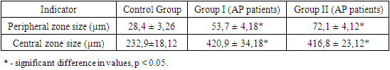
The examined patients reported a range of clinical complaints: 100% of patients noted halitosis, and bleeding was observed during routine daily oral hygiene procedures. Clinical examination revealed pathological tooth mobility and changes in tooth position. The clinical findings showed that 45% of patients exhibited no visible signs of microcirculatory response, such as edema or mixed hyperemia of gingival tissues. However, despite the mild clinical presentation of inflammation, patients with AP had an average periodontal pocket depth of 7.5 mm, with some cases exceeding 8.5 mm. In Group II, 36% of patients exhibited grade II–III tooth mobility. No purulent exudate was detected.
Orthopantomographic data from Group II patients revealed massive generalized destruction of alveolar bone, including both vertical and horizontal types. Bone loss reached up to 2/3 of tooth root length, with grade III furcation defects and osteoporosis. In Group I, bone destruction was also observed but was less pronounced.
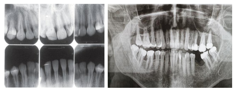
Figure 1. Radiographs of patients from Group I and Group II
In addition, a large number of areas with reduced density were detected, which serves as a radiological criterion for bone structure destruction in AP. Unlike chronic generalized periodontitis, aggressive periodontitis is characterized by a discrepancy between clinical symptoms and radiological findings, particularly noticeable in cases with mild to moderate inflammatory processes. In severe forms of the disease, this discrepancy becomes less pronounced. However, the use of index-based methods for assessing the condition of periodontal tissues makes it possible to identify such deviations even at later stages of pathology development.
In the control group, the values for the Muhlemann bleeding index, PMA, and PI indices were considered "0," and the OHI-S index corresponded to a satisfactory level of oral hygiene. The results of the index calculations are presented in Table 1.
Table 1. Indicators of periodontal tissue condition and oral hygiene level (M ± m)
     
Statistical analysis of the OHI-S index revealed no significant difference between Group I and Group II (p > 0.05) but demonstrated a significant difference compared to the control group (p < 0.05). Consequently, oral hygiene in both groups was rated as poor. However, it is noteworthy that despite similar OHI-S index values, there were differences in PMA, PI, and Muhlemann bleeding indices, with statistically significant differences in the PMA index between the groups. All other indices showed significant differences compared to the control group (p < 0.05).
The results confirm significant differences in the clinical and radiological characteristics of various forms of AP, supported by data from numerous scientific studies. According to J. Albandar and T. Rams [1], AP is characterized by early onset, rapid progression, and significant destruction of periodontal tissues, which correlates with our findings. Despite minimal clinical signs of inflammation, AP patients exhibit a high degree of tissue destruction, indicating a disproportion between localized inflammatory processes and systemic tissue destruction.
A comparative analysis of the OHI-S index revealed a comparable level of oral hygiene among patients with different forms of AP. This finding aligns with data from Kinane [6], which demonstrated that oral hygiene status alone is not always a key factor in disease progression. However, despite similar hygiene levels, patients with generalized AP exhibited greater bone destruction, emphasizing the role of additional factors such as immune system hyperactivity and specific microbial biofilm composition, which may exacerbate the inflammatory process and lead to rapid tissue destruction [4].
Radiological studies showed that Group II patients predominantly exhibited generalized bone loss with deep periodontal pockets and furcation defects. These findings corroborate Kornman’s work [7], which notes that AP is associated with both vertical and horizontal bone loss types, accompanied by pronounced alveolar ridge destruction. Conversely, Group I patients more frequently exhibited localized changes, consistent with descriptions in [4], which highlight that localized pathological processes in periodontal tissues are primarily driven by bacterial aggression, while systemic auto-aggression becomes the dominant mechanism in generalized forms.
The differences identified between the groups in this study are of great clinical significance. Despite similar hygiene levels, destructive processes in the generalized form of AP progress much faster, necessitating early intervention and the use of personalized treatment approaches. These approaches should include not only microbial control but also modulation of the immune response [2,4].
Currently, the distribution of organic and inorganic molecules, proteins, and glycoproteins during the crystallization of mixed saliva is well-documented. The center of the crystallization pattern typically contains an accumulation of small inorganic salt molecules forming crystals, while the periphery predominantly comprises large protein molecules and glycoproteins. According to contemporary scientific data, the "morphological" pattern of mixed saliva in healthy individuals is characterized by distinctly formed crystals, commonly referred to as "fern leaves" or "coral branches." These crystals are uniformly distributed across the entire droplet. The findings of this study in the control group of healthy patients align fully with the literature. In these patients, light microscopy revealed the characteristic "fern leaf" pattern (Figure 2).
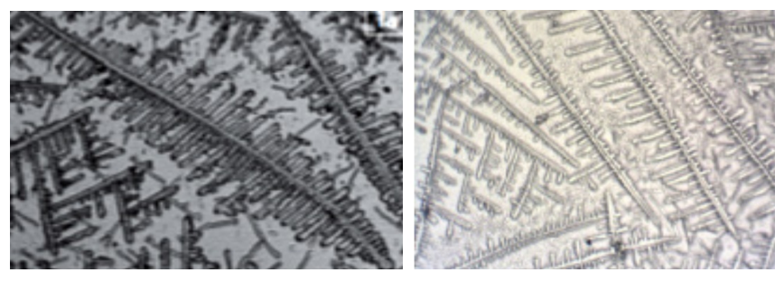
Figure 2. Light microscopy image of mixed saliva from the control group
The central zone occupied the largest area and most frequently appeared as a lattice structure with characteristic patterns resembling a "fern leaf." In rare cases, the central zone of the facies displayed star-shaped structures. The ratio between the central and peripheral zones in the control group was approximately 70% to 30%, respectively. The peripheral zone in this group was clearly visualized and was most often represented by an amorphous protein structure.
Analysis of light microscopy results in patients with AP revealed significant morphological changes in the OCS. In both Groups I and II, light microscopy showed an altered ratio between the central and peripheral zones: 55–60% to 35–45% in Group I and 70–80% to 15–20% in Group II, respectively (p < 0.05) (Figure 3).
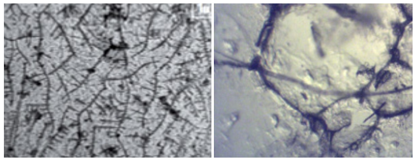
Figure 3. Light microscopy of mixed saliva in patients with aggressive periodontitis
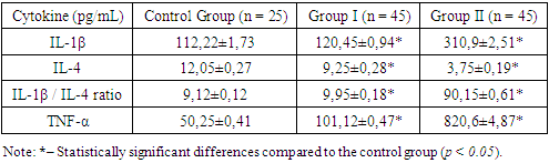
For Group II, a disruption of clear radial orientation was characteristic, with numerous star-shaped crystals observed. Significant decystallization of the central zone of the facies was reliably visualized in patients from Group II (p < 0.05). The peripheral zone lost its distinct contours, merging with the central zone, and contained a small number of tiny crystals showing signs of disaggregation.
Table 2. Presents the results of morphometric analysis in patients with aggressive periodontitis (measured in μm)
     
A comparative statistical analysis of quantitative morphometric indicators revealed significant differences between the control group and patients with AP across all parameters. However, comparisons between Group I and Group II showed a significant difference only in the size of the peripheral crystallization zone of saliva (p < 0.05), while the indicators for the central zone were not statistically significant (p > 0.05).
Thus, our findings suggest that alterations in the crystallization of mixed saliva in patients with generalized AP may serve as a marker of a malignant progression of the pathological process, further leading to decompensation. This represents an additional risk factor for the severity of oral cavity damage.
One of the primary results is the significant increase in levels of IL-1β and TNF-α in the gingival crevicular fluid (GCF) of patients with AP. IL-1β is a central inflammatory mediator that activates immune cells, stimulates the production of other cytokines, and exacerbates periodontal tissue destruction by activating osteoclasts. TNF-α, as a potent pro-inflammatory cytokine, further contributes to tissue degradation by activating matrix metalloproteinases and inducing apoptosis of periodontal cells [11,14].
As shown in Table 3, significant differences were identified in patients with AP (p < 0.05). For instance, the mean concentrations of IL-1β in the GCF of AP patients were three times higher than those in the control group, while TNF-α levels were 16.7 times higher, indicating pronounced inflammatory activity characteristic of AP.
Table 3. Levels of key pro-/anti-inflammatory cytokines in gingival crevicular fluid (M ± m)
     
IL-4, in contrast, is an anti-inflammatory cytokine that regulates the immune response by inhibiting the production of pro-inflammatory mediators and promoting tissue regeneration. Its concentration in GCF was significantly reduced, highlighting an imbalance in the cytokine profile and suppression of mechanisms capable of mitigating the progression of inflammation.
These findings are consistent with previously published studies, which noted that cytokine hyperactivity is an important marker of AP severity [1,2,13].
In this study, a more detailed analysis of the IL-1β/IL-4 ratio was performed, which not only confirmed the cytokine profile imbalance but also demonstrated its potential as a diagnostic marker [7]. The analysis revealed an increase in the IL-1β/IL-4 ratio by more than 150%, confirming a pronounced imbalance between pro-inflammatory and anti-inflammatory mechanisms. The high value of this index indicates that inflammation in AP is a systemic and hyperactive process, likely exceeding the scope of a localized tissue response in the periodontium. This ratio may be used as an indicator of disease severity and the effectiveness of therapeutic interventions.
This approach to studying the cytokine profile is of great clinical significance [6,9]. It can be used for the early diagnosis of AP and for monitoring the effectiveness of therapy. The application of cytokine analysis in gingival crevicular fluid, as a minimally invasive method, represents a promising tool for real-time assessment of the inflammatory status. Future research could focus on studying the dynamics of cytokine profile changes during treatment, including the use of modern anti-inflammatory drugs and biological agents targeting specific cytokine pathways. Such research will improve personalized therapy for AP, aiming not only to suppress inflammation but also to restore regenerative mechanisms in the periodontium.
Thus, the results of this study confirm that cytokine profile analysis plays a key role in understanding the pathogenesis of aggressive periodontitis and in developing effective diagnostic and therapeutic strategies.

4. Conclusions

1. The evaluation of quantitative indicators of mixed saliva crystallization demonstrated a statistically significant imbalance in patients with the generalized form of aggressive periodontitis.
2. Quantitative diagnosis of mixed saliva crystallization serves as a clinical and diagnostic marker for the severity of pathological processes in the oral cavity in aggressive periodontitis.
3. Patients with aggressive periodontitis exhibit a significant increase in pro-inflammatory cytokine levels (IL-1β, TNF-α) and a decrease in the anti-inflammatory cytokine IL-4 in gingival crevicular fluid.
4. The IL-1β/IL-4 ratio can be used as a marker of inflammatory process activity.
5. Cytokine profile assessment is crucial for diagnosing and monitoring the prognostic significance of aggressive periodontitis.
6. The generalized form of aggressive periodontitis is characterized by significant bone tissue destruction with relatively mild inflammatory signs, indicating a complex pathogenic mechanism of the disease.
7. The OHI-S index values indicate a low level of oral hygiene among patients, which may highlight the etiological significance of the microbial factor in the disease's pathogenesis.
8. For accurate diagnosis and a personalized approach to managing patients with aggressive periodontitis, it is essential to correlate the results of clinical examination with radiological findings of bone structure changes.

References

[1]  Dzyuba E. V., Nagaeva M. O., Zhdanova E. V. The role of immunological processes in the development of inflammatory periodontal diseases and the possibilities of their correction // Problems of Dentistry. 2019. No. 2. Pp. 25–31.
[2]  Pritulina Yu. G., Krivoruchko I. V., Shentsova V. V., et al. Practical significance of cytokine profile analysis in a number of infectious diseases // Actual Infectology. 2014. No. 1 (2). Pp. 38–42.
[3]  Yudina N. A., Yakovleva-Malykh M. O. Cytokine profile of gingival fluid of patients with various forms of periodontal diseases // Modern Dentistry. 2021. No. 2 (83). Pp. 58–62. Liu Y., Zhao R., Reda B., Yang W. Profiling of cytokines, chemokines and growth factors in saliva and gingival crevicular fluid. Cytokine. 2021; 142: 155504.
[4]  Green, J. C., Vermillion, J. R. The Simplified Oral Hygiene Index. Journal of the American Dental Association, 1964; 68: 7–13.
[5]  Pihlstrom, B. L., Michalowicz, B. S., Johnson, N. W. Periodontal diseases. Lancet, 2005; 366(9499): 1809–1820.
[6]  Graves, D. Cytokines that promote periodontal tissue destruction. Journal of Periodontology, 2008; 79(8): 1585–1591.
[7]  Kornman, K. S. Interleukin-1 genetics, inflammatory mechanisms, and nutrigenetic opportunities to modulate diseases of aging. American Journal of Clinical Nutrition, 2006; 83(2): 475–483.
[8]  Genco, R. J., Borgnakke, W. S. Risk factors for periodontal disease. Periodontology 2000, 2013; 62(1): 59–94.
[9]  Bhardwaj, A., Bhardwaj, S. V. Role of cytokines in periodontal disease: A review. Annals of Dental Specialty, 2014; 2(2): 45–51.
[10]  Kinane, D. F., Stathopoulou, P. G., Papapanou, P. N. Periodontal diseases. Nature Reviews Disease Primers, 2017; 3: 17038.
[11]  Albandar, J. M., Rams, T. E. Global epidemiology of periodontal diseases: An overview. Periodontology 2000, 2020; 82(1): 13–28.
[12]  Bartold, P. M., Van Dyke, T. E. Host modulation: Control of inflammation to promote periodontal regeneration. Journal of Clinical Periodontology, 2013; 40(Suppl 14): S1–S16.
[13]  Dinarello, C. A. Overview of the interleukin-1 family of ligands and receptors. Seminars in Immunology, 2013; 25(6): 389–393.
[14]  Albandar J. M., Rams, T. E. Global epidemiology of periodontal diseases: An overview. Periodontology 2000. 82(1). 13–28.
[15]  Bartold P. M., Van Dyke, T. E. Host modulation: Control of inflammation to promote periodontal regeneration. Journal of Clinical Periodontology. 2013. 40(Suppl 14). S1–S16.
[16]  Genco R. J., Borgnakke, W. S. Risk factors for periodontal disease. Periodontology 2000. 2013. 62(1). 59–94.
[17]  Graves D. T. Cytokines that promote periodontal tissue destruction. Journal of Periodontology. 2008. 79(8). 1585–1591.
[18]  Green J. C., Vermillion, J. R. The Simplified Oral Hygiene Index. Journal of the American Dental Association. 1964. 68. 7–13.
[19]  Kinane D. F., Stathopoulou, P. G., Papapanou, P. N. Periodontal diseases. Nature Reviews Disease Primers. 2017. 3. 17-38.
[20]  Kornman K. S. Interleukin-1 genetics, inflammatory mechanisms, and nutrigenetic opportunities to modulate diseases of aging. American Journal of Clinical Nutrition. 2006. 83(2). 475–483.
[21]  Lindhe J., Ranney, R., Lamster, I., et al. Consensus report: Chronic periodontitis. Annals of Periodontology. 1999. 4(1). 38.
[22]  Pihlstrom B. L., Michalowicz, B. S., Johnson, N. W. Periodontal diseases. Lancet. 2005. 366(9499). 1809–1820.
[23]  Page R. C., Kornman, K. S. The pathogenesis of human periodontitis: An introduction. Periodontology 2000. 1997. 14(1). 9–11.