-
Paper Information
- Previous Paper
- Paper Submission
-
Journal Information
- About This Journal
- Editorial Board
- Current Issue
- Archive
- Author Guidelines
- Contact Us
American Journal of Medicine and Medical Sciences
p-ISSN: 2165-901X e-ISSN: 2165-9036
2025; 15(4): 982-986
doi:10.5923/j.ajmms.20251504.28
Received: Mar. 6, 2025; Accepted: Apr. 3, 2025; Published: Apr. 8, 2025

Morphological Changes of the Adrenal Gland of Infants Who Died of Pneumonia Between the Ages of Six Months and One Year
 Abstract
 Reference
 Full-Text PDF
 Full-text HTML
Orınbayev Jumabay Tileubayevich
Assistant Professor, Department of Pathology, Karakalpakstan State Medical Institute, Uzbekistan
Correspondence to: Orınbayev Jumabay Tileubayevich, Assistant Professor, Department of Pathology, Karakalpakstan State Medical Institute, Uzbekistan.
Copyright © 2025 The Author(s). Published by Scientific & Academic Publishing.
This work is licensed under the Creative Commons Attribution International License (CC BY).
http://creativecommons.org/licenses/by/4.0/

In pneumonia in infants up to one year of age, the normality of the immune system is maintained due to the integration of the morphologically formed adrenal gland and thymus. In pneumonia at 3-6 months of age, secondary immunodeficiency is observed due to the fact that hyperproduction of the adrenal gland depresses the thymus and prematurely causes apoptosis of the produced lymphocytes. In this case, hyperplasia of the adrenocortical zone and the appearance of foci of hemorrhage are sometimes manifested by adrenal insufficiency, which can lead to sudden death in infants. The formation of intermediate tumors in the adrenal gland tissues confirms the development of a hyperfunctional state of the adrenocortical and adrenocortical zones in this studied group.
Keywords: Pathomorphology, Thymus, Adrenal gland, Pneumonia, Immunodeficiency, Depression, Immune system, Hyperfunctional state
Cite this paper: Orınbayev Jumabay Tileubayevich, Morphological Changes of the Adrenal Gland of Infants Who Died of Pneumonia Between the Ages of Six Months and One Year, American Journal of Medicine and Medical Sciences, Vol. 15 No. 4, 2025, pp. 982-986. doi: 10.5923/j.ajmms.20251504.28.
Article Outline
1. Introduction
- In the world, pneumonia in infants under one year of age occurs on average at 8.2-9.75% per 1000 live births [1], due to intrauterine infections and nosocomial infections, as well as various influencing factors in the postnatal period. In developed countries, the USA and Europe, this figure is on average 4-5 per 1000 live births, and in the 3-6 month period, it is 11.72% per 1000 live births [2]. The highest rate in the world, in the period of newborns under 1 month, is in Turkmenistan, one of the Central Asian countries, with 45 cases per 1000 live births, while in our country this figure is 16 cases per 1000 live births [3]. This indicates that the full screening of extragenital diseases during intrauterine development or the tests that should be performed during pregnancy have not been fully implemented. Death in early infancy mainly develops in conjunction with infectious and non-infectious etiological processes, and is manifested in the morphological aspects of the integral connection of the adrenal gland and thymus in pneumonia of various etiologies in infants.Namely, the high mortality rate from pneumonia in the period up to 1 year of age, the incomplete implementation of screening and preventive work in the first and second link systems that monitor pregnant women, and the neglect of maternal and child protection, and the lack of specific scientific and practical recommendations and evidence-based medical recommendations during this period, and this process requires the need for constantly updated scientific and practical recommendations, once again confirming the urgency of the problem.
2. The Purpose of the Study
- To study the pathomorphological characteristics of the adrenal gland in children who died from pneumonia in the neonatal period and died with a diagnosis of pneumonia.
3. The Materials and Methods
- The materials and methods of the study were the autopsy medical documents, adrenal gland and thymus tissue of 101 infants who died with a diagnosis of pneumonia in the Republic of Karakalpakstan in 2020-2023. The thymus tissue of infants who died with a diagnosis of pneumonia was analyzed using the morphological method.
4. Results and Discussion
- It should be noted that in this group of infants, the thymus and adrenal glands developed in direct proportion to each other due to the age-related morphological formation and the mutual integrative function of these two organs [4]. The adrenal glands in the group from 6 months to 12 months had the following weights:left – 2.46±0.15 g.; right – 2.63±0.15 g.The average weight of both adrenal glands was 5.10±0.30 g., i.e., lower than the age-related norm. However, the relative weight of the adrenal glands to body weight was higher (0.00086±0.00006) than the norm (0.00060±0.00001), and this difference was statistically significant (p<0.05).
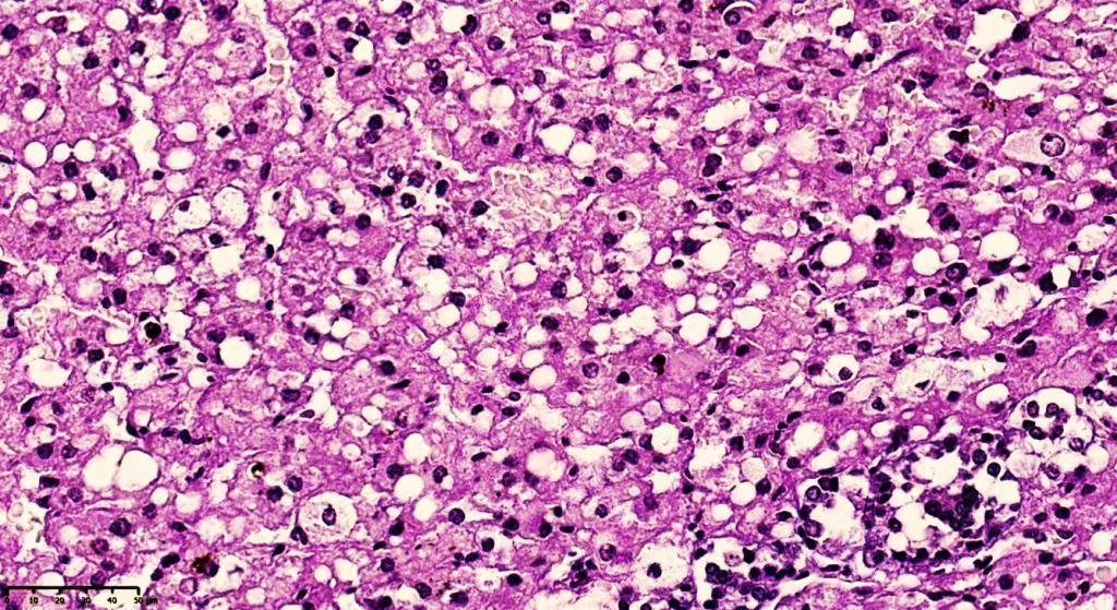
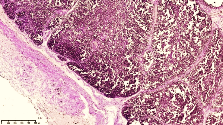
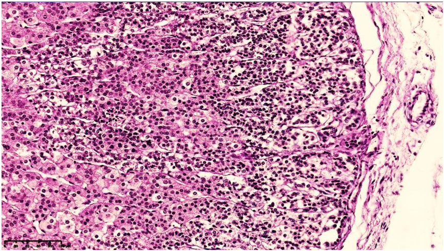 | Figure 1. Adrenal gland of an 8-month-old infant who died of pneumonia, protocol 21 DI. Morphologically mature adrenal cortex. The glomerular layer is hypercellular, with sparse fibrous structures at the perimeter of the developing glomerular structures (1). In the spongiotic zone, spongiocytes of various sizes are present, and the largest ones are cells rich in vacuolar dystrophy and homogeneous pink eosinophilic inclusions (2). Stained with G.E. Size 10x10 |
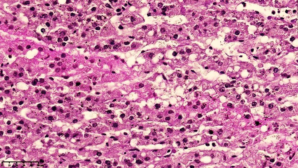
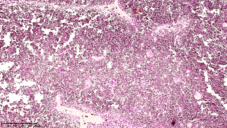
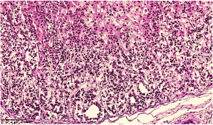 | Figure 2. Adrenal gland of a 10-month-old infant who died of pneumonia Report 56DI.. Morphologically determined adrenal cortical layer. The glomerular layer is hypercellular, and sparse fibrous structures are detected at the perimeter of developing glomerular structures (1). Paint G.E. The size is 10x10 |
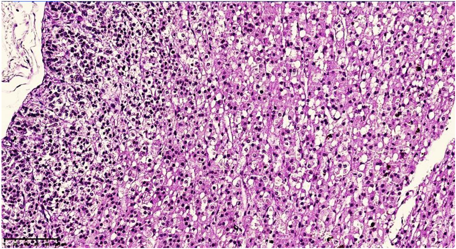 | Figure 3. Adrenal gland of a 12-month-old infant who died of pneumonia Report 55DI. In the area of the tumor, spongiocytes of various sizes are detected, the largest of which are rich in lipid inclusions and homogeneous pink eosinophilic inclusions (2). Staining GE. Size 10x10 |
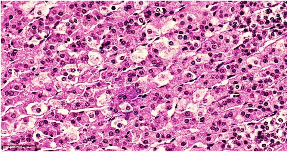 | Figure 4. Adrenal gland of an 11-month-old infant who died of pneumonia Report 55DI.. In the spongiotic area, spongiocytes of various sizes are detected, and the largest ones have undergone vacuolar dystrophy (1). Staining GE. Size 10x10 |
 | Figure 5. Adrenal gland of a 12-month-old infant who died of pneumonia Report 55DI. In the tufted area, spangiocytes, cells rich in lipid inclusions of different sizes are identified (1). Dye G.E. The size is 10x10 |
 | Figure 6. Adrenal gland of a 12-month-old infant who died of pneumonia Report 55DI. Focal necrosis foci and advanced fibrotic tissue are detected in spangiocytes in the tufted area (1). Dye G.E. The size is 10x10 |
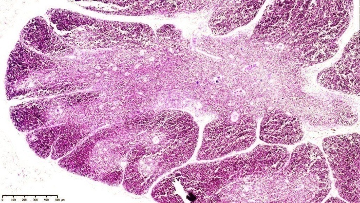 | Figure 7. 8-month-old baby diaper. The boundaries of the corticomedullary layers of the thymus cortex and medulla are clear, and small numbers of Gassal corpuscles are identified. Pieces of different sizes. Paint G.E. Size 4x10 |
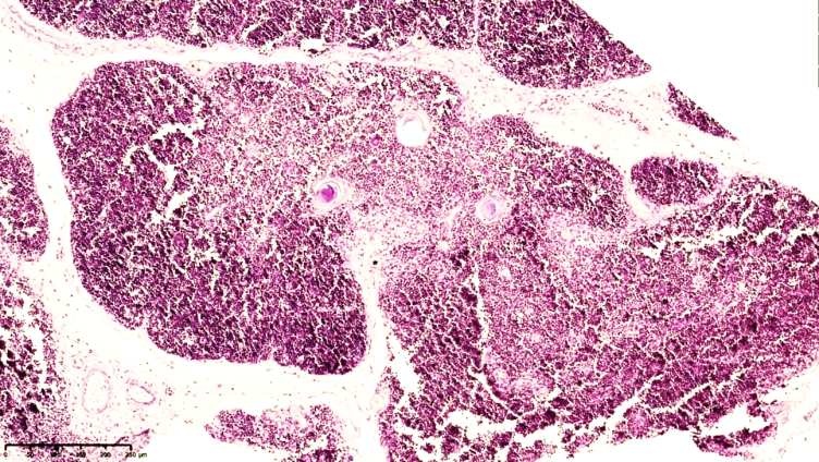 | Figure 8. An 11-month-old infant's salivary gland tissue. Fragments have decorticated foci. In the preserved fragment, the medullary layer is reduced and its borders are unclear. Paint G.E. Size 4x10 |
 | Figure 9. Thymus tissue of a 12-month-old baby. Interlobular barrier fibrotic tissue is thickened. The boundaries of the medullary layer of the lobes are unclear and of different widths. The thymus capsule and thickened vessels appear plump. Paint G.E. Size 4x10 |
 | Figure 10. A 12-month-old infant's salivary gland tissue. Gassal corpuscles of various sizes in the medulla, signs of fullness are detected in small-caliber blood vessels. Paint G.E. Size 4x10 |
 | Figure 11. A 12-month-old infant's salivary gland tissue. Foci of developing Gassal bodies, reticular and fibrous structures are identified in the medulla. Paint G.E. The size is 40x10 |
5. Conclusions
- Thus, the number of mature lymphocytes in the cortical layer of the thyroid gland is small, the foci of apoptosis are massive, the postcapillary venules are full, interstitial tumors are formed in the intermediate stroma, macrophages are mainly located on the perimeter of reticuloepithelial cell growths, and a large number of small lymphocytes are found. The medullary layer is characterized by a sharp decrease in lymphocytes around reticuloepithelial cells, dendritic cells, and reticular cells, and the presence of Hassall bodies of various sizes, and in the focus of reticuloepithelial cells, vacuolar dystrophy, and some of them are of various shapes due to protein dystrophy.