Yuldashov Sarvarkhon1, Akhmedova Sayyora2, Nozimov Akhmadjon3
1Doctoral Student of the Department of Human Anatomy and OSTA Tashkent Medical Academy, Tashkent, Uzbekistan
2Doctor of Medical Sciences, Professor, Tashkent Medical Academy, Tashkent, Uzbekistan
3PhD, Republican Specialized Scientific and Practical Medical Center of Eye Microsurgery, Tashkent, Uzbekistan
Correspondence to: Yuldashov Sarvarkhon, Doctoral Student of the Department of Human Anatomy and OSTA Tashkent Medical Academy, Tashkent, Uzbekistan.
| Email: |  |
Copyright © 2025 The Author(s). Published by Scientific & Academic Publishing.
This work is licensed under the Creative Commons Attribution International License (CC BY).
http://creativecommons.org/licenses/by/4.0/

Abstract
This study aimed to investigate the morphological changes in the extraocular muscles, periorbital tissues, and choroid layer of rats subjected to experimental thyrotoxicosis. A total of 28 Wistar laboratory rats were divided into two groups (сalled the experimental group and control group). The experimental group received oral administration of levothyroxine sodium at a dosage of 0.3 mg/kg. Morphological analysis revealed hyperplasia of the extraocular muscles, densification of collagen fibers, and swelling of the periorbital tissues in the rats from the experimental group. Additionally, there were signs of vascular dilation, increased blood perfusion, and an increase in the number of capillaries in the choroid layer. These alterations are attributed to the effects of thyroid hormones on eye tissues and provide important insights for understanding the experimental model of ophthalmopathy that occurs alongside thyrotoxicosis. The findings of this study are significant for further exploration of ophthalmological issues related to thyrotoxicosis and for developing therapeutic strategies aimed at their prevention.
Keywords:
Thyrotoxicosis, Extraocular muscles, Periorbital tissues, Choroid, Levothyroxine, Morphological changes, Endocrine ophthalmopathy, Experimental research
Cite this paper: Yuldashov Sarvarkhon, Akhmedova Sayyora, Nozimov Akhmadjon, Morphological Features of the Visual Organ in Experimental Thyrotoxicosis, American Journal of Medicine and Medical Sciences, Vol. 15 No. 4, 2025, pp. 956-960. doi: 10.5923/j.ajmms.20251504.23.
1. Introduction
Thyrotoxicosis is a pathological condition caused by excessive production of thyroid hormones, which has a significant impact on various body tissues, including the ocular system [1,4]. Ophthalmological changes associated with thyrotoxicosis primarily manifest as Graves' ophthalmopathy or thyroid orbitopathy, characterized by forward displacement of the eyeball (exophthalmos), periorbital edema, hyperplasia of the extraocular muscles, and orbital inflammatory processes [2,5,9]. Experimental studies play a crucial role in investigating the effects of thyroid hormones on ocular tissues. Research using laboratory animals, particularly rats, allows for identifying morphological and functional changes and provides a deeper understanding of pathological mechanisms [3,6]. Although previous studies have confirmed that thyrotoxicosis induces changes in the eyeball and some components, Morphological alterations in the extraocular muscles, periorbital tissues, and choroid layer have not been sufficiently explored [7,8].This study aims to determine the morphological changes in the extraocular muscles, periorbital tissues, and choroid layer of rats under experimental thyrotoxicosis. The findings may offer more detailed insights into the pathogenesis of ophthalmological changes associated with thyrotoxicosis and establish a scientific foundation for developing of effective treatment strategies for this condition.
2. Purpose of the Research
To identify changes in the morphometric parameters of the eye in laboratory rats under experimental thyrotoxicosis and assess their impact on pathological processes.
3. Materials and Methods
Forty-eight healthy, sexually mature male Wistar rats weighing between 200 and 250 g were selected as subjects for this study. The rats were kept under standard laboratory conditions (temperature: 22–24°C, light/dark cycle: 12 h/12 h) throughout the experiment and were provided with a balanced diet, along with free access to food and drinking water. The experiment adhered to the international recommendations set forth by the European Convention for the Protection of Vertebrate Animals. Thyroid hormone levels, including T3, T4, and TSH, were measured using immunoenzymatic analysis conducted by DRG International Inc. in Germany. The parameters assessed included uT3 (total triiodothyronine), uT4 (total thyroxine), and TSH (thyroid-stimulating hormone). The data were statistically analyzed utilizing Microsoft Office Excel 2020 on a Lenovo Core i3 personal computer.The animals were randomly divided into two groups:1. Control Group (n = 24): Rats in this group were not exposed to any chemical treatments; they were maintained under standard conditions and later used for morphological analysis after the experiment.2. Experimental Group (n = 24): A model of experimental thyrotoxicosis was induced in this group. To achieve this, the rats were administered levothyroxine sodium orally at a dosage of 0.3 mg/kg daily. This dosage was selected based on previous studies and was found to be adequate to induce thyrotoxicosis under experimental conditions.Morphometric analysis: At the end of the study, all rats were euthanized under general anesthesia (using ketamine at 80 mg/kg and xylazine at 10 mg/kg) via decapitation. The eyeballs were removed along with the periorbital tissues. These tissues were fixed in a 10% neutral formalin solution for 24 hours, embedded in paraffin blocks, and sectioned into 5 μm-thick slices using a microtome. The sections were stained using hematoxylin-eosin to assess general histological structure, tissue integrity, and the nuclear-cytoplasmic ratio, as well as Masson's trichrome to evaluate the density of connective tissue and collagen fibers.
4. Results and Discussion
Analysis of the results from the experimental thyrotoxicosis indicated that by the seventh day of the experiment, the levels of TSH and T4 had changed slightly but did not differ significantly from those in the control group (Table 1).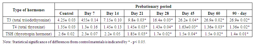 | Table 1. Indicators of thyroid hormone levels in the blood of rats with an experimentally induced thyrotoxicosis model were examined over time |
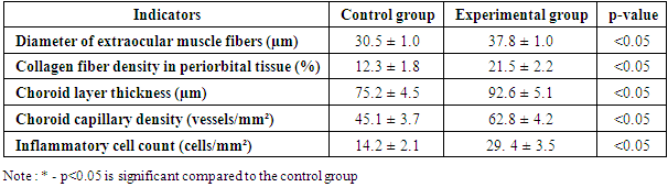
Histological examination of enucleated rat eyes under conditions of experimental thyrotoxicosis revealed pathological changes in various ocular tissues. A detailed analysis of the morphological results, presented in the table, showed significant differences between the experimental and control groups. In the experimental group, the diameter of extraocular muscle fibers demonstrated a functional decrease compared to the control group. The density of collagen fibers in the periorbital tissues was measured at 12.3 ± 1.8% in the control group, while it significantly increased to 21.5 ± 2.2% in the experimental group, highlighting a notable rise in collagen fiber density. Additionally, the thickness of the choroid layer increased by 23.1% in the experimental group, and the density of choroidal capillaries rose by 39.3%.A significant increase in the inflammatory cell count was observed in the experimental group, which rose by 107.0% compared to the control group, indicating a marked intensification of inflammatory processes (p < 0.05).Thyrotoxicosis induces dynamic changes characteristic of hyperplasia in the extraocular muscles. On day 28 of the study, peak hyperplasia of the extraocular muscles was observed in the experimental group. However, between days 60 and 90, a decrease in the number of muscle fibers was noted, alongside the onset of fibrosis (Table 2).Table 2. Comparison of morphological results of experimental and control groups (day 28)
 |
| |
|
Hyperplasia and hypertrophy of extraocular muscle fibers were observed in the experimental group. The diameter of the muscle fibers in this group was significantly larger compared to the control group (p < 0.05). Morphological changes in the periorbital tissues were characterized by swelling that appeared in the experimental group early in the study. These infiltrative changes primarily affected the adipose and connective tissues. By day 60, there was evidence of blood vessel dilation, along with the presence of lymphocytes and macrophages, indicating an ongoing inflammatory process. Additionally, an increase in the density of collagen fibers and the number of fibroblasts was noted (Figures 1, 2).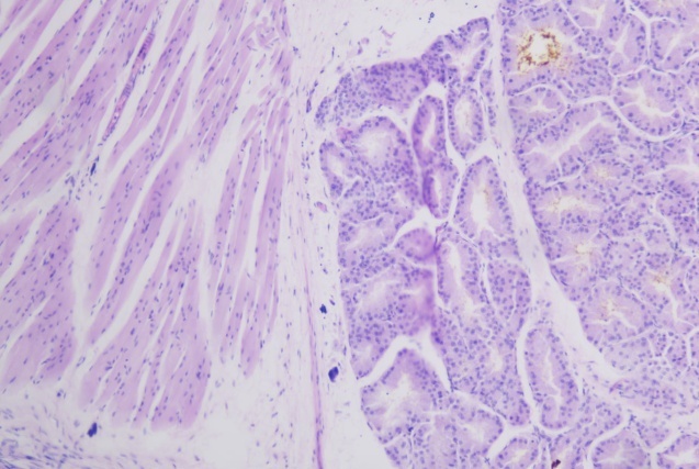 | Figure 1. Periorbital tissue edema in Wistar rats induced by thyrotoxicosis on the 60th day of the experiment. Staining method : Hematoxylin-eosin. Size 20x10 |
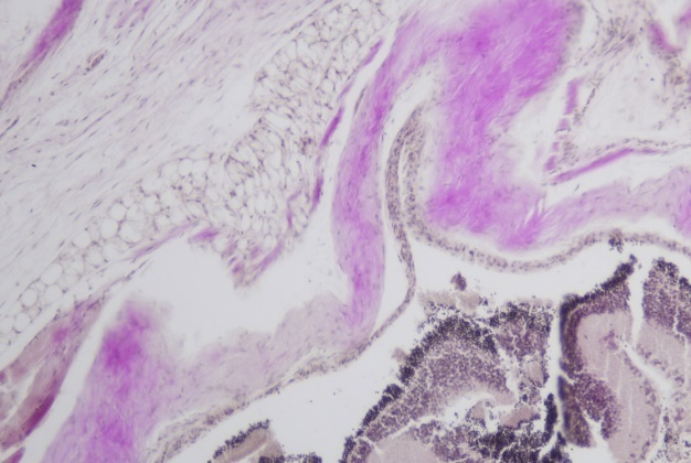 | Figure 2. Focal edema in the peripheral zones of the cornea in Wistar rats induced by thyrotoxicosis on the 60th day of the experiment. Accumulation of lymphocytes and plasma cells. Staining method: Hematoxylin-eosin. Size 20x10 |
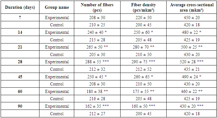
During this period, swelling of the corneal epithelium and focal dystrophic changes were detected, which included vacuolar and balloon dystrophy, as well as signs of desquamation in the dystrophically altered cells. The collagen fibers appeared swollen, fused, and deformed. There was also focal subfluid infiltration of lymphocytes and plasma cells observed in the peripheral zones of the cornea. For the first time, a small number of newly formed blood vessels were detected growing from the limbus into the cornea.In this study, veins and capillaries were significantly dilated, showing signs of stasis. Beneath this fluid, foci of lymphoplasmacytic infiltration were observed within the eye. According to the histological analysis conducted by day 90 of the study, an irregular arrangement of extraocular muscle fibers and areas of atrophy were detected. Additionally, there was an increase in connective tissue between muscle fibers and a higher number of fibroblasts.During the early months of experimental thyrotoxicosis, notable morphological changes occurred in the extraocular muscles, periorbital tissues, and the choroid. Hyperplasia of the extraocular muscles and an increase in muscle fiber diameter indicated the progression of ophthalmopathy associated with thyrotoxicosis (see Table 3). Edema and thickening of collagen fibers in the periorbital tissues suggested activation of inflammatory and fibrotic processes in that area. Vasodilation and elevated inflammatory markers in the choroid indicated that thyrotoxicosis could contribute to changes in ocular blood supply and immune responses. The primary pathological effects of thyrotoxicosis appear to be driven by inflammation, autoimmune reactions, and oxidative stress.Table 3. Morphometric parameters characteristic of extraocular muscle fiber hyperplasia in rats with experimental thyrotoxicosis
 |
| |
|
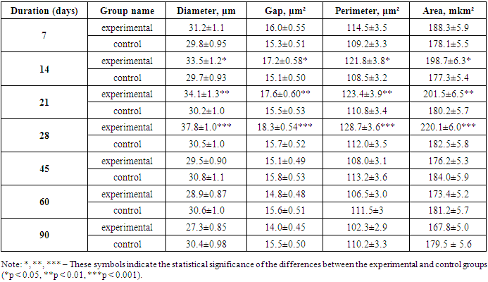
Analysis of Results:• On day 14, there was a slight increase in the number and density of muscle fibers compared to the control group. This change is associated with the initial effects of thyrotoxic hormones on the muscles.• Between days 21 and 28, the number, density, and cross-sectional area of muscle fibers increased significantly, indicating that the hyperplasia process had reached its peak.• From days 28 to 45, there was a decrease in both the number and density of muscle fibers, suggesting the onset of degenerative and fibrotic processes.• Between days 45 and 60, the decline in the number of fibers and other indicators continued.• By day 90, the number of muscle fibers and related indicators had approached levels similar to those of the control group, indicating intensified muscle atrophy and fibrosis.Immunomodulators, antioxidants, and hormonal therapy may be effective treatment options. Additionally, leukocyte infiltration has been observed, confirming the presence of an ongoing inflammatory process. Specifically, the accumulation of neutrophilic granulocytes, lymphocytes, and macrophages was detected in the choroid and periorbital tissues. These immune cells secrete inflammatory mediators that further enhance processes leading to changes in morphometric parameters. The histological results obtained demonstrate significant alterations in ocular tissues under conditions of experimental thyrotoxicosis, providing a scientific basis for more in-depth studies of the disease's pathogenesis. Key parameters—including diameter, gap, perimeter, and area—reflect the hyperplastic processes in the extraocular muscle fibers of rats under experimental thyrotoxicosis. During the first month of the study, the formation of new muscle fibers due to hyperplasia resulted in a significant increase in these parameters in the experimental group compared to the control group (Table 4).Table 4. Morphometric parameters of extraocular muscle fibers of rats with experimental thyrotoxicosis
 |
| |
|
The table displays the average morphometric values (± standard deviation) of the extraocular muscles in both the experimental group (thyrotoxicosis) and the control group of rats. In the experimental group, there was a progressive decrease over time in the diameter, area, perimeter, and field size of the muscles. In contrast, these parameters remained stable in the control group.By day 90 of the experiment, changes in the extraocular muscle fibers had intensified, and alterations in the layers of the eyeball were also observed. Swelling and thickening of the corneal epithelium were noted, along with the accumulation of mucous edema fluid beneath the epithelium. Epithelial cells underwent dystrophic changes, including vacuolar and balloon dystrophy, with their nuclei showing signs of pyknosis. Small vesicles were present in the epithelial layer, surrounded by atrophied epithelial cells. Beneath the epithelium, lymphoplasmacytic infiltration was observed, featuring small foci containing eosinophils. The stroma became saturated with edema fluid, and collagen fibers appeared interconnected. The corneal membranes showed swelling, scattered areas, and partial disruption, leading to the formation of uniform amorphous masses. The corneal endothelium exhibited severe dystrophic changes, with most of it undergoing desquamation. By this stage of the experiment, scleral thickening was evident, accompanied by vascular hyperemia and edema. Increased edema and hyperemia were also observed in the pigmented layer, which was associated with the destruction of the pigment cell layer. Diffuse mucoid edema, along with swelling and proliferation of connective tissue structures, was detected in the ciliary body. Some epithelial cells were destroyed, resulting in desquamation and deepithelialization. The study results demonstrated that under experimental thyrotoxicosis, significant morphological changes occurred in the extraocular muscles, periorbital tissues, and choroid layer. Hyperplasia of the extraocular muscles and an increase in muscle fiber diameter indicated the progression of ophthalmopathy associated with thyrotoxicosis. Edema and thickening of collagen fibers in the periorbital tissues suggested the activation of inflammatory and fibrotic processes. Vasodilation and increased inflammatory markers in the choroid layer suggested that thyrotoxicosis may lead to alterations in ocular blood supply and immune responses.The primary pathological effects of thyrotoxicosis appear to be driven by inflammation, autoimmune reactions, and oxidative stress. The presence of leukocyte infiltration confirmed an ongoing inflammatory process, specifically an accumulation of neutrophilic granulocytes, lymphocytes, and macrophages in the choroid and periorbital tissues. These immune cells secrete inflammatory mediators, further enhancing processes that lead to changes in morphometric parameters. The obtained histological results demonstrate significant alterations in ocular tissues under conditions of experimental thyrotoxicosis, providing a scientific foundation for an in-depth study of the disease's pathogenesis.
5. Conclusions
The findings of this study indicate that significant morphological changes occur in ocular tissues under experimental thyrotoxicosis. Alterations in the extraocular muscles, periorbital tissues, and choroid layer suggest ophthalmopathic processes associated with thyrotoxicosis, including hyperplasia, collagen fiber densification, choroid layer thickening, and increased inflammatory activity. These pathological changes severely disrupt the morphological and functional integrity of the ocular system, negatively impacting eye movement, blood supply, and visual processes. Furthermore, the increased inflammatory and autoimmune responses, the development of oxidative stress, and enhanced leukocyte infiltration necessitate a deeper investigation into the progression of these processes and the overall pathogenesis of the disease. This study suggests that immunomodulators, antioxidants, and therapies aimed at restoring hormonal balance may be effective in mitigating the pathogenesis of experimental thyrotoxicosis.
References
| [1] | Maheshwari R, Weis E. Thyroid associated orbitopathy. Indian Journal of Ophthalmology 2012; 60: 87–93. pmid: 22446901. |
| [2] | Clement SC, Peeters RP, Ronckers CM, Links TP, van den Heuvel-Eibrink MM, et al. Intermediate and long-term adverse effects of radioiodine therapy for differentiated thyroid carcinoma-A systematic review. Cancer Treat Rev 2015; 10: 00165–6. |
| [3] | Czarnywojtek A, Zgorzalewicz-Stachowiak M, Budny B, Wasko R, Florek E, et al. The influence of radioiodine therapy on ocular changes and their relation to urine cotinine level in patients with Graves' Ophthalmopathy. Neuro Endocrinol Lett 2013; 34: 241–248. pmid: 23685424. |
| [4] | Gerding MN, van der Meer JW, Broenink M, Bakker O, Wiersinga WM, Prummel MF. Association of thyrotrophin receptor antibodies with the clinical features of Graves' ophthalmopathy. Clin Endocrinol (Oxf). 2000; 52: 267–271. |
| [5] | Kroll AJ, Kuwabara T. Dysthyroid ocular myopathy. Anatomy, histology, and electron microscopy. Arch Ophthalmol. 1966; 76: 244–257. pmid: 5945177. |
| [6] | Konuk EB, Konuk O, Misirlioglu M, Menevse A, Unal M. Expression of cyclooxygenase-2 in orbital fibroadipose connective tissues of graves' ophthalmopathy patients. Eur J Endocrinol. 2006; 155: 681–685. pmid: 17062883. |
| [7] | Vondrichova T, de Capretz A, Parikh H, Frenander C, Asman P, Aberg M, et al. COX-2 and SCD, markers of inflammation and adipogenesis, are related to disease activity in graves' ophthalmopathy. Thyroid. 2007; 17: 511–517. pmid: 17614770. |
| [8] | Kim SE, Yoon JS, Kim KH, Lee SY. Increased serum interleukin-17 in Graves' ophthalmopathy. Graefes Arch Clin Exp Ophthalmol. 2012; 250: 1521–1526. pmid: 22752189. |
| [9] | Mogulkoc, R., Baltaci, AK, Oztekin, E., Sivrikaya, A., and Aydin, L. (2006). Effects of hyperthyroidism induced by L-thyroxin administration on lipid peroxidation in various rat tissues. Acta. Biol. Hung. 57, 157–163. |

 Abstract
 Reference
 Full-Text PDF
 Full-text HTML