-
Paper Information
- Next Paper
- Previous Paper
- Paper Submission
-
Journal Information
- About This Journal
- Editorial Board
- Current Issue
- Archive
- Author Guidelines
- Contact Us
American Journal of Medicine and Medical Sciences
p-ISSN: 2165-901X e-ISSN: 2165-9036
2025; 15(4): 893-895
doi:10.5923/j.ajmms.20251504.09
Received: Mar. 8, 2025; Accepted: Mar. 28, 2025; Published: Apr. 6, 2025

Intrafetal Cytokine Levels in Fetuses with Hemolytic Disease Depending on Gestational Age
 Abstract
 Reference
 Full-Text PDF
 Full-text HTML
U. U. Jabborov1, M. N. Abdullayeva2, S. A. Abdullayev3
1Professor, Doctor of Medical Sciences, Republican Perinatal Center of the Ministry of Health of the Republic of Uzbekistan
2Independent Researcher, Central Asian Medical University, Uzbekistan
3Assistant, Ferghana Medical Institute of Public Health, Uzbekistan
Copyright © 2025 The Author(s). Published by Scientific & Academic Publishing.
This work is licensed under the Creative Commons Attribution International License (CC BY).
http://creativecommons.org/licenses/by/4.0/

Objective: to study the cytokine status in fetuses with hemolytic disease depending on the gestational age in pregnancies with Rh immunization. Materials and methods: The studies were conducted at the RPC in 2024. Sixty pregnant women participated, who were divided into three groups. The first main group consisted of 20 pregnant women with Rh immunization in the second trimester, the second comparison group included 20 pregnant women with Rh immunization in the third trimester, and the third control group comprised 20 healthy pregnant women in both the second and third trimesters. All of them underwent transabdominal cordocentesis for diagnostic purposes during the antenatal period to collect blood from the fetal umbilical vein. All immunological studies were conducted at the Institute of Immunology of the Academy of Sciences of the Republic of Uzbekistan. Results: IL-18 is significantly increased in the second trimester by 1.6 times and in the third trimester by 1.7 times. IL-6 in the fetus is elevated 1.7 times in the second trimester and 1.8 times in the third trimester. TNF-alpha is also elevated in the second trimester by 2.4 times and in the third trimester by 2.7 times. The smallest increase was observed in IL-4, by 1.5 times in the second trimester and by 1.4 times in the third trimester compared to the control group. Conclusion: The activity of cytokine regulation and nonspecific immunity in the fetus indicates the protective activity of immunity through the production of pro-inflammatory and anti-inflammatory cytokines in hemolytic disease. Specifically, the increase in the chemokine IL-18 has a direct correlation with the increase of a whole cascade of inflammatory reactions and transmits inflammation to other cytokines.
Keywords: Rh immunization, Hemolytic disease of the fetus, Cytokines, TNF-alpha, IL-4, IL-6, and IL-18, Second and third trimesters
Cite this paper: U. U. Jabborov, M. N. Abdullayeva, S. A. Abdullayev, Intrafetal Cytokine Levels in Fetuses with Hemolytic Disease Depending on Gestational Age, American Journal of Medicine and Medical Sciences, Vol. 15 No. 4, 2025, pp. 893-895. doi: 10.5923/j.ajmms.20251504.09.
Article Outline
1. Introduction
- A pregnant woman is sensitized to the RhD antigen if anti-D antibodies are present in the serum [1]. Maternal IgG anti-D antibodies can be actively transported across the placenta in RhD-negative women carrying an RhD-positive fetus [2]. Destruction of fetal erythrocytes in the spleen, mediated by alloantibodies to erythrocytes, can lead to anemia [3]. Hemolysis of fetal erythrocytes leads to an increase in bilirubin levels. Since bilirubin passes through the placenta, the excess bilirubin is eliminated through the maternal bloodstream during pregnancy [4]. After birth, the hemolytic process continues, but the relatively immature liver of the newborn cannot adequately conjugate the excess bilirubin. This can lead to severe hyperbilirubinemia and irreversible damage to the central nervous system if left untreated [5]. Moise et al. reported back in 2012 that maternal Rh antibody titers are not a predictor of the degree of fetal anemia, especially in subsequent pregnancies, but this old method is still widely used in our country [6]. To this day, the issue of Rh immunization remains relevant in our country. According to domestic authors, in Uzbekistan from 2016 to 2018, there was a trend of increasing incidence of hemolytic disease of the newborn (HDN) [7]. The WHO currently recommends delayed cord clamping for newborns within the first minute after birth, the benefits of which have been demonstrated for both term and preterm infants in terms of their hemoglobin levels at birth, the need for transfusions, and other newborn treatments [8]. It has been shown that delayed cord clamping in newborns with HDFN increases hemoglobin levels at birth and reduces the need for postnatal exchange transfusions in the infant, as well as not requiring increased phototherapy [9]. In 2020, for the first time in the country, two fetal cytokines in hemolytic disease were studied: IL-1 beta and IL-8 [10]. Considering this and the fact that more than 5 years have passed since those studies, we decided to study the cytokine status in fetuses with hemolytic disease in more detail and on a broader scale, depending on the gestational age.The aim of the study was to assess the cytokine status in fetuses, such as TNF-alpha, IL-4, IL-6, and IL-18, in the umbilical cord blood of fetuses with hemolytic disease due to Rh immunization in pregnant women, depending on the gestational age.
2. Research Materials
- The studies were conducted in the Republican Perinatal Center of the Ministry of Health of the Republic of Uzbekistan in 2024. Sixty pregnant women participated, who were divided into three groups. The first main group consisted of 20 pregnant women with Rh immunization in the second trimester, the second comparison group included 20 pregnant women with Rh immunization in the third trimester, and the third control group comprised 20 healthy pregnant women in both the second and third trimesters. All of them underwent transabdominal cordocentesis for diagnostic purposes during the antenatal period to collect blood from the fetal umbilical vein.
3. Immunological Research Methods
- All studies on the cytokine status of the fetus in umbilical cord blood were conducted at the Fundamental Immunology Laboratory of the Institute of Immunology and Human Genome of the Academy of Sciences of the Republic of Uzbekistan. The determination of cytokine levels was conducted using enzyme-linked immunosorbent assay (ELISA) with commercial "Human" test systems produced in Germany, 2023. The test systems are based on the sandwich method of solid-phase enzyme-linked immunosorbent assay using horseradish peroxidase as the indicator enzyme. The reagent kits represent a set, the main reagents of which are monoclonal antibodies to the studied cytokines, adsorbed onto the surface of the wells of a disposable polystyrene plate. The kits are designed for the quantitative determination of human cytokines in peripheral blood serum and biological fluids. The optical density in each well was measured using an automatic microplate photometer at a wavelength of 450 nm by the enzyme-linked immunosorbent assay method on a "Stat-Fax" analyzer (USA).Statistical processing of the results was conducted using parametric and non-parametric analysis methods. Accumulation, correction, systematization of the initial information, and visualization of the obtained results were carried out in Microsoft Office Excel 2018 spreadsheets. Statistical analysis was conducted using the IBM SPSS Statistics v.26 software (developer: IBM Corporation). When comparing means in normally distributed populations of quantitative data, the Student's t-test was calculated. The obtained values of the Student's t-test were evaluated by comparing them with critical values. Differences in indicators were considered statistically significant at a significance level of p<0.05.
4. Results and Discussions
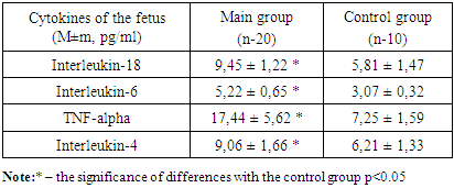
- Cytokines do not function separately from each other, but in close interrelation, ensuring a certain state of the immune system and maintaining the body's homeostasis. In various pathological processes, individual cytokines can perform different functions. Often, researchers find opposing changes in cytokine levels in the same pregnancy complications. It is likely that the course of pregnancy and the fetus is influenced not only by the specific properties of cytokines and their concentrations in the body at systemic and local levels, but also by their dynamic equilibrium.Below, we present an analysis of the values of key cytokines or interleukins, such as TNF-alpha, IL-4, IL-6, and IL-18, which play an important role in the functioning of the immune system of the fetus and the mother. Regarding the fetus, we studied umbilical cord blood from the vein of a fetus with hemolytic disease at gestational ages between 24 and 28 weeks in women with Rh conflict. As seen in Table 1, which presents the main cytokines and their values, it is evident that almost all values of the studied cytokines were elevated in the fetus during the second trimester of pregnancy compared to the control group.
|
|
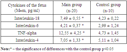
5. Conclusions
- 1. In hemolytic disease during the second trimester of pregnancy, the synthesis of all studied cytokines in the fetuses increases compared to the control group. IL-18 is significantly increased by 1.6 times, IL-6 by 1.7 times, TNF-alpha by 2.4 times, and IL-4 by 1.5 times compared to the control group.2. In the third trimester of pregnancy with hemolytic disease, the synthesis of all studied cytokines in the fetuses increases compared to the control group. IL-18 is significantly elevated by 1.7 times, IL-6 by 1.8 times, TNF-alpha by 2.7 times, and IL-4 by 1.4 times compared to the control group.3. The activity of cytokine regulation and nonspecific immunity is a result of the fetal immune response. This indicates the protective activity of the fetal immune system through the production of pro-inflammatory and anti-inflammatory cytokines in hemolytic disease. Specifically, the increase in the chemokine IL-18 has a direct correlation with the increase of a whole cascade of inflammatory reactions and transmits inflammation to other cytokines.