-
Paper Information
- Next Paper
- Previous Paper
- Paper Submission
-
Journal Information
- About This Journal
- Editorial Board
- Current Issue
- Archive
- Author Guidelines
- Contact Us
American Journal of Medicine and Medical Sciences
p-ISSN: 2165-901X e-ISSN: 2165-9036
2025; 15(3): 806-809
doi:10.5923/j.ajmms.20251503.66
Received: Mar. 2, 2025; Accepted: Mar. 19, 2025; Published: Mar. 22, 2025

Features of Morpho-Functional States of the Adrenal Glands in Rats with Nephrolithiasis Against the Background of Androgen Deficiency
 Abstract
 Reference
 Full-Text PDF
 Full-text HTML
Tosheva Zebuniso1, Akhmedov Kamoliddin2, Shonazarova Dilnoza1
1Assistant, Department of Propaedeutics of Internal Diseases, Rehabilitation, Folk Medicine and Endocrinology, Termez Branch of Tashkent Medical Academy, Surkhondarya, Uzbekistan
2Associate Professor, Department of Normal and Pathological Physiology, Termez Branch of Tashkent Medical Academy, Surkhondarya, Uzbekistan
Correspondence to: Tosheva Zebuniso, Assistant, Department of Propaedeutics of Internal Diseases, Rehabilitation, Folk Medicine and Endocrinology, Termez Branch of Tashkent Medical Academy, Surkhondarya, Uzbekistan.
| Email: |  |
Copyright © 2025 The Author(s). Published by Scientific & Academic Publishing.
This work is licensed under the Creative Commons Attribution International License (CC BY).
http://creativecommons.org/licenses/by/4.0/

The features of morpho-functional states of the adrenal glands of rats with nephrolithiasis against the background of androgen deficiency. Objective: To study the dynamics of structural and functional changes in the adrenal glands of rats with nephrolithiasis against the background of androgen deficiency. The experiments were carried out on 30 white outbred male rats of a mixed population with an initial weight of 180-200 g, kept in a laboratory diet in vivarium conditions. For morphological examination, pieces of kidneys of the control and experimental groups of experimental animals were selected, from which pieces of 1.0 cm in size were cut out and placed in a fixing solution of 15% formalin for 24 hours, then the pieces were fixed in wire alcohols and chloroform. As a result of the morphological study of the adrenal gland of the intact group, no significant pathological changes were revealed; only weakly expressed vacuolization of focal cells was noted.
Keywords: Experiment, Adrenal gland, Rats, Nephrolithiasis, Cortisol
Cite this paper: Tosheva Zebuniso, Akhmedov Kamoliddin, Shonazarova Dilnoza, Features of Morpho-Functional States of the Adrenal Glands in Rats with Nephrolithiasis Against the Background of Androgen Deficiency, American Journal of Medicine and Medical Sciences, Vol. 15 No. 3, 2025, pp. 806-809. doi: 10.5923/j.ajmms.20251503.66.
Article Outline
1. Introduction
- Urolithiasis is a polyetiological disease that occupies an important place in the practice of a urologist. It is widespread in the world and can occur in all age groups. The main approach to the treatment of urolithiasis is to find the cause of the disease and competent correction of disorders, which will help to avoid recurrence of stone formation. In the prevention and treatment of this disease, an interdisciplinary approach should be used approach [2]. Urolithiasis (nephrolithiasis) is a common renal disease characterized by the deposition of stones in the renal cavity and urinary tract and can occur at any age. The incidence of nephrolithiasis currently accounts for 1-2% of the total disease structure [4]. In the structure of the terminal stage of chronic kidney disease, nephrolithiasis accounts for up to 3% [1,3]. The incidence of androgen deficiency and metabolic syndrome increases with age, which is explained mainly by endogenous mechanisms that lead to impaired testosterone production and increased insulin resistance. The influence of environmental factors on the development of these diseases is known, but they receive much less attention than they deserve. However, we should not forget that the body of a modern person is constantly affected not only by endogenous, but also paraecological pathogenic factors, which, in addition to those associated with the individual's activities - stress, bad habits, eating disorders - also include anthropogenic environmental factors - chemical compounds, medications, etc. [6,7].
2. Purpose of the Study
- To study the dynamics of structural and functional changes in the adrenal glands of rats with nephrolithiasis against the background of androgen deficiency.
3. Materials and Methods
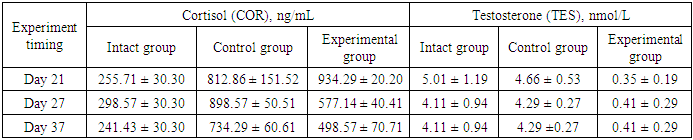
- The experiments were conducted on 30 white mongrel male rats of a mixed population with an initial weight of 180-200 g, kept on a laboratory diet in vivarium conditions. After receipt from the nursery, the experimental rats underwent a 14-day quarantine period in the quarantine block of the vivarium in order to exclude animals with somatic and / or infectious pathology from the experiment. The study was carried out in accordance with the ethical principles of handling animals, observed in accordance with the "European Convention for the Protection of Vertebral Animals Used for Experimental and Other Scientifi c Purposes. CETS No. 123". All animals were divided into 3 groups: 1) intact group (10 rats), 2) control group (10 rats without castrated males), 3) experimental group (10 castrated rats, which were castrated before the 5th day of the experiment). The animals of the control and experimental groups, against the background of the general viviparous diet, received 1% aqueous solution of ethylene glycol instead of drinking water for 21 days with free access, which induced the development of experimental oxalate nephrolithiasis. The condition of the animals was observed daily for 21 days.Rats were given standard species-appropriate food and drinking water. On the 21st, 27th and 37th days, blood was collected from the animals (3-4 ml) from the cardiac region under ether anesthesia (inhalation) for laboratory research.For morphological study, kidney pieces of the control and experimental groups of experimental animals were selected, from which pieces measuring 1.0 cm were cut out and placed in a fixing solution of 15% formalin for 24 hours, then the pieces were fixed in conduction alcohols and chloroform. Then they were loaded into a thermostat at 37 degrees for 1-2 hours, and 57 degrees for impregnation for 1 hour, after hardening, paraffin blocks were cut out. Serial sections were prepared from the finished blocks. Glasses were smeared with protein and pierced with an alcohol lamp, the cut materials were fixed on the glass and stained with hematoxylin and eosin. The finished preparations were viewed under a BIO BLUE binocular microscope with an adapter and a Euromex Microscopen BV camera.Enzyme immunoassays were performed on serum obtained after centrifugation of blood samples at 3000 rpm for 8 minutes. The serum was stored at -20°C until analysis for enzyme immunoassay parameters such as Luteinizing Hormone (LH), Cortisol (Cor) and Testosterone (Tes). For the analysis of enzyme immunoassay parameters, a semi-automatic Elisa microplate reader "Scitek EMLR-112" and enzyme immunoassay kits were used.The results were processed using the ANOVA variation statistics at a significance level of p=0.05, using the GraphPad Prism version 8.0.0 program for Windows, GraphPad Software, San Diego, California, USA, www.graphpad.com [5].
4. Results and Discussion
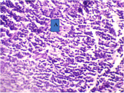
- For morphological study, pieces of adrenal glands were selected from intact, control and experimental groups of experimental animals, from which pieces measuring 1.0 cm were cut out and placed in a fixing solution of 15% formalin for 24 hours, then the pieces were fixed in conductive alcohols and chloroform. Then they were loaded into a thermostat at 37 degrees for 1-2 hours, and 57 degrees for impregnation for 1 hour, after hardening, paraffin blocks were cut out. Serial sections were prepared from the finished blocks. Glasses were smeared with protein and pierced with an alcohol lamp, the cut materials were fixed on the glass and stained with hematoxylin and eosin. The finished preparations were viewed under a BIO BLUE binocular microscope with an adapter and a Euromex Microscopen BV camera.For an experimental study of the androgenic effect on the development of nephrolithiasis, we selected outbred mice, which were divided into 3 groups by the terms of the 22nd day. The first group is an intact group, the 2nd group is a control group (castrated rats) and the 3rd experimental group (males with nephrolithiasis).As a result of the morphological study of the adrenal gland of the intact group, no significant pathological changes were revealed; only weakly expressed vacuolization of focal cells was noted (Fig. 1).
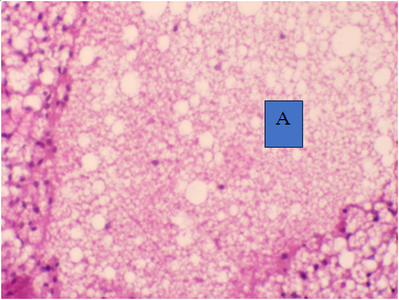 | Figure 1. Adrenal gland with vacuolization of cells - A, amongchromaffinocytes increase in the number of cells with light cytoplasm-B. Intact, day 22. Hematoxylin and eosin staining. Magnification 100 |
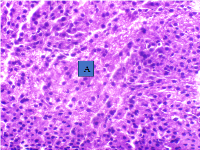 | Figure 2. Under high magnification of the objective lens in the adrenal medulla there is mucous edema of the stroma and macrophage-mononuclear infiltration - A. Day 22, nephrolithiasis. Hematoxylin and eosin staining. Magnification 400 |
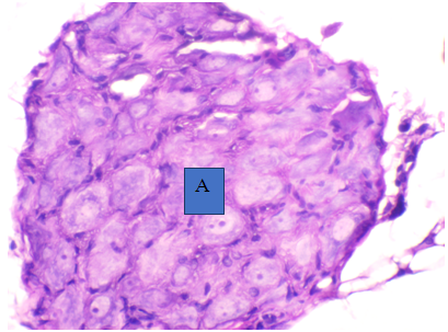 | Figure 3. Dystrophy of parenchyma cells, necrotic changes in the adrenal gland. Day 22. Castrated rat. Hematoxylin and eosin staining. Magnification 400 |
 | Figure 4. Under high magnification of the objective lens, adrenocorticocytes in small quantities, in the adrenal medulla, mucous edema of the stroma - A. 37 days, nephrolithiasis. Hematoxylin and eosin staining. Magnification 400 |
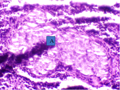
 | Figure 5. Adrenal adrenocorticocytes are closely located, their nuclei are darkly stained and rounded –A of the adrenal gland. Day 37. Castrated rat. Hematoxylin and eosin staining. Magnification 400 |
|
5. Conclusions
- 1. In experimental nephrolithiasis against the background of androgen deficiency, morphological changes in the adrenal glands worsen with the duration of the pathological process as dystrophy of parenchymal cells,2. In experimental nephrolithiasis, cortisol concentrations in experimental groups (castrated rats) increase slightly compared to control groups. This is probably due to a decline in the compensatory capacity of the adrenal gland against the background of androgen deficiency.