-
Paper Information
- Next Paper
- Previous Paper
- Paper Submission
-
Journal Information
- About This Journal
- Editorial Board
- Current Issue
- Archive
- Author Guidelines
- Contact Us
American Journal of Medicine and Medical Sciences
p-ISSN: 2165-901X e-ISSN: 2165-9036
2025; 15(3): 803-805
doi:10.5923/j.ajmms.20251503.65
Received: Feb. 25, 2025; Accepted: Mar. 18, 2025; Published: Mar. 22, 2025

Morphological Changes in the Adrenal Glands in Experimental Nephrolithiasis Depending on Individual Characteristics of the Nervous System
 Abstract
 Reference
 Full-Text PDF
 Full-text HTML
Ostanaqulov Sherzod1, Akhmedov Kamoliddin2, Urokova Shahlo1, Boymurodova Nilufar1
1Assistant, Department of Normal and Pathological Physiology, Termez Branch of Tashkent Medical Academy, Surkhondarya, Uzbekistan
2Associate Professor, Department of Normal and Pathological Physiology, Termez Branch of Tashkent Medical Academy, Surkhondarya, Uzbekistan
Correspondence to: Ostanaqulov Sherzod, Assistant, Department of Normal and Pathological Physiology, Termez Branch of Tashkent Medical Academy, Surkhondarya, Uzbekistan.
| Email: |  |
Copyright © 2025 The Author(s). Published by Scientific & Academic Publishing.
This work is licensed under the Creative Commons Attribution International License (CC BY).
http://creativecommons.org/licenses/by/4.0/

The morphological changes in the adrenal glands in experimental nephrolithiasis depending on the individual characteristics of the nervous system. Objective: To study the dynamics of structural changes in the adrenal glands in experimental nephrolithiasis depending on the individual characteristics of the nervous system. For the morphological study, pieces of the adrenal glands of highly emotional and low emotional groups of experimental animals were selected, from which pieces of 1.0 cm in size were cut out and placed in a fixing solution of 15% formalin for 24 hours, then the pieces were fixed in conductive alcohols and chloroform. Then they were loaded into a thermostat at 37 degrees for 1-2 hours, and 57 degrees for impregnation for 1 hour, after hardening, paraffin blocks were cut out.
Keywords: Morphology, Nephrolithiasis, Experiment, Adrenal gland
Cite this paper: Ostanaqulov Sherzod, Akhmedov Kamoliddin, Urokova Shahlo, Boymurodova Nilufar, Morphological Changes in the Adrenal Glands in Experimental Nephrolithiasis Depending on Individual Characteristics of the Nervous System, American Journal of Medicine and Medical Sciences, Vol. 15 No. 3, 2025, pp. 803-805. doi: 10.5923/j.ajmms.20251503.65.
Article Outline
1. Introduction
- Urolithiasis (UCD) is a common urological disease, which in the modern world, according to statistics, affects about 10-15% of the population of developed countries [1,3]. The main nosological subunit of ICD is calcium oxalate nephrolithiasis, since, according to the modern mineralogical classification, about 60% of all kidney stones consist mainly of calcium oxalate biominerals: we wellie (CaCrO2xH2O) and wedellite (CaC2O4x2H2O) [5]. The most adequate and used model is the experimental oxalate nephrolithiasis model, since in 75-85% of cases of urolithiasis in humans, calcium stones are found, most of them (more than 70-85%) are oxalate [4].
2. Purpose of the Study
- To study the dynamics of structural changes in the adrenal glands in experimental nephrolithiasis depending on the individual characteristics of the nervous system.
3. Materials and Methods
- The experiments were conducted on 50 white mongrel male rats of a mixed population with an initial weight of 180-200 g, kept on a laboratory diet in vivarium conditions. After receipt from the nursery, the experimental rats underwent a 14-day quarantine period in the quarantine block of the vivarium in order to exclude animals with somatic and/or infectious pathology from the experiment. The study was carried out in accordance with the ethical principles of handling animals, observed in accordance with the "European Convention for the Protection of Vertebral Animals Used for Experimental and Other Scientific Purposes. CETS No. 123".The study used the most widely used ethylene glycol model of urolithiasis (adding 1% ethylene glycol solution to drinking water for 21 days), which induces the development of experimental oxalate nephrolithiasis. Ethylene glycol is slowly oxidized in the body to form oxalic acid, which is then excreted by the kidneys. This model is generally accepted and most adequately reproduces human nephrolithiasis [6].To assess the state of the nervous system, the "Open Field" test was conducted. The experiment involved 50 heads of white mongrel male rats, the open field method was performed according to the method of Buresh Ya.I., Bureshov O. [2]50 male rats were divided into two groups of 25 each: 1) control group; 2) low-emotional group. Rats of the 1st group received tap water for drinking, experimental nephrolithiasis was not reproduced, this group was used as a control. Animals of the 2nd group, against the background of a general viviparous diet, received 1% aqueous ethylene glycol solution instead of drinking water for 21 days with free access, which induced the development of oxalate experimental nephrolithiasis. The condition of the animals was observed daily for 21 days.
4. Results and Discussion
- Pieces were selected for morphological examination. Adrenal glands of the highly emotional and low emotional groups experimental animals, from which pieces measuring 1.0 cm were cut out and placed in a fixing solution of 15% formalin for 24 hours, then the pieces were fixed in conductive alcohols and chloroform. Then they were loaded into a thermostat at 37 degrees for 1-2 hours, and 57 degrees for impregnation for 1 hour, after hardening, paraffin blocks were cut out. Serial sections were prepared from the finished blocks. Glasses were smeared with protein and pierced with an alcohol lamp, the cut materials were fixed on the glass and stained with hematoxylin and eosin. The finished preparations were examined under a BIO BLUE binocular microscope with an adapter for a Euromex microscopen BV camera (Fig. 1 and 2).
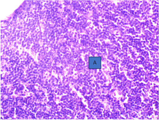
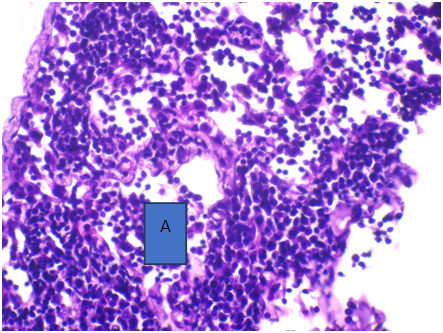 | Figure 1. Infiltration of the adrenal medulla with macrophages and histiocytes -A. high emotional 22 days. Hematoxylin and eosin staining. Magnification 400 |
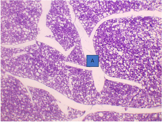 | Figure 2. In the interlobular spaces there is pronounced edema and vacuolar degeneration of the adrenal parenchyma cells. low emotional 22 days. Hematoxylin and eosin staining. Magn. 40 |
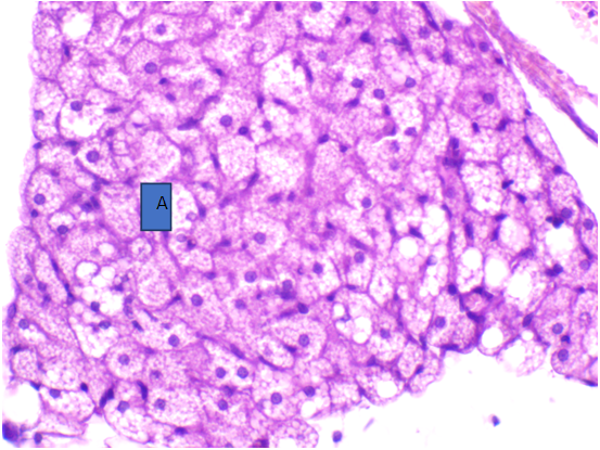
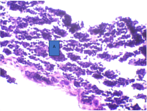 | Figure 3. Adrenal gland with areas of destruction, adrenocorticocytes with a light rim of cytoplasm and hyperchromic kernels- A, high emotional 37 days. Hematoxylin and eosin staining. Magn. 400 |
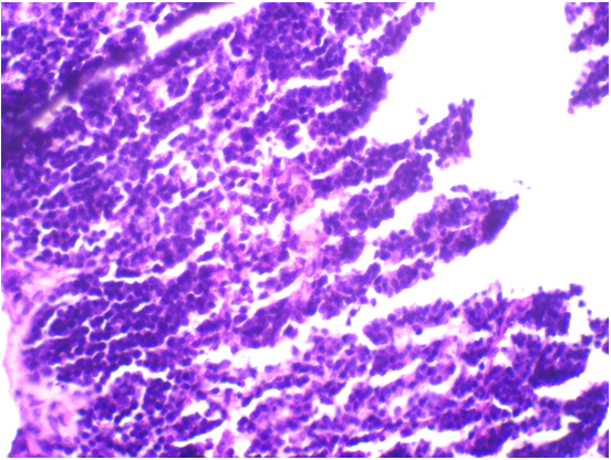 | Figure 4. Adrenocorticocytes with dark-stained nuclei -A, a small number of light cells -B. 37th day, low emotional adrenal gland. Hematoxylin and eosin staining. Magnification 100 |
 | Figure 5. Light cells with granular cytoplasm in the adrenal medulla -A low emotional 37 day. Hematoxylin and eosin staining. Magn. 400 |
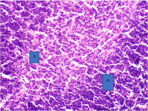 | Figure 6. In the adrenal cortex, the cells of the glomerular zone have round dark-stained nuclei - A, in the medulla - infiltration by macrophages, -B. high emotional 53 days. Hematoxylin and eosin staining. Magnification 400 |
 | Figure 7. In the reticular and glomerular zones of the adrenal gland, adrenocorticocytes with a round nucleus and a light rim of cytoplasm, stromal edema. low emotional 53 days. Hematoxylin and eosin staining. Magn. 40 |
5. Conclusions
- In experimental nephrolithiasis dynamics, structural changes in the adrenal glands of the low emotional group are more pronounced with congestive hyperemia and dystrophic changes than in the high emotional group. In our opinion, these changes are related to their low reactivity of the nervous system.