American Journal of Medicine and Medical Sciences

p-ISSN: 2165-901X    e-ISSN: 2165-9036

2025;  15(3): 722-731

doi:10.5923/j.ajmms.20251503.51

Received: Feb. 9, 2025; Accepted: Feb. 26, 2025; Published: Mar. 15, 2025


Method for Increasing the Effectiveness of Treatment of Peripheral Type of Trigeminal Neuralgia

Abdullaeva Muborak Bekkulovna

Associate professor, Tashkent state dental institute, Tashkent, Uzbekistan

Correspondence to: Abdullaeva Muborak Bekkulovna, Associate professor, Tashkent state dental institute, Tashkent, Uzbekistan.

Copyright © 2025 The Author(s). Published by Scientific & Academic Publishing.

This work is licensed under the Creative Commons Attribution International License (CC BY).
http://creativecommons.org/licenses/by/4.0/

Abstract

Trigeminal neuralgia (TN) is widely recognized as one of the most painful conditions and is a chronic, non–inflammatory disorder of the peripheral nervous system characterized by sharp, lightning–like facial pain that is often nearly uncontrollable. This pain results from a disruption in the transmission of sensory signals from the skin of the face and head and the mucous membrane of the mouth to the brain. These guidelines present a method for complex pharmacotherapy targeting the peripheral type of trigeminal neuralgia, developed and tested by our team, which demonstrates objectively and subjectively greater effectiveness compared to standard treatments.

Keywords: Method, Effectiveness, Treatment, Peripheral type, Trigeminal neuralgia, Non–inflammatory disorder, Laser therapy, Sensory signals, Painful conditions, Brain, Mouth, Nervous system, Mucous membrane, Radiofrequency denervation, Anticonvulsants

Cite this paper: Abdullaeva Muborak Bekkulovna, Method for Increasing the Effectiveness of Treatment of Peripheral Type of Trigeminal Neuralgia, American Journal of Medicine and Medical Sciences, Vol. 15 No. 3, 2025, pp. 722-731. doi: 10.5923/j.ajmms.20251503.51.

1. Introduction

Trigeminal neuralgia (TN) is one of the most debilitating neuropathic conditions, characterized by sudden, severe, and recurrent facial pain. As a chronic disorder of the peripheral nervous system, TN affects the transmission of sensory signals from the facial skin and mucous membranes to the brain, leading to excruciating episodes of pain. Despite various pharmacological and surgical interventions, achieving long-term pain relief remains a clinical challenge.
The peripheral type of TN, distinct from its central variant, often requires targeted therapeutic approaches that enhance nerve function while minimizing adverse effects. Traditional treatments primarily rely on anticonvulsants, such as carbamazepine and gabapentin, which, while effective, may not provide sustained relief and are associated with side effects. In recent years, advancements in pharmacotherapy and neuromodulation techniques have opened new avenues for managing TN with improved patient outcomes.
This study evaluates a novel method of complex pharmacotherapy for peripheral TN, incorporating standard treatment protocols alongside supplementary interventions aimed at enhancing treatment efficacy. By analyzing patient responses to this combined approach, we aim to determine its effectiveness in reducing pain severity, improving quality of life, and minimizing recurrence rates. The findings provide valuable insights into optimizing therapeutic strategies for managing TN with greater precision and patient-centered care.
Relevance of the problem: Trigeminal neuralgia (TN) is widely recognized as one of the most painful conditions and is a chronic, non–inflammatory disease of the peripheral nervous system. It is characterized by sharp, lightning–like pains in the face that is often nearly uncontrollable due to disrupted transmission of sensory signals from the skin of the face and head and the mucous membrane of the mouth to the brain [11,24].
The incidence of TN in Europe is 10–15 people per 100,000 population annually. In the United States, it was estimated that 12 out of 100,000 people develop TN annually [24]. In England, the prevalence is higher–27 per 100,000 population [21,22]. The average annual incidence of TN is 4–13 per 100,000 population [15], with more than 90% in people over 40 years of age and often women (60–70%) [8,10]. However, the exact prevalence rates of TN vary by region, research methodology and other factors.
Aim of the study: To improve the effectiveness of treatment of patients with different duration of peripheral TN.

2. Materials and Methods of Research

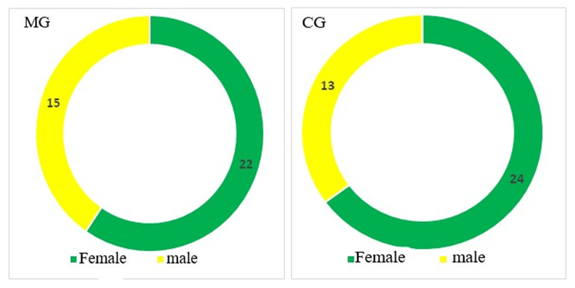
Clinical observations were conducted at the Tashkent State Dental Institute (Rector–MD, Professor Khaidarov N.K.) on the basis of the multidisciplinary clinic of the Tashkent Medical Academy (Chief Physician–Rustamova D.A.). To achieve the goal of the study, 74 patients (28 men and 46 women) with TN aged 19–68 years (mean age–47.76±14.87 years) were examined for the period 2016–2023. Women predominated 1.64:1.
We divided the patients into a main group (MG) and a comparison group (CG) by random sampling (random distribution) until the groups were evenly filled according to the number of subjects.
The MG included 37 patients with peripheral TN aged 19–68 years (22 women and 15 men) (mean age–47.84±15.03 years).
The CG included 37 patients with peripheral TN aged 19–65 years (24 women and 13 men) (mean age–46.58±14.65 years).
The social and living conditions of all subjects were satisfactory. Among the examined patients, the majority were able–bodied and socially active people.
The diagnoses were verified according to standard criteria based on complaints, a carefully collected anamnesis, the duration of the process, and an objective neurological examination.
In our study, we used anamnestic, clinical, laboratory, instrumental and statistical research methods.
The anamnestic method of examination involved active collection of anamnesis with an emphasis on the duration of TN, the frequency of attacks and their severity, the treatment received and its effectiveness.
Figure 1. Gender composition of the study groups
All patients underwent a standard clinical–neurological examination and somatic examination, with an emphasis on the syndromes and symptoms characteristic of TN, the condition of the skin, visible mucous membranes and blood pressure (BP).
Using a visual analogue scale (VAS) interpreted as a digital rating scale (DRS), the patients subjectively determined the severity of TN and the intensity of the pain syndrome [17]. We also used the Hospital Anxiety and Depression Scale (HADS) [9]. Our subjects completed the Spielberger–Hanin test (the “Spielberger STAI Test”) [15]. We also dynamically applied the Beck Anxiety Scale to all patients [11].
The blood flow in the common and internal carotid arteries (CCA and ICA), vertebral arteries (VA) (in V2 segments), and middle cerebral arteries (MCA) was studied using the color duplex scanning (CDS) method on the Sonomed–325 apparatus from Spectromed using sensors generating ultrasound waves with a frequency of 4 MHz and 8 MHz.
MRI was performed on the GE SIGNA Creator (USA) with a magnetic field strength of 1.5 Tesla. Tomograms were performed using the standard technique in axial, sagittal and coronal projections with T1 and T2 pulse sequences, FLAIR and DWI programs with subsequent post-processing in the MR navigation mode in the FIESTA–C program.
All subjects underwent stimulation electroneuromyography (ENMG) of the TN. Laboratory examination consisted of standard and specific analyses (TNF–α, IL–lβ, IL–8, substance P, vitamin D, estradiol, luteinizing hormone (LH), follicle–stimulating hormone (FSH)) on the Microplate Reader RT 2100 C device and with Vector–Best Baltic reagents.
The study group included 74 patients with peripheral TN, which was divided into two subgroups. CG included 37 patients who received standard treatment, including: to relieve paroxysmal pain, Carbamazepine was prescribed 200–600 mg per day; to improve the trophism of the peripheral nerve–vitamin B complex (Zifort 3.0 intramuscularly for 10 days, followed by a transition to NatB, 1 drop 3 times a day for 1–3 months); for dehydration purposes: L–Lysine–Escinate 10.0+NaCl 0.9% 100.0 intravenously, drops for 5 days, followed by a transition to Veroshpiron 50 mg, 1 drop in the morning on an empty stomach every other day for 20 days; for anti–inflammatory purposes: Dioxaflex B12 3.0 i/m 6 days, then Goofen 400 mg 1 drop 2 times a day for 20 days; to improve myelination of the nerve–Nucleo–CMF 2.0 i/m for 6 days, then Nucleo–CMF forte 1 drop twice a day for 1 month.
The MG included 37 patients with peripheral TN who received, in addition to standard treatment, vitamin D at 1000 IU daily for a long time (3 months) and the multivitamin complex “Demoton D”. The contents of 1 vial were diluted with 5 ml of water for injection and the powder was dissolved by shaking, after which the resulting solution was dissolved in a 5% glucose solution or saline solution for 10 days.
Statistical processing of the research results was carried out using the methods of variation statistics using Microsoft Office Excel–2019 programs with the calculation of the mean square deviation and the average arithmetic error by the method of moments (M±m), the mean square deviation (σ). The statistical reliability of the comparison of the results of the significance study (P) at a 95% confidence interval in our work had 4 levels: high–P<0.001, medium–P<0.01, low (limit) – P<0.05, insignificant (insignificant) – P>0.05.

3. Results of the Research

The duration of the course of conservative treatment of peripheral TN in both groups was 12–14 days, after which we compared their immediate effectiveness for all studied parameter, the remote effectiveness of the course in a comparative aspect was assessed by us after 6 months of follow–up observation.
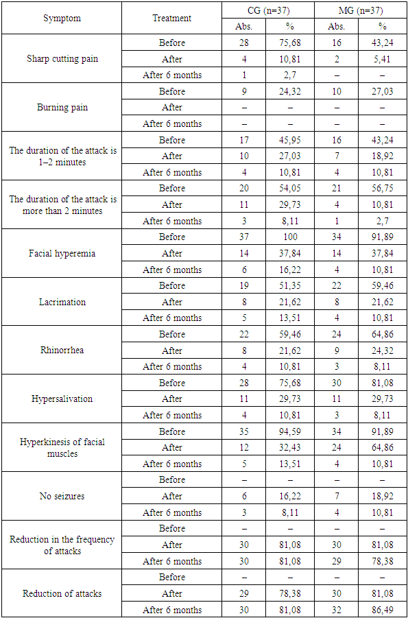
Thus, the analysis of the dynamics of acute cutting pain during an attack of TN demonstrated in the CG subgroup a sevenfold decrease in the frequency of this symptom from 28 (75.68%) to 4 (10.81%) at the end of treatment, after six months–in 1 (2.7%) in the MG subgroup–an eightfold decrease from 16 (43.24%) to 2 (5.41%) patients at the end of treatment and was completely absent after six months (Table 1).
Table 1. The dynamics of TN attacks by treatment groups
     
Burning pain in an attack before treatment in the CG subgroup was noted by 9 patients (24.32%), after treatment it was completely relieved, an identical tendency also characterizes the MG subgroup–10 (27.03%) before treatment and absence after. After 6 months, this type of pain was not noted by any of the studied patients (Table 1).
In CG, the duration of attacks of 1–2 minutes before treatment was noted in 17 (45.95%) patients and 10 (27.03%) patients after treatment with a decrease of 1.7 times, after 6 months it decreased by 2.5 times to 4 (10.81%) patients (Table 1).
In the MG, before treatment, the duration of attacks of 1–2 minutes was recorded in 16 (43.24%) patients, and after treatment–in 7 (18.92%) patients, i.e. the frequency decreased by 2.29 times, after 6 months–decreased by 1.75 times to 4 (10.81%) patients (Table 1).
The duration of attacks over 2 minutes before treatment was recorded in 20 (54.05%) patients in the CG, in 11 (29.73%) patients after treatment with a decrease of 1.82 times, after 6 months–decreased by 3.67 times to 3 (8.11%) patients (Table 1).
In the MG, before treatment, the duration of TN attacks over 2 minutes was in 210 (56.75%) patients, and after treatment–4 (10.81%) patients, i.e. it decreased by 2.5 times, after 6 months–it decreased fourfold to 1 (2.7%) patient (Table 1).
Such a symptom of an attack of TN as facial hyperemia bothered all 37 (100%) patients in the CG before treatment and 14 (37.84%) patients after treatment with a decrease of 2.64 times, after 6 months its frequency decreased by 2.33 times to 6 (16.22%) patients (Table 1).
In the MG, before treatment, this symptom bothered 34 (91.89%) patients, and after treatment–14 (37.84%) patients, i.e. it decreased by 2.43 times, after 6 months its frequency decreased by 3.5 times to 4 (10.81%) patients (Table 1).
Lacrimation during an attack bothered 19 (51.35%) patients in the CG before treatment and 8 (21.62%) patients after treatment with a decrease of 2.38 times; after 6 months, its frequency decreased by 1.6 times to 5 (13.51%) patients (Table 1).
In the MG, before treatment, this symptom bothered 22 (59.46%) patients, and after treatment–8 (21.62%) patients, i.e. it decreased by 2.75 times, after 6 months its frequency decreased twofold to 4 (10.81%) patients (Table 1).
Rhinorrhea during an attack of TN bothered 22 (59.46%) patients in the CG before treatment and 8 (21.62%) patients after treatment with a decrease of 2.75 times; after 6 months, its frequency decreased to 7 (18.92%) patients (Table 1).
In the MG, before treatment, this symptom bothered 24 (64.86%) patients, and after treatment–9 (24.32%) patients, i.e. it decreased by 2.67 times; after 6 months, its frequency decreased threefold to 3 (8.11%) patients (Table 1).
Hypersalivation during an attack of TN bothered 28 (75.68%) patients in the CG before treatment and 11 (29.73%) patients after treatment with a decrease of 2.55 times; after 6 months, its frequency decreased by 2.75 times to 4 (10.81%) patients (Table 1).
In the MG, before treatment, this symptom bothered 30 (81.08%) patients, and after treatment–11 (29.73%) patients, i.e. it decreased by 2.73 times; after 6 months, its frequency decreased by 3.67 times to 3 (8.11%) patients (Table 1).
Hyperkinesis of the facial muscles during an attack of TN was characterized in the CG before treatment in 35 (94.59%) patients and 12 (32.43%) patients after treatment with a decrease of 2.92 times; after 6 months, its frequency decreased by 2.4 times to 5 (13.51%) patients (Table 1).
In the MG, before treatment, this symptom bothered 34 (91.89%) patients, and after treatment–24 (64.86%) patients, i.e. it decreased by 1.42 times; after 6 months, its frequency decreased six fold to 4 (10.81%) patients (Table 1).
Such a progressive symptom, reflecting the effectiveness of the treatment as the complete disappearance of attacks of TN in the CG was recorded in 6 (16.22%) patients after treatment, after 6 months–in 3 (8.11%), in the MG–in 7 (18.92%) patients immediately after treatment, after 6 months–in 4 (10.81%) (Table 1).
A decrease in the frequency of attacks of TN in the MG and CG at the end of treatment characterized 30 (81.08%) patients, after 6 months–30 (81.08%) and 29 (78.38%) patients, respectively (Table 1).
We found a decrease in the intensity of TN attacks in 30 (89.19%) patients in the MG and in 29 (78.38%) patients in the CG at the end of treatment; after six months, these figures were 32 (86.49%) and 30 (81.08%) patients, respectively (Table 1).
Thus, the symptoms during an attack of TN in our study after treatment decreased relative to the initial level in the CG by 2.97 times, in the MG–by 3.1 times.
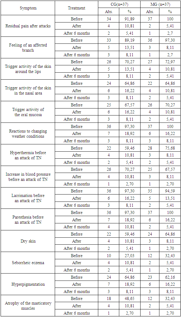
We also took into account the symptoms of TN outside of attacks (Table 2). Residual pain after attacks of TN was characterized in the CG before treatment in 34 (91.89%) patients and 4 (10.81%) patients after treatment with a decrease of 8.5 times, after 6 months their frequency decreased twofold to 2 (5.41%) patients (Table 2).
Table 2. The dynamics of symptoms outside attacks of TN in the context of treatment groups
     
In the MG, before treatment, this symptom bothered all 37 (100%) patients, and after treatment–2 (5.41%) patients, i.e. it decreased by 18.5 times, after 6 months its frequency decreased twofold to 1 (2.7%) patient (Table 2).
The sensation of the affected branch after an attack of TN was characterized in the CG before treatment in 33 (89.19%) patients and 5 (13.51%) patients after treatment with a decrease of 6.6 times; after 6 months, their frequency decreased to 3 (8.11%) patients (Table 2).
In the MG, before treatment, this symptom bothered 36 (97.30%) patients, and after treatment – 3 (8.11%) patients, i.e. it decreased 12 times, after 6 months their frequency decreased threefold to 1 (2.7%) patient (Table 2).
The treatment we carried out allowed us to establish a decrease in the activity of all trigger zones for attacks of TN, so the skin around the lips reduced the frequency of the trigger value in the CG from 26 (70.27%) patients before treatment and 5 (13.51%) patients after treatment with a decrease of 5.2 times, after 6 months this symptom remained in 3 (8.11%) patients (Table 2).
In the MG, before treatment, this symptom bothered 27 (72.97%) patients, and after treatment–4 (10.81%) patients, i.e. it decreased by 6.75 times; after 6 months, the frequency decreased twofold to 2 (5.41%) patients (Table 2).
The skin in the nasal area as a trigger zone for TN was characterized in the MG before treatment in 22 (59.46%) patients and after treatment in 4 (10.81%) patients with a 5.5–fold decrease; after 6 months, the frequency decreased twofold to 2 (5.41%) patients (Table 2).
In the CG, before treatment, 24 (64.86%) patients had this symptom, and after treatment–6 (16.22%) patients, i.e. it decreased fourfold; after 6 months, the frequency decreased twofold to 3 (8.11%) patients (Table 2).
In the MG, 26 (70.27%) patients had trigger properties for an attack of TN in the oral mucosa before treatment and 4 (10.81%) patients after treatment with a 6.5–fold decrease; after 6 months, the frequency decreased twofold to 2 (5.41%) patients (Table 2).
In the CG, before treatment, this symptom bothered 25 (67.57%) patients, and after treatment–6 (16.22%) patients, i.e. it decreased by 4.17 times, after 6 months the frequency decreased twofold to 3 (8.11%) patients (Table 2).
The frequency of reaction to changes in weather conditions also decreased–in the MG, 37 (100%) patients had this symptom before treatment and 6 (16.22%) patients after treatment with a decrease of 6.17 times, after 6 months the frequency decreased twofold to 3 (8.11%) patients (Table 2).
In the CG, before treatment, 36 (97.30%) patients had this symptom, and after treatment–7 (18.92%) patients, i.e. it decreased by 5.14 times; after 6 months, the frequency decreased by 2.5 times to 3 (8.11%) patients (Table 2).
Of the clinical symptoms preceding the attack of vegetative aura, all symptoms underwent regression to varying degrees. Thus, hyperthermia before the attack of TN bothered 22 (59.46%) patients in the CG before treatment and 4 (10.81%) patients after treatment with a decrease of 5.5 times, after 6 months the frequency decreased twofold to 2 (5.41%) patients (Table 2).
In the MG, before treatment, this symptom bothered 28 (75.68%) patients, and after treatment–3 (8.11%) patients, i.e. it decreased by 9.33 times; after 6 months, the frequency decreased by one and a half times to 2 (5.41%) patients (Table 2).
An increase in blood pressure as a vegetative aura preceding an attack of TN was characterized in the MG before treatment in 25 (67.57%) patients and in 3 (8.11%) patients after treatment with a decrease of 8.33 times; after 6 months, their frequency decreased threefold to 1 (2.70%) patient (Table 2).
In the CG, before treatment, this symptom bothered 26 (70.27%) patients, and after treatment–4 (10.81%) patients, i.e. it decreased by 6.5 times, after 6 months their frequency decreased fourfold to 1 (2.70%) patient (Table 2).
Such a symptom–precursor of an attack of TN as lacrimation was characterized in the MG before treatment in 35 (94.59%) patients and 5 (13.51%) patients after treatment with a sevenfold regression; after 6 months, the frequency decreased by 2.5 times to 2 (5.41%) patients (Table 2).
In the CG, before treatment, this symptom bothered 36 (97.30%) patients, and after treatment–6 (16.22%) patients, i.e. it decreased six fold, after 6 months the frequency decreased twofold to 3 (8.11%) patients (Table 2).
Paresthesia as a vegetative aura preceding an attack of TN was characterized in the CG before treatment in 36 (97.30%) patients and 7 (18.92%) patients after treatment with a decrease of 5.14 times; after 6 months, the frequency decreased by 1.75 times to 4 (10.81%) patients (Table 2).
In the MG, before treatment, this symptom bothered all 37 (100%) patients, and after treatment–6 (16.22%) patients, i.e. it decreased by 6.17 times; after 6 months, the frequency decreased threefold to 2 (5.41%) patients (Table 2).
Our analysis of the dynamics of skin and muscle manifestations of TN in the patients studied by subgroups allowed us to study these manifestations of the pathology under study. Thus, dry skin of the MG before treatment was 24 (64.86%) patients and 3 (8.11%) patients after treatment with an eight–fold regression, after 6 months the frequency decreased threefold to 1 (2.7%) patient (Table 2).
In the CG, before treatment, this symptom bothered 22 (59.46%) patients, and after treatment–4 (10.81%) patients, i.e. it decreased by 5.5 times, after 6 months the frequency decreased twofold to 2 (5.41%) patients (Table 2).
Seborrheic eczema was a problem in 12 (32.43%) patients in the MG before treatment and 2 (5.41%) patients after treatment with a six–fold decrease; after 6 months, the frequency decreased twofold to 1 (2.7%) patient (Table 2).
In the CG, before treatment, this symptom bothered 10 (27.03%) patients, and after treatment–4 (10.81%) patients, i.e. it decreased by 2.5 times; after 6 months, the frequency decreased twofold to 2 (5.41%) patients (Table 2).
Hyperpigmentation in patients with TN was a concern in the CG before treatment in 24 (64.86%) patients and 7 (18.92%) patients after treatment with a decrease of 3.43 times, after 6 months the frequency decreased twofold to 3 (8.11%) patients (Table 2).
In the MG, before treatment, this symptom bothered 23 (62.16%) patients, and after treatment–6 (16.22%) patients, i.e. it decreased by 3.83 times; after 6 months, the frequency decreased twofold to 2 (5.41%) patients (Table 2).
Atrophy of the masticatory muscles in patients with TN was a concern in the CG before treatment in 18 (48.65%) patients and in 4 (10.81%) patients after treatment with a 4.5–fold decrease; after 6 months, their frequency decreased fourfold to 1 (2.7%) patient (Table 2).
In the MG, before treatment, this symptom bothered 12 (32.43%) patients, and after treatment–2 (5.41%) patients, i.e. it decreased six fold; after 6 months, their frequency decreased twofold to 1 (2.7%) patient (Table 2).
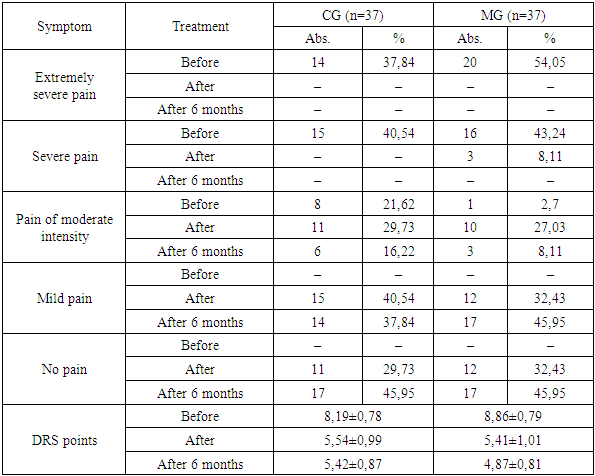
When conducting a comparative analysis of the effectiveness of various methods of treating TN by groups in terms of the presence of pain in subjective perception in the CG before treatment, moderate pain was noted by 8 (21.62%) patients, severe pain by 15 (40.54%) and extremely severe pain by 14 (37.84%) patients, i.e. 78.38% suffered from TN pain, the average score on the DRS was 8.19±0.78. After the treatment, no patient reported severe or extremely severe pain–all 78.38% of patients in this subgroup reported the absence of excruciating pain, with 11 (29.73%) patients reporting moderate pain, 15 (40.54%) patients reporting mild pain, and 11 (29.73%) patients reporting its absence, with the average score being 5.54±0.99. Thus, in the CG, 78.38% of patients had excruciating pain before treatment, while after treatment, 26 (70.27%) patients had mild pain or no pain at all (Table 3).
Table 3. The dynamics of pain in attacks of TN according to the Central DRS in the context of treatment groups
     
In the GS, 6 months after treatment, pain of moderate intensity was noted by 6 (16.22%) subjects, mild pain–by 14 (37.84%), absence of pain was noted by 17 (45.95%) patients with TN, the average DRS score was 5.42±0.87 points (Table 3).
A comparative analysis of the dynamics of pain syndrome in TN in the MG demonstrated the following changes: before treatment, 1 (2.7%) patient reported moderate pain, 16 (43.24%) patients reported severe pain, and 20 (54.05%) patients reported extremely severe pain, i.e. 97.3% of the group had excruciating pain, the average score was 8.86±0.79. After treatment, none of the patients reported extremely severe pain, severe pain was noted by 3 (8.11%) patients, moderate pain was noted by 10 (27.03%) patients, mild pain and its absence were noted by 12 (32.43%) patients each, and the average score was 5.41±1.01. Thus, in the MG before treatment, 97.3% of patients had excruciating pain, and after treatment, 24 (64.86%) patients had mild pain or no pain at all, and pain of moderate intensity was present in 10 (27.03%) patients (Table 3).
In the MG, 6 months after treatment, moderate pain was noted by 3 (8.11%) subjects with TN, mild pain and no pain were noted by 17 (45.95%) patients, the average DRS score was 4.87±0.81 points (Table 3).
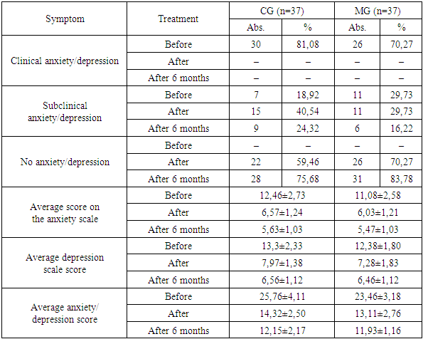
In a comparative analysis of the dynamics of subjective assessment of anxiety/depression on the HADS scale in the studied patients with TN from the CG, we found that before treatment, 30 (81.08%) patients had a clinical level of anxiety/depression and 7 (18.92%) had a subclinical level, the average sum of points on the anxiety scale was 12.46±2.73 points, on the depression scale–13.3±2.33 points, and the total sum was 25.76±4.11 points, i.e. pronounced anxiety/depression.
At the end of treatment in the CG, no patient had a clinical level of anxiety/depression, subclinical anxiety/depression was found in 15 (40.54%) patients, the absence of significantly expressed symptoms of anxiety and depression was found in 22 (59.46%) patients, the average score on the anxiety scale was 6.57±1.24 points, on the depression scale–7.97±1.38 points, and the total score was 14.32±2.50 points, i.e. the absence of significant symptoms of anxiety against the background of subclinical depression.
Thus, during the treatment, signs of clinical anxiety and depression were relieved in 30 (81.08%) patients, anxiety with residual subclinical signs of depression was completely eliminated in 15 (40.54%) patients.
After 6 months, subclinical anxiety/depression in the CG persisted in 9 (24.32%) patients, the absence of symptoms of anxiety and depression–in 28 (75.68%) patients, the average score on the anxiety scale was 5.63±1.03 points, on the depression scale–6.56±1.12 points, and the total score was 12.15±2.17 points.
In the MG, 11 patients (29.73%) had subclinical anxiety/depression before treatment, the remaining 26 (70.27%) patients were characterized by signs of clinical anxiety and depression, the average score on the anxiety scale was 11.08±2.58 points, on the depression scale–12.38±1.80 points, and the total score was 23.46±3.18 points. After the treatment, no patient was found to have a clinical level of anxiety/depression, subclinical anxiety/depression–in 11 patients (29.73%), no symptoms of anxiety and depression–in 26 patients (70.27%), the average score on the anxiety scale was 6.03±1.21 points, on the depression scale–7.28±1.83 points, and the total score was 13.11±2.76 points, i.e. absence of significant symptoms of anxiety against the background of some subclinical depression.
Thus, during the treatment, signs of clinical anxiety and depression were relieved in 26 (70.27%) patients, anxiety with residual subclinical signs of depression was completely eliminated in 26 (70.27%) patients.
After 6 months, subclinical anxiety/depression in the MG persisted in 6 (16.22%) patients, the absence of symptoms of anxiety and depression–in 31 (83.78%) patients, the average score on the anxiety scale was 5.47±1.03 points, on the depression scale–6.46±1.12 points, and the total score was 11.93±1.16 points (Table 4).
Table 4. The dynamics of HADS scores across treatment groups
     
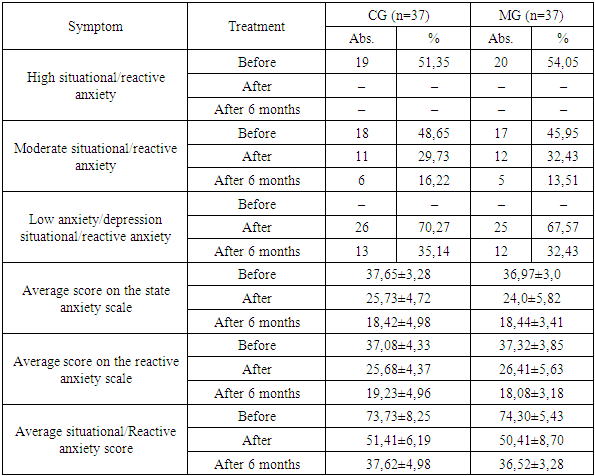
In the CG before treatment, the average degree of situational and reactive anxiety according to the results of the Spielberger STAI test was found in 18 (48.65%) patients and 19 (51.35%) patients had a high degree of situational and reactive anxiety, the average sum of points of situational anxiety was 37.65±3.28 points, reactive anxiety–37.08±4.33 points, and the total sum–73.73±8.25 points. After the treatment, none of the patients were diagnosed with a high degree of situational and reactive anxiety, the average degree of situational and reactive anxiety was found in 11 (29.73%) patients, the low degree of situational and reactive anxiety was found in 26 (70.27%) patients, the average total score of situational anxiety was 25.73±4.72 points, reactive anxiety was 25.68±4.37 points, and the total score was 51.41±6.19 points, which was statistically significantly different from the levels before treatment at p≤0.05 (Table 5).
Table 5. The dynamics of Spielberger STAI test scores across treatment groups
     
After 6 months in the CG, the average score of state anxiety of the Spielberger STAI test was 18.42±4.98 points, the average score of reactive anxiety was 19.23±4.96 points, the average sum of anxiety scores was 37.62±4.98 points, the average degree of anxiety was in 6 (16.22%) patients, low anxiety was in 13 (35.14%) patients, i.e. after 6 months, anxiety was completely eliminated in 18 (48.65%) patients of this subgroup (Table 5).
In the MG, before treatment, 17 (45.95%) patients had a moderate degree of situational and reactive anxiety, and 20 (54.05%) patients had a high degree of situational and reactive anxiety, the average sum of points of situational anxiety was 36.97±3.0 points, reactive anxiety–37.32±3.85 points, and the total sum was 74.30±5.43 points. After the treatment, none of the patients had a high degree of situational and reactive anxiety, the average degree of situational and reactive anxiety was found in 12 (32.43%) patients, the low degree of situational and reactive anxiety was found in 25 (67.57%) patients, the average total score of situational anxiety was 24.0±5.82 points, reactive anxiety was 26.41±5.63 points, and the total score was 50.41±8.70 points, which was statistically significantly different from the level before treatment (p≤0.05) (Table 5).
Thus, during the treatment process, signs of a high degree of situational and reactive anxiety were relieved in 20 (54.05%) patients, and a low degree of situational and reactive anxiety was achieved in 25 (67.57%) patients (Table 5).
After 6 months in the MG, the average score of state anxiety of the Spielberger STAI test was 18.44±3.41 points, the average score of reactive anxiety was 18.08±3.18 points, the average sum of anxiety scores was 36.52±3.28 points, the average degree of anxiety was in 5 (13.51%) patients, low anxiety was in 12 (32.43%) patients, i.e. after 6 months, anxiety was completely eliminated in 20 (54.05%) patients of this subgroup (Table 5).
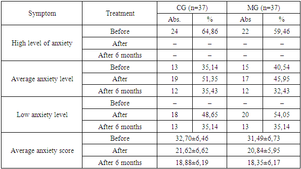
In the MG, before treatment, the average degree of anxiety according to the Beck scale was found in 15 patients (40.54%) and 22 patients (59.46%) had a high degree of anxiety, the average sum of points according to the Beck scale was 31.49±6.73 points. After the treatment, no patient had a high degree of anxiety, the average degree of anxiety was diagnosed in 17 (45.95%), low anxiety–in 20 (54.05%) patients, the average sum of points was 20.84±5.95 points. Thus, during the treatment, signs of a high degree of anxiety were relieved in 22 (59.46%) patients, a low degree of anxiety was achieved in 20 (54.05%) patients (Table 6).
Table 6. The dynamics of scores on the Beck scale in the context of treatment groups
     
After 6 months in the MG, average anxiety according to the Beck scale was diagnosed in 12 (32.43%) patients, low anxiety – in 13 (35.14%) patients, the average sum was 18.35±6.17 points, i.e. anxiety was leveled in 12 (32.43%) patients (Table 6).
In the CG, before treatment, 13 patients (35.14%) had a moderate anxiety level according to the Beck Anxiety Scale, and 24 patients (64.86%) had a high anxiety level, the average anxiety score was 32.70±6.46 points. After the treatment, no patients had a high anxiety level, 19 patients (51.35%) had a moderate anxiety level, and 18 patients (48.65%) had a low anxiety level, the average anxiety score was 21.62±6.62 points. Thus, during the treatment, severe anxiety was eliminated in 24 patients (64.86%), and a low anxiety level was achieved in 18 patients (48.65%) (Table 6).
After 6 months in the CG, according to the Beck scale, moderate anxiety was diagnosed in 12 (35.43%) patients, low anxiety–in 13 (35.14%) patients, the average sum was 18.88±6.19 points, i.e. anxiety was leveled in 12 (32.43%) patients (Table 6).
Throughout the entire follow-up observation period, the blood flow indices of the studied patients with TN according to the CDS of the extra–cranial sections of the BCA–CCA, ECA, ICA and VA on both sides in all patients did not show any dynamics, as did the results of EEG and ENMG in all branches of the TN.
The dynamics of the immunological biochemical analysis parameters before the treatment of IL–1 and TNF–β showed that the levels of TNF–α, IF–γ and SP were within the normal range, exceeding the norm without statistical significance, after the treatment the studied immunological biochemical parameters also remained within the reference values. In the context of the groups, we also did not find statistically significant differences from the levels before the treatment, although all the parameters remained within the normal range.
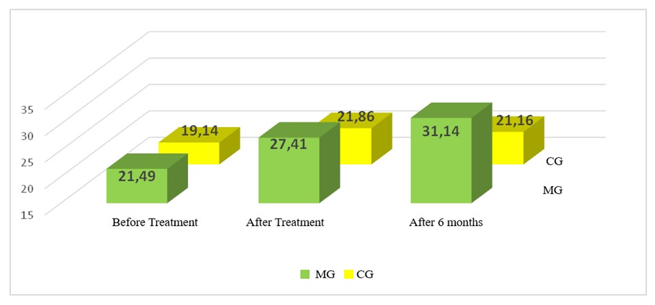
Vitamin D before treatment in the MG was 21.49±7.23 ng/ml, after treatment–27.41±6.58 ng/ml, after six months–31.14±1.39 ng/ml; in the CG it was 19.14±5.87 ng/ml before treatment and 21.81±6.14 ng/ml after treatment, after six months–21.16±6.28 ng/ml, which indicated its deficiency in the CG and normalization of the level in the MG six months after treatment, taking into account the norm–30–35 ng/ml.
Figure 2. The dynamics of serum vitamin D concentration by treatment groups, ng/ml
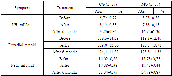
In women with CG, the LH level before treatment was 5.72±0.77 mIU/ml, and at the end of treatment–8.12±0.33 mIU/ml with the norm of 7.7–58.5 mIU/ml, i.e. it returned to normal, the serum concentration of estradiol before treatment was 119.5±14.38 pmol/l, and after treatment–129.8±12.86 pmol/l with the norm of 127–138 pmol/l, i.e. the average concentration approximately corresponded to the upper permissible limit of the norm. The FSH level in the blood serum of female HS patients before treatment was 16.02±0.86 mIU/ml, and after treatment–19.38±0.38 mIU/ml, while the norm is 25.8–134.8 mIU/ml, i.e. normalization was not achieved.
Table 7. The dynamics of scores on the Beck scale in the context of treatment groups
     
In women with MG, the LH level before treatment was 5.78±0.78 mIU/ml, and at the end of treatment it was 7.88±0.15 mIU/ml with the norm of 7.7–58.5 mIU/ml, i.e. it returned to normal. The serum concentration of estradiol before treatment was 118.8±12.40 pmol/l, and after treatment it was 128.3±3.71 pmol/l with the norm of 127–138 pmol/l, i.e. the average concentration approximately corresponded to the upper permissible limit of the norm. The FSH level in the blood serum of MG patients before treatment was 15.78±0.75 mIU/ml, and after treatment–19.43±0.44 mIU/ml, while the norm is 25.8–134.8 mIU/ml, i.e. normalization was not achieved.

4. Conclusions

The treatment carried out in the context of groups allowed us to conduct a comparative analysis of its effectiveness–the complex treatment carried out by the MG turned out to be more effective, which in addition to the standard treatment of the peripheral type of TN included vitamin D at 1000 IU daily for a long time (3 months) and the multivitamin complex “Demoton D”.

References

[1]  Akkalaeva, I. A., & Nigkolova, D. E. (2023). Trigeminal neuralgia. Nauchny Lider, (4[102]), 44–46.
[2]  Byvaltsev, V. A., Kalinin, A. A., Okoneshnikova, A. K., & Egorov, A. V. (2021). Comparative analysis of the clinical effectiveness of laser and radiofrequency denervation in patients with primary trigeminal neuralgia. Journal of Neurology and Psychiatry Named After S.S. Korsakov, 121(4), 38–43.
[3]  Gabidullin, A. F., Rogozhin, A. A., & Abdullaeva, F. R. (2021). Trigeminal neuralgia: Experience in the use of Gasserian ganglion destruction and microvascular decompression of the trigeminal nerve root. Proceedings of the IX All-Russian Congress of Neurosurgeons, 85–86.
[4]  Dzo, M. D., Hwang, S. G., & Yi, H. D. (2021). Use of carbamate for the prevention or treatment of trigeminal neuralgia. Patent No. RU 2751504 C2.
[5]  Zhurkin, A. N., Semenov, A. V., Sorokovikov, V. A., & Bartul, N. V. (2021). Historical aspects of the problem of treating trigeminal neuralgia and the role of neurosurgical methods in its resolution (Literature review). Acta Biomedica Scientifica, 6(4), 123–126.
[6]  Kolycheva, M. V., Bezborodova, T. Yu., Shimansky, V. N., Strunina, Yu. V., & Gadzhieva, O. A. (2022). Carbamazepine in the treatment of trigeminal neuralgia. Russian Journal of Pain, 20(4), 28–34.
[7]  Maksimova, M. Yu. (2021). Trigeminal neuralgia. Pharmateka, 28(3), 106–112.
[8]  Markin, S. P. (2005). Comprehensive treatment of patients with facial neuropathy and trigeminal neuralgia. Med-Practice.
[9]  Sadykova, Kh. K., Tursunov, M. K., Khudayberdiev, K. T., & Babokhujaev, A. S. (2020). Surgical treatment of trigeminal neuralgia using the method of radiofrequency rhizotomy of the Gasserian ganglion. Stomatologiya, 1, 28–30.
[10]  Sturov, N. V., Pereverzev, A. P., & Rogozhina, A. V. (2008). Trigeminal neuralgia. Difficult Patient, 6(1), 37–41.
[11]  Shimansky, V. N., Konovalov, A. N., Tanyashin, S. V., & Poshataev, V. K. (2023). Vascular decompression in hyperfunction of cranial nerves (trigeminal neuralgia, hemifacial spasm, glossopharyngeal neuralgia). ABV-Press Publishing House.
[12]  Allam, A. K., Sharma, H., Larkin, M. B., & Viswanathan, A. (2023). Trigeminal neuralgia: Diagnosis and treatment. Neurologic Clinics, 41(1), 107–121.
[13]  Araya, E. I., Claudino, R. F., Piovesan, E. J., & Chichorro, J. G. (2020). Trigeminal neuralgia: Basic and clinical aspects. Current Neuropharmacology, 18(2), 109–119.
[14]  Cameron, M. H. (2013). Physical agents in rehabilitation: From research to practice (4th ed.). Elsevier Health Sciences.
[15]  Cruccu, G., Finnerup, N. B., Jensen, T. S., & Scholz, J. (2016). Trigeminal neuralgia: New classification and diagnostic grading for practice and research. Neurology, 87(2), 220–228.
[16]  Dong, B., Xu, R., & Lim, M. (2023). The pathophysiology of trigeminal neuralgia: A molecular review. Journal of Neurosurgery, 139(5), 1471–1479.
[17]  Ellis, H. (2019). Neurectomy for trigeminal neuralgia. Journal of Perioperative Practice, 29(4), 102–103.
[18]  Fothergill, J. (1773). On a painful affliction of the face. Medical Observations and Inquiries, 5, 129–142.
[19]  Rose, F. C. (1999). Trigeminal neuralgia. Archives of Neurology, 56, 1163–1164.
[20]  Ganz, J. C. (2022). Trigeminal neuralgia and other cranial pain syndromes. Progress in Brain Research, 268(1), 347–378.
[21]  Gerwin, R. (2020). Chronic facial pain: Trigeminal neuralgia, persistent idiopathic facial pain, and myofascial pain syndrome—An evidence-based narrative review and etiological hypothesis. International Journal of Environmental Research and Public Health, 17(19), 7012–7021.
[22]  Hall, G. C., Carroll, D., Parry, D., & McQuay, H. J. (2006). Epidemiology and treatment of neuropathic pain: The UK primary care perspective. Pain, 122, 156–162.
[23]  Koopman, J. S., Dieleman, J. P., Huygen, F. J., de Mos, M., Martin, C. G., & Sturkenboom, M. C. (2009). Incidence of facial pain in the general population. Pain, 147(1–3), 122–127.
[24]  Maarbjerg, S., Di Stefano, G., Bendtsen, L., & Cruccu, G. (2017). Trigeminal neuralgia—Diagnosis and treatment. Cephalalgia, 37(7), 648–657.
[25]  Maarbjerg, S., Gozalov, A., Olesen, J., & Bendtsen, L. (2014). Trigeminal neuralgia—A prospective systematic study of clinical characteristics in 158 patients. Headache, 54(10), 1574–1582.
[26]  Molina-Olier, O., Marsiglia-Pérez, D., & Alvis-Miranda, H. (2022). Surgical treatment of trigeminal neuralgia in adults. Cirugía, 90(4), 548–555.