-
Paper Information
- Next Paper
- Previous Paper
- Paper Submission
-
Journal Information
- About This Journal
- Editorial Board
- Current Issue
- Archive
- Author Guidelines
- Contact Us
American Journal of Medicine and Medical Sciences
p-ISSN: 2165-901X e-ISSN: 2165-9036
2025; 15(3): 575-576
doi:10.5923/j.ajmms.20251503.18
Received: Feb. 3, 2025; Accepted: Feb. 25, 2025; Published: Mar. 8, 2025

Assessment of Morphological Changes in the Testes on the Third Day After Experimental Traumatic Brain Injury
 Abstract
 Reference
 Full-Text PDF
 Full-text HTML
Rakhimova Gulnоz Shamsiyevna, Teshayev Shuxrat Jumayevich
Bukhara State Medical Institute named after Abu Ali ibn Sina, Bukhara, Uzbekistan
Correspondence to: Rakhimova Gulnоz Shamsiyevna, Bukhara State Medical Institute named after Abu Ali ibn Sina, Bukhara, Uzbekistan.
| Email: |  |
Copyright © 2025 The Author(s). Published by Scientific & Academic Publishing.
This work is licensed under the Creative Commons Attribution International License (CC BY).
http://creativecommons.org/licenses/by/4.0/

54 to 60 million people worldwide suffer from traumatic brain injury (TBI) every year. Traumatic brain injury stands out among other injuries because it often leads to death or permanent disability, especially in young and healthy people. Scientific sources indicate that brain injuries damage most organs. In the experiment, we studied morphological changes in the testicles of male rats caused by brain damage. In the study, 90-day-old 150 white outbred male rats weighing from 115 to 150 g were selected, examined on the 1st, 3rd, 7th and 21st days after brain injury and compared with control groups. Traumatic brain injury affected the macroscopic and histomorphological data of the testicles on the third day after the injury.
Keywords: Testis, Traumatic brain injury, Albuminous membrane, Convoluted seminiferous tubules
Cite this paper: Rakhimova Gulnоz Shamsiyevna, Teshayev Shuxrat Jumayevich, Assessment of Morphological Changes in the Testes on the Third Day After Experimental Traumatic Brain Injury, American Journal of Medicine and Medical Sciences, Vol. 15 No. 3, 2025, pp. 575-576. doi: 10.5923/j.ajmms.20251503.18.
Article Outline
1. Introduction
- After a traumatic brain injury, complex systemic pathomorphological and regulatory changes develop in various brain centers, which, in turn, affect a single functional system consisting of mental, neurohumoral, erectile and ejaculatory processes [1]. While the sympathoadrenal part of the autonomic nervous system is responsible for mild forms of traumatic brain injury, stable changes occur in moderate and severe forms due to hormonal imbalance (increased prolactin and decreased testosterone) [2]. It was found that the testicles morphologically change under the influence of various external factors, including changes in traumatic brain injury [3]. Scientific sources provide evidence that as a result of injury and ischemia, vacuolization, necrosis, and loss of germinal cells of the convoluted seminal tubules can occur in the testes [4]. In the laboratory of the Bukhara State Medical Institute, studies have been conducted on the effect of traumatic brain injury on the morphological parameters of the testis, which deepens the understanding of the mechanisms of the testis' adequate response to the effects of exogenous factors and determines the order of possible sequential changes in the morphological parameters of this organ.The aim of the study: to study morphological changes in the testes of 90-day-old white outbred male rats on the third day after a simulated acute traumatic brain injury.
2. Materials and Methods of the Study
- The study was conducted on 90-day-old 150 white outbred male rats weighing from 115 to 150 g. The animals were kept in standard conditions. All animal experiments were conducted with the approval of the local ethics committee. A model of a "traffic accident" was modeled on 80 rats and an experiment was conducted on a device in the form of a vehicle (patent no. FAP 02271) [5]. 3 days after the experiment, 18 control rats (group 1b) and 18 rats with traumatic brain injury (group 2b) were decapitated, their testis were extracted and examined macroscopically, as well as histologically.
3. The Results of the Study
- In group 1b during macroscopic examination, the testis have a normal shape, soft and elastic density. During histological examination in group 1b rats, the tunica albuginea in the stromal part is thin, collagen, elastic fibers, and connective tissue cells are visible in it. In group 2b, the color of the capsule and parenchyma of the testes did not visually change upon macroscopic examination, but the consistency was somewhat loose compared to the testes of intact rats.During histological examination in group 2b, the tunica albuginea in the stromal part is swollen, loose. There is a slight increase in the number of fibroblasts, mast cells and histiocytes. Due to the looseness of the collagen fibers, the boundary between the outer and inner layers is not clearly distinguishable. Foci of fresh hemorrhage are detected in the tunica albuginea under the capsule and in the stroma of the testes. The wall of the blood vessel is slightly fibrous (Fig. 1). The diameter of the blood vessels under the capsule is similar to that of the control group, we see an increase in the thickness of the wall of blood vessels by 26%. Leydig cells are outlined, some are swollen, the nuclei are reduced in size, have an oval or rounded shape. The number of Leydig cells decreased by 6.8%.
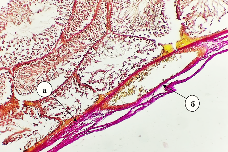 | Figure 1. The tunica albuginea of the testes of an experimental group of white outbred rats on the 3rd day after traumatic brain injury: a-loose and swollen; b-the walls of blood vessels are fibrous. Staining: Van Gieson. Size 10х10 |
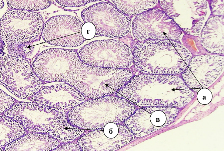 | Figure 2. Convoluted seminiferous tubules of testes of an experimental group of white outbred rats on the 3rd day after traumatic brain injury: the tubule cavity is dilated, a-pear-shaped, b-bean-shaped and c-doubled seminiferous tubules; d- rupture of seminiferous tubules and immature cells at the rupture site. Staining: H&E. Size 10х4 |
4. Conclusions
- Histological examination on the 3rd day after experimental traumatic brain injury shows pathomorphological changes in the testicles of rats, morphometric studies show an increase in morphometric parameters in the testicular stroma as a result of degenerative and inflammatory processes, and a decrease in the number of spermatogenic cells. These results require further research to identify the mechanisms of the emerging pathology and develop the necessary therapeutic measures.