-
Paper Information
- Next Paper
- Paper Submission
-
Journal Information
- About This Journal
- Editorial Board
- Current Issue
- Archive
- Author Guidelines
- Contact Us
American Journal of Medicine and Medical Sciences
p-ISSN: 2165-901X e-ISSN: 2165-9036
2025; 15(3): 532-534
doi:10.5923/j.ajmms.20251503.08
Received: Feb. 3, 2025; Accepted: Feb. 26, 2025; Published: Mar. 8, 2025

Comparative Description of Liver Morphometric Indicators During Pregnancy in Experimental Chronic Kidney Diseases
Rakhmonkulova N. G.
Bukhara State Medical Institute named after Abu Ali ibn Sina, Bukhara, Uzbekistan
Correspondence to: Rakhmonkulova N. G., Bukhara State Medical Institute named after Abu Ali ibn Sina, Bukhara, Uzbekistan.
| Email: |  |
Copyright © 2025 The Author(s). Published by Scientific & Academic Publishing.
This work is licensed under the Creative Commons Attribution International License (CC BY).
http://creativecommons.org/licenses/by/4.0/

Investigation of morphological and morphometric alterations in the liver of pregnant white rats following chronic renal failure. Examination of the normal morphological and morphometric characteristics of the liver in pregnant white rats. Evaluation of changes in liver tissue based on morphometric analysis one month after treatment in 7-month-pregnant and experimentally induced chronic kidney disease-affected non-purebred pregnant white rats. Evaluation of changes in liver tissue based on morphometric analysis one month after treatment in 7-month-pregnant and experimentally induced chronic kidney disease-affected non-purebred pregnant white rats. Analysis of the anatomical features of the liver in purebred pregnant rats and its reactive changes after induced chronic renal failure. Comparison of pregnancy-related histo-topographic modifications in the liver of purebred rats with chronic renal failure to those of healthy rats. A comparative assessment of morphometric liver changes in pregnant rats after treatment with Juyzar waters in experimental chronic renal failure.
Keywords: Experimental chronic kidney disease, Pregnancy, Morphometric indicators, Comparative description, Hepatocytes, Histological changes, Functional disorders, Histotopographic changes
Cite this paper: Rakhmonkulova N. G., Comparative Description of Liver Morphometric Indicators During Pregnancy in Experimental Chronic Kidney Diseases, American Journal of Medicine and Medical Sciences, Vol. 15 No. 3, 2025, pp. 532-534. doi: 10.5923/j.ajmms.20251503.08.
1. Introduction
- Pregnancy induces a variety of physiological changes in the maternal body, with the liver playing a key role in managing metabolic demands, detoxification, and hormonal regulation. The liver undergoes structural and functional adaptations during pregnancy to support both the mother and the developing fetus. However, these adaptive mechanisms can be significantly altered in the presence of chronic kidney disease (CKD). CKD, a progressive condition marked by a gradual decline in kidney function, triggers systemic metabolic disturbances, inflammation, and oxidative stress, which can have a profound impact on liver morphology and function, especially during pregnancy. In a healthy pregnancy, the liver adjusts to the increased demands by modifying its metabolic pathways, enlarging hepatocytes, and enhancing blood flow. However, in the context of CKD, these normal processes are disrupted [1]. The kidneys and liver are closely linked in maintaining overall bodily function, particularly in processes like waste elimination and metabolic balance. Renal dysfunction can therefore lead to significant liver morphological changes, including hepatocyte enlargement, fibrosis, sinusoidal alterations, and changes in liver vascularization, which may impair the liver’s ability to support pregnancy [3].The morphological changes in the liver during pregnancy in the presence of CKD are a subject of increasing interest in experimental models. Using experimental chronic kidney disease models in pregnant rats, researchers can observe how renal dysfunction affects liver morphology during pregnancy. These models often involve inducing CKD through nephrotoxic agents, ischemic injury, or unilateral nephrectomy, allowing for a controlled examination of liver changes under renal stress. For example, in an experimental model, pregnant rats with induced CKD may exhibit hepatocyte hypertrophy (increased cell size), indicative of metabolic stress due to renal failure. Furthermore, liver fibrosis may develop, characterized by the deposition of collagen around the liver lobules, reflecting ongoing liver damage due to inflammation. [2,4]. Sinusoidal dilation and vascular changes could also occur, indicating impaired blood flow and oxygenation of liver tissue, which is crucial for maintaining the increased demands of pregnancy. The structural alterations may lead to impaired liver function, making it less effective at detoxifying waste products and regulating metabolic processes, thus complicating pregnancy.In this study, we aim to comparatively characterize liver morphometric indicators in pregnant rats with experimental CKD and healthy pregnant rats. By analyzing hepatocyte size, lobular architecture, fibrosis, and vascular changes, we will explore the specific morphological changes induced by chronic kidney disease during pregnancy. The goal is to understand how CKD impacts liver function and morphology during pregnancy, providing insights into potential therapeutic strategies to manage liver and kidney dysfunction in pregnant women with CKD.Through a better understanding of these morphological changes, we can develop more effective treatment strategies for pregnant women suffering from CKD, ensuring improved maternal and fetal health outcomes [5,6].The purpose of the scientific work: Studying the normative morphological and morphometric indicators of the liver in pregnant non-purebred rats; Investigating the anatomical parameters of the liver in non-purebred rats during pregnancy and its reactive changes after experimental chronic kidney disease.
2. Materials and Methods
- Pregnancy is 7 months, 1 month after experimental chronic renal failure, a total of 150 white mongrel rats. Histological examination of the cellular structure of the liver after experimental chronic renal failure in non-white pregnant rats. General morphological examination by staining with hematoxylin and eosin; Morphometric - examination of the size of hepatocytes; statistical methods are used. Hepatocytes are the main cells of the liver. The cellular structure of hepatocytes is cubic or polygonal. The nucleus is located in the center of the cell, round in shape - in most cases it consists of two nuclei. The cytoplasm is stained with eosinophils. Its cytoplasm is rich in endoplasmic reticulum (organelles synthesizing plasma proteins) and a large amount of granular endoplasmic reticulum (organelles synthesizing toxins, bilirubin and bile). The following surfaces are distinguished in hepatocytes. The sinusoidal surface of hepatocytes. proteins.
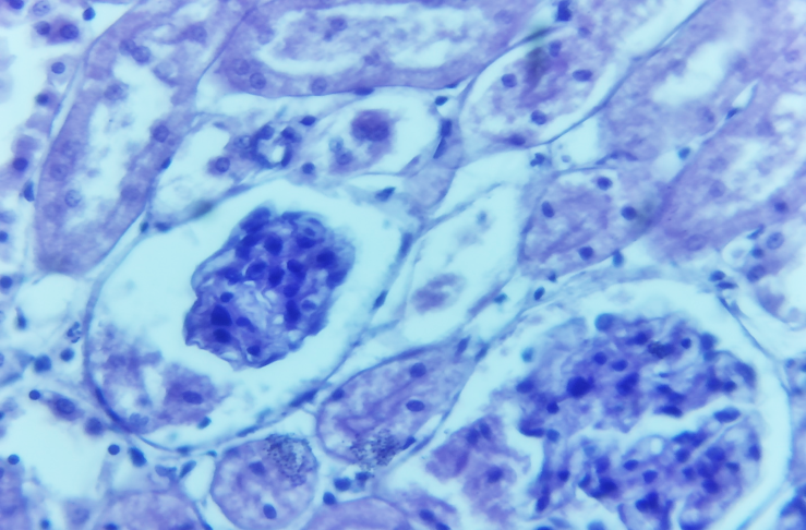
 | Figure 1 |
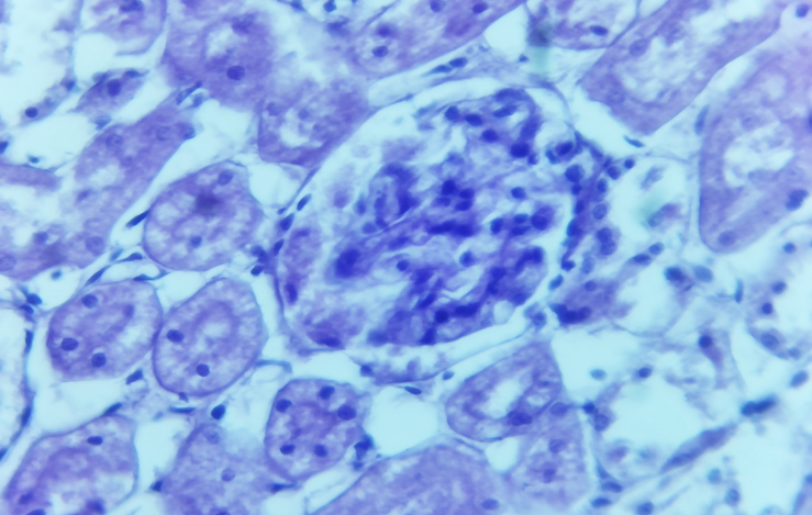
 | Figure 2 |
3. Conclusions
- The study of liver hepatocyte morphology and structural changes provides insight into the complex mechanisms underlying liver processes following experimental chronic renal failure. These findings contribute to expanding theoretical knowledge about the digestive system. Since a living organism is composed of approximately 70% water, and 81 of the 92 naturally occurring elements are found in the human body, proper water composition is crucial. Ideally, one liter of drinking water should contain specific amounts of essential trace elements. This research evaluates the development of preventive measures for pregnancy-related liver structural changes in experimental chronic renal failure. It also aims to improve the effectiveness of treatment methods, facilitate early diagnosis of liver pathologies associated with chronic renal failure, and assess the therapeutic potential of Juzar water. Chronic renal failure, particularly in the context of chronic glomerulonephritis, results from various glomerulopathies, including acute glomerulosclerosis, membranous, and membranoproliferative glomerulonephritis. In such cases, the kidneys remain symmetrically positioned with a granular surface due to a lack of mobility. Our experiment, involving white breed rats with pronounced chronic renal insufficiency, revealed several histopathological changes. Microscopic examination identified a reduction in glomerular size, sclerotic alterations, hyalinosis, enlarged cavities, and infiltration. Additionally, narrowing of afferent and efferent arterioles disrupted blood flow, leading to secondary glomerular damage. Thickening of the Shmulyansky-Bauman capsule was observed. Ischemic processes were found to cause focal karyolysis and karyorrhexis in the nuclei of distal and proximal tubular epithelial cells. These processes also led to protein dystrophy (including hydropic and hyaline degeneration) in the cytoplasm, interstitial and intermediate tissue edema, and fibrosis, resulting in tissue thickening. Moreover, the walls of medium- and small-caliber arteries thickened, and narrowing of blood flow pathways contributed to secondary hypertension and renal parenchymal atrophy.Anatomical and morphological liver changes during pregnancy were also examined in the same group of rats. Macroscopic analysis revealed an enlarged liver with a smooth surface, while the capsule had a strained, nutmeg-like appearance. Microscopic findings showed deformation and sclerotic changes in the central venous wall, reduced lumen size, and inflammatory infiltration around the veins. Hepatocytes exhibited hyperchromatic protein dystrophy, characterized by hydropic and hyaline degeneration. Additionally, binucleated hepatocytes increased, suggesting a compensatory response. The hepatocyte nuclei displayed reduced basophilic staining, while their cytoplasm contained vacuoles of varying sizes, indicating fatty degeneration. Sinusoidal spaces and the perisinusoidal (Disse) area were enlarged and swollen, suggesting impaired metabolism and slowed physiological processes. These findings highlight the structural and functional liver alterations that occur in response to chronic renal failure and pregnancy, emphasizing the need for targeted treatment strategies and preventive measures.
 Abstract
 Reference
 Full-Text PDF
 Full-text HTML