Berdiev A. Sh., Akhmedov F. K.
Bukhara State Medical Institute named after Abu Ali ibn Sina, Bukhara, Uzbekistan
Copyright © 2025 The Author(s). Published by Scientific & Academic Publishing.
This work is licensed under the Creative Commons Attribution International License (CC BY).
http://creativecommons.org/licenses/by/4.0/

Abstract
The purpose of the study: Study of biochemical markers and indicators of rheumatic factor in physiological and pathological late pregnancy. 100 pregnant women were followed, 70 of them were in the main group and 30 were in the control group. Thus, our study demonstrated that the use of NT-proBNP and troponin I for the diagnosis of cardiac disease in the maternity ward has an important prognostic value. In our opinion, the exact mechanism of the increase in plasma levels of these markers may be a decrease in diastole, followed by hypertrophy and ischemia. To our knowledge, this is the first report of a marker strategy (NT-proBNP and troponin I) aimed at diagnosing rheumatic diseases in pregnant women. We have shown that routine assessment of NT-proBNP and troponin I can be used to identify patients with rheumatic mitral valve insufficiency in the obstetric pathology department of perinatal centers. The results of this study, namely NT-proBNP and troponin I, can be implemented in clinical practice as diagnostic markers.
Keywords:
Pregnancy, Rheumatism, NT-proBNP, Troponin I, Mitral valve insufficiency
Cite this paper: Berdiev A. Sh., Akhmedov F. K., Evaluation of Biochemical Markers and Indicators of Rheumatic Factor in Physiological and Pathological Late Pregnancy, American Journal of Medicine and Medical Sciences, Vol. 15 No. 2, 2025, pp. 462-465. doi: 10.5923/j.ajmms.20251502.41.
1. Introduction
Rheumatic heart disease is a systemic immune-mediated disease caused by beta-hemolytic streptococci, with recurrent attacks leading to stiffness and deformation of the heart valves, resulting in mitral valve regurgitation and stenosis. In the literature, it is reported that in 50-60% of cases, rheumatic diseases affect only the heart valves [1,3].Mitral valve regurgitation is the most common form of rheumatic heart disease in pregnant women, and rheumatic heart disease is one of the most common causes of mitral valve regurgitation worldwide. According to statistics, rheumatic heart disease causes at least 200,000-250,000 premature deaths each year. It is also one of the main causes of death from cardiovascular diseases in young people and pregnant women in countries with poorly developed health care systems [2,4].It is known that SRO is an acute phase protein. Its main task is to activate the body's complement system and strengthen phagocytosis processes [5,6]. SRO is synthesized in the liver under the influence of IL-6, has a half-life of 19 hours, and after the end of the injury process, its amount approaches normal values. SRO has high sensitivity and increases mainly in rheumatic diseases. In general, due to the development of SRO placental microcirculatory endotheliitis to a certain degree and carditis in the heart, we detected SRO protein in the blood serum of pregnant women in the study.The purpose of the study: Study of biochemical markers and indicators of rheumatic factor in physiological and pathological late pregnancy.
2. Research Methods and Materials
During 2022-2024, 100 pregnant women with mitral valve insufficiency of rheumatic etiology who applied to the Bukhara branch of the State Institution of the Republican Specialized Scientific and Practical Medical Center for Maternal and Child Health were studied, of which 70 were in the main group and 30 were in the control group, whose pregnancies were without complications. During the study of pregnant women included in our study, biomaterials (blood and plasma) were collected based on their verbal consent. In pregnant women included in the study, natriuretic peptide (NT-proBNP), rheumatology test (ASL-O - antistreptolysin O), rheumatoid factor, C-reactive protein, troponin -I, HbA1c - glycated hemoglobin, were studied.We studied pregnant women who participated in the study and divided them into 3 groups:Group 1 – hospitalized with clinical manifestations of rheumatic processes (n=35);Group 2 – pregnant women who were examined from the early stages of pregnancy and received appropriate preventive treatment in a timely manner (n=35).Control group – a group of pregnant women with a physiological pregnancy (n=30);All pregnant women under observation underwent a complete clinical examination at the above-mentioned medical institution, and based on clinical laboratory results, their complaints, and additional examination methods, the pregnant women were diagnosed with mitral valve insufficiency of rheumatic origin.
3. Results and Analyses
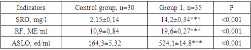
The conducted studies were 2.15±0.14 mg/l in physiological pregnancy (see table 1). This is in accordance with the information presented in the literature, and it can increase 1.5-2 times compared to the normal values during pregnancy.Table 1. Rheumatic factor levels in the serum of pregnant women included in the study, M±m
 |
| |
|
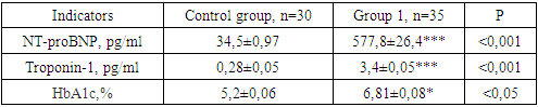
The data presented in the table show that in women with active clinical manifestations of rheumatic processes, the level of CRP in the blood serum increased statistically significantly by 6.6 (P<0.001) times, reaching 14.2±0.34 mg/l, compared to women with physiological pregnancy.As reported in the literature, it is possible to determine the gradation of the amount of SPO in blood plasma and, accordingly, the level of infiltrative inflammatory processes [Parsanathan R., Jain S.K., 2020]. The results obtained revealed a moderate increase in the amount of SPO in all groups (10-25 mg/l). This indicates the presence of chronic inflammatory processes in the body of pregnant women, including in the placenta and heart.In recent years, the literature has expressed different opinions about the role of rheumatoid factor in the development of rheumatism. Rheumatoid factors (RF) are found not only in rheumatism, but also in a wide range of pathologies, including other autoimmune and non-autoimmune diseases. Rheumatoid factors are antibodies of various isotypes and affinity directed against the Fc part of immunoglobulin G. Although other types of immunoglobulins, such as IgG and IgA, are rarely found, the most frequently mentioned rheumatoid factor (RF IgM) is [Yunmei Liang., Dingle Yu., 2023].Based on the above, we also determined the amount of rheumatoid factor (RO IgM) in blood plasma. In our study, its amount in physiologically active pregnancy was 10.9±0.84 IU/ml. Compared to the control group, the rheumatoid factor in pregnant women of group 1 was 1.79 (P<0.001) times higher, equal to 19.6±0.27 IU/ml, which was statistically significant.According to the literature, rheumatoid factors are usually not detected in the bloodstream without immunogenic stimulation. They form immune complexes, which are subsequently phagocytosed by inflammatory cells. These rheumatoid factors (RF) are low-affinity, transient, and polyclonal antibodies produced by germinal centers [Vivekanand Tiwari., Jagmohan S., 2023].Antistreptolysin O (ASTO) is an antibody against streptolysin O, a toxic enzyme produced by group A Streptococcus bacteria. ASLO and anti-DNase B are the most common types of antibodies produced by the body's immune system in response to group A streptococcal infection. Despite the recent decrease in the incidence of rheumatic mitral valve insufficiency, the ASLO test is often performed as a screening test for the diagnosis of rheumatic diseases. Among the pregnant women included in our study, the level of the control group was 164.3 ± 5.32 U / ml, while in our pregnant women included in group 1, it was 3.19 (P < 0.001) times higher than in the control group, equal to 524.1 ± 14.8 U / ml, which was statistically significant.Thus, an increase in rheumatic factors in the blood serum of pregnant women indicates a risk of mitral valve damage. Scientists believe that determining the amount of rheumatic factors in the 1st and 2nd trimesters of pregnancy can be used as a criterion for predicting the risk of developing mitral valve disease of rheumatic origin [Victoria J., Brookes., Caitlin E., 2024]. The NT-proBNP marker has been proven by scientists to be a useful marker for distinguishing pregnant women presenting with cardiovascular disease and those complicated by rheumatism in obstetric complexes, and has been noted as a strong prognostic marker for pregnant women with heart failure [Siegmund A.S., Pieper P.G., Bouma B.J., 2021].The NT-proBNP marker is considered by the European Society of Cardiology to be useful for diagnosing heart failure and providing prognostic potential, and it has also been found that this marker helps predict the complications it may cause, even in the absence of symptoms, in pregnant women complicated by mitral valve insufficiency of cardiac and rheumatic origin.Based on the above data, we also determined the amount of NT-proBNP in blood serum (see Table 2).Table 2. Biochemical markers in the blood serum of pregnant women included in the study, M±m
 |
| |
|
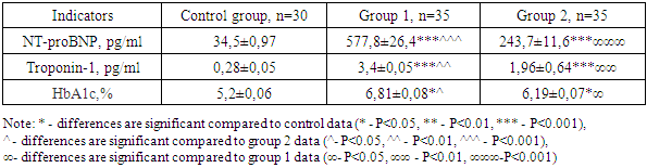
In pregnant women included in the control group, it was 34.5±0.97 pg/ml, while in group 1, that is, women with active clinical manifestations of rheumatic processes, the level of NT-proBNP in the blood serum increased statistically significantly by 16.7 (P<0.001) times compared to women with physiological pregnancy, reaching 577.8±26.4 pg/ml.Cardiac troponin-I, a sensitive and specific marker of myocardial cell injury, is a useful marker in the diagnosis and evaluation of heart and mitral valve insufficiency, and acute coronary syndromes. Studies have shown that troponin-I is elevated in heart failure and may be an important biomarker for predicting adverse outcomes [Aly M., Nayel M., Salama A., 2020]. Troponins are proteins involved in the regulation of cardiac and skeletal muscle contraction. The presence of cardiac troponins in serum indicates myocardial damage or loss of cell membrane integrity. In addition, this marker is an effective biomarker for assessing the state of central and intracardiac hemodynamics in pregnant women [Wong Y-K., Cheung C.Y., Tang C.S., 2019].Table 2 shows that in group 1, that is, in women with active clinical manifestations of rheumatic processes, the level of troponin-I in the blood serum increased statistically significantly by 12.1 (P<0.001) times compared to women with physiological pregnancy, reaching 3.4±0.05 pg/ml. In the control group, this indicator was 0.28±0.05 pg/ml.According to the literature, there are various studies that show that patients with rheumatic fever are at increased risk of developing diabetes mellitus (DM). In addition, insulin resistance is an independent risk factor for cardiovascular disease, which has also been well documented in studies [Lurati A., Laria A., Mazzocchi D., 2021]. Based on the above data, we decided to determine HbA1c in the serum of pregnant women involved in the study, which was 5.2±0.06% in the control group, and 6.81±0.08% in our pregnant women in group 1. From the point of view of statistical analysis, no significant difference was detected between the groups (P<0.05).If the SRO, RO, ASLO indicators in pregnant women of group 1 increased by 1.78, 1.25, 1.37 times, respectively, compared to pregnant women of group 2 who received timely medical treatment, then they increased by 6.6, 1.79, 3.2 times, respectively, compared to the indicators of women of the control group.In group 2, that is, in pregnant women who received medical treatment and were under control, CRP was 7.94±0.17 mg/l, while it was 3.7 (P<0.01) times higher than in pregnant women in the control group.In many diseases, the amount of CRP increases, especially in rheumatic heart disease, an increase in the level of this indicator may indicate an increased inflammatory process and the nature of the pathology. This marker was also observed to increase in pregnant women involved in our study, indicating the presence of an inflammatory process.At the same time, the levels of RO and ASLO in the blood plasma of pregnant women in group 2 increased by 1.44 (P<0.01) and 2.33 (P<0.001) times compared to women in the control group, and their levels were 15.7±0.49 IU/ml and 382.2±3.8 IU/ml, respectively.It has been shown in the literature that elevated levels of NT-proBNP are a response to increased angiotensin II and sympathetic nervous system tone. In addition, elevated levels of NT-proBNP are associated with left ventricular hypertrophy and dysfunction. This marker is also increased in infectious-inflammatory heart diseases and increased left ventricular ejection fraction [Cui K., Huang W., Fan J., Lei H., 2018]. NT-proBNP causes vasodilation, increases diuresis, and suppresses renin-aldosterone production.Table 3. Biochemical markers in the blood serum of pregnant women included in the study, M±m
 |
| |
|
As can be seen from Table 3, we observed even stronger changes in the group of pregnant women with rheumatic mitral valve insufficiency. In particular, in this group of pregnant women, a statistically significant increase in serum NT-proBNP levels was observed by 16.8 (P<0.001) times, reaching 577.8±26.4 pg/ml [995.5-398.4 pg/ml]. These indicators were 2.37 (P<0.001) times higher than those of pregnant women in group 2. The level of NT-proBNP in pregnant women in group 2 was 243.7±11.6 pg/ml [412.7-206.5 pg/ml], which was 7.1 (P<0.001) times higher than that of the control group. At the same time, the level of troponin-I in the blood serum of pregnant women in this group was 12.2 (P<0.001) times higher than in the control group, reaching 3.4±0.05 pg/ml [3.78-2.8 pg/ml], and a tendency to decrease was detected compared to the indicators of group 2, reaching 1.73 (P<0.01) times higher. The level of troponin-I in pregnant women in group 2 was 1.96±0.64 pg/ml [2.3-1.1 pg/ml], which was 7.0 (P<0.001) times higher than in pregnant women in the control group. In the pregnant women included in the study, the HbA1c level in the control group was 5.2±0.06%, with a range of [5.81-4.4%]. In pregnant women in groups 1 and 2, its level was 6.81±0.08% [7.8-5.5%] and 6.19±0.07% [6.6-4.5%], respectively, with no statistically significant difference.The authors believe that increasing levels of NT-proBNP and troponin-I can be used as an early diagnostic criterion. The results obtained in our study also indicate that the determination of NT-proBNP and troponin-I in the blood serum of women in the 1st and 2nd trimesters of pregnancy is also of prognostic significance, especially since an increase of NT-proBNP by 16.8 times and troponin-I by 7.0 times (P<0.001) was observed, and these indicators continued to increase thereafter.Currently, most scientists and most European countries use NT-proBNP and tropanin-I levels to predict the risk of developing rheumatic fever [Ghomian N., Vakilian F., 2019].Our study also showed that increased NT-proBNP and tropanin-I were accompanied by an increase in heart rate and total peripheral vascular resistance, and it is recommended to use these markers to increase attention to pregnant women with a history of rheumatic fever when they present to the maternity ward. In addition, we showed that increased plasma NT-proBNP levels are an independent predictor of rheumatic mitral valve insufficiency. Studies have shown that natriuretic peptides NT-proBNP are markers of myocardial wall stress and have been used as markers for several types of heart disease. When the heart rate exceeds 100 beats per minute, the amount of NT-proBNP in the blood plasma increases [Kim Y.S., Karisa N., Jeon W.Y., Lee H., 2019]. In accordance with these studies, we preferred tropanin-I and NT-proBNP as diagnostic markers. In addition, we observed that NT-proBNP could be used as a diagnostic marker to identify patients complicated by tachycardia and preeclampsia in the maternity ward, since pregnant women with rheumatic mitral valve insufficiency included in our study were complicated by preeclampsia.Similar to our results, there are several reports of troponin elevation in patients with various rheumatic diseases. Cinzia Trumello et al. (2021). Troponin levels were elevated in patients with supraventricular tachycardia without evidence of ischemic heart disease. All pregnant women in groups 1 and 2 included in our study had elevated troponin-I levels and underwent Doppler echocardiography, with some pregnant women showing cardiac hypertrophy. In this study, we also observed that mean troponin-I values were significantly higher in patients with rheumatic mitral valve insufficiency compared with controls.Thus, our study demonstrated that the use of NT-proBNP and troponin I for the diagnosis of cardiac disease in the maternity ward has an important prognostic value. In our opinion, the exact mechanism of the increase in plasma levels of these markers may be a decrease in diastole, followed by hypertrophy and ischemia. To our knowledge, this is the first report of a marker strategy (NT-proBNP and troponin I) aimed at diagnosing rheumatic diseases in pregnant women. We have shown that routine assessment of NT-proBNP and troponin I can be used to identify patients with rheumatic mitral valve insufficiency in the obstetric pathology department of perinatal centers. The results of this study, namely NT-proBNP and troponin I, can be implemented in clinical practice as diagnostic markers.
References
| [1] | Akhmedov, F.K., Negmatullaeva, M.N., Tuksanova, D.I. Features of renal function in women with complicated preeclampsia International Journal of Current Research and Review, 2021, 13(1), страницы 70–74. |
| [2] | Masharipova R. T. The course of rheumatism in pregnant women of fertile age in the Khorezm region / / Science, technology and education. 2022. № 1 (84). |
| [3] | Mitchell K, Kaul M, Clowse ME. The management of rheumatic diseases in pregnancy. Scand J Rheumatol. 2020 Mar; 39(2): 99-108. |
| [4] | Chang SA, Khakh P, Janzen M, Lee T, Kiess M, Rychel V, Grewal J. Trending Cardiac Biomarkers During Pregnancy in Women with Cardiovascular Disease. Circ Heart Fail. 2022 Aug; 15(8): e009018. Epub 2022 Jul 29. PMID: 35904022. |
| [5] | Siegmund AS, Pieper PG, Bouma BJ, Rosenberg FM, Groen H, Bilardo CM, van Veldhuisen DJ, Dickinson MG. Early N-terminal pro-B-type natriuretic peptide is associated with cardiac complications and function during pregnancy in congenital heart disease. Neth Heart J. 2021 May; 29(5): 262-272. |
| [6] | Victoria J. Brookes, Caitlin E. Henning, Kate A. Worthing, Chris Degeling/ Eliminate all risks: A call to reexamine the link between canine scabies and rheumatic heart disease, PLOS Neglected Tropical Diseases, 18, 5, (e0012115), (2024). |

 Abstract
 Reference
 Full-Text PDF
 Full-text HTML