-
Paper Information
- Previous Paper
- Paper Submission
-
Journal Information
- About This Journal
- Editorial Board
- Current Issue
- Archive
- Author Guidelines
- Contact Us
American Journal of Medicine and Medical Sciences
p-ISSN: 2165-901X e-ISSN: 2165-9036
2025; 15(2): 307-310
doi:10.5923/j.ajmms.20251502.08
Received: Jan. 16, 2025; Accepted: Feb. 5, 2025; Published: Feb. 8, 2025

Characteristics and Types of Vitiligo in Patients with Skin of Color
 Abstract
 Reference
 Full-Text PDF
 Full-text HTML
Mahdi M. A. Shamad1, Mohammed T. M. Siddig2
1College of Medicine, University of Bahri, Sudan
2Khartoum Dermatology and Venereology Teaching Hospital, Khartoum, Sudan
Correspondence to: Mahdi M. A. Shamad, College of Medicine, University of Bahri, Sudan.
| Email: |  |
Copyright © 2025 The Author(s). Published by Scientific & Academic Publishing.
This work is licensed under the Creative Commons Attribution International License (CC BY).
http://creativecommons.org/licenses/by/4.0/

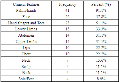
Vitiligo is a common acquired skin disorder which results from the destruction of melanocytes, which results in hypopigmented or/and depigmented macules and patches of the skin, mucosae, or both, and is sometimes associated with Leukotrichia. Vitiligo is divided into segmental and non-segmental forms, and the two clinical forms behave differently. After some time, segmental vitiligo usually stabilizes, whereas non-segmental vitiligo can have an unpredictable course, with inactive and progressive phases. Mixed vitiligo (MV) is other form of vitiligo that is defined as the association of a characteristic segmental involvement followed by the onset of bilateral vitiligo patches. There is a relationship between segmental vitiligo (SV) and nonsegmental vitiligo (NSV); it is suggested that NSV is a continuum of SV, this is explained by combination of these two types to form MV. There are limited data on characteristics of vitiligo in skin of color. The current study will provide data that will aid in a better understanding of the disease in dark skin, ultimately leading to better management and treatment of vitiligo. The purpose of this paper is to identify the characteristics of vitiligo in patients with skin of color. This is a descriptive, cross-sectional hospital-based study, conducted in a phototherapy center, in the period from September 2022 to March 2023, included all patients with vitiligo who agreed to be enrolled in the study. A total of 45 patients with skin of color presented to study area during the period of the study and formed the study population. All patients were subjected to clinical examination and findings were recorded in a specially designed spreadsheet. A total of 45 vitiligo patients were included in this study, most of patients were young aged being 12 years and less. More than half of the study population (53.3%) had a disease onset less than 5 years duration, 37.8% of the patients had a disease onset 5-10 years duration, and 8.9% had a disease onset greater than 10 years. Regarding gender, 60% of vitiligo patients were females and 40% were males. Concerning types of vitiligo, most of the patients (71.1%) presented with non-segmental vitiligo, 20% patients with segmental vitiligo, and 8.9% patients presented with mixed vitiligo. Palm/hand were the affected sites in 91.1% of patients, face in 57.8%, hand fingers and toes in 51.1%, lower limbs in 33.3%, abdomen in 31.1%, upper limbs affected in 31.1%, lips in 22.2%, neck in 15.6%, scalp in 11.1%, back in 11.1%, and sole/feet in 8.9%. Oral mucosa was involved in 11.1% and genital mucosa involved in 8.9% of patients. In conclusion, this study highlights the clinical characteristics of vitiligo, with a predominance of non-segmental vitiligo and frequent involvement of sun-exposed and friction-prone areas. The significant association between lesion distribution and vitiligo type underscores the importance of clinical evaluation in diagnosing and classifying the disease.
Keywords: Vitiligo types, Segmental vitiligo, Non-segmental vitiligo, Vitiligo, Skin of color, Dark skin
Cite this paper: Mahdi M. A. Shamad, Mohammed T. M. Siddig, Characteristics and Types of Vitiligo in Patients with Skin of Color, American Journal of Medicine and Medical Sciences, Vol. 15 No. 2, 2025, pp. 307-310. doi: 10.5923/j.ajmms.20251502.08.
1. Introduction
- Vitiligo is an acquired pigmentary condition characterized by destruction of melanocytes that causes depigmented areas of the skin, and/or mucosae, and occasionally with Leukotrichia [1]. Affected individuals' performance and quality of life are significantly impacted by the condition, especially those with darker skin [2]. There are two clinical types of vitiligo: segmental and non-segmental, and they exhibit distinct behaviours [3]. While non-segmental vitiligo might have an unpredictable course with inactive and progressing periods, segmental vitiligo often stabilizes after some time [4]. Another type of vitiligo is mixed vitiligo (MV), which is characterized by initial segmental involvement appearance then followed by bilateral distinctive vitiligo patches. Segmental vitiligo (SV) and nonsegmental vitiligo (NSV) are related, indicating a continuum between the two subtypes that previously considered as opposed categories. Additionally, the shift from SV to MV is a stronger expected if the affected segment is initially on the trunk [5]. The SV/NSV continuum in MV may be explained by a combination of immunological dysregulation and somatic mosaicism [6]. It has been reported that presence of leukotrichia and halo nevi at the onset of SV is a risk factors for the development of mixed vitiligo [7]. Segmental vitiligo and nonsegmental vitiligo may associate to form mixed vitiligo (MV). MV have been neglected until now and it is not yet part of the conventional classification and needs more in-depth researches [8].Information regarding the features and characteristics of vitiligo in people with of color is scarce. Data from the current study will help us better understand vitiligo in people with dark skin, which will ultimately improve how the condition is managed and treated. The purpose of this paper is to identify the characteristics of vitiligo in patients with skin of color.
2. Methodology
- This is a descriptive, cross-sectional hospital-based study, conducted in Phototherapy Center at Khartoum Dermatology and Venereology Teaching Hospital - Sudan, in the period from September 2022 to March 2023, included all patients with vitiligo who agreed to be enrolled in the study. A total of 45 patients of Fitzpatrick skin types IV to VI with different types of vitiligo presented to study area during the period of the study and formed the study population. After obtaining ethical clearance from the relevant bodies, privacy of data collection was considered, and written informed consent was taken from all participants. All subjects were clinically examined and results were recorded in a specially designed questionnaire. The questionnaire consisted of demographical and clinical data filled out directly form patients who attended the Phototherapy center. The collected data were analyzed by Statistical Packages for Social Sciences (SPSS) 23.0 software with Chi-square test, as appropriate P-value < 0.05 was considered statistically significant (Confidence Interval: CI 95%).
3. Results
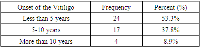
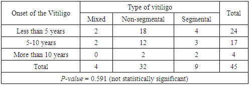
- A total of 45 vitiligo patients were included in this study, 12 (26.7%) patients aged less than 18 years, 11 (24.4%) in the age group of 18-35 years, 10 (22.2%) in the age group 36-50 years age group, 9 (20%) in age group 51-65 years, and 3 (6.7%) were aged more than 65 years. More than half of the study population, (24, 53.3%) had a disease onset less than 5 years duration, 17 (37.8%) of the patients had a disease onset 5 to10 years duration, and 4 (8.9%) of the patients had a disease onset greater than 10 years [Table 1].
|
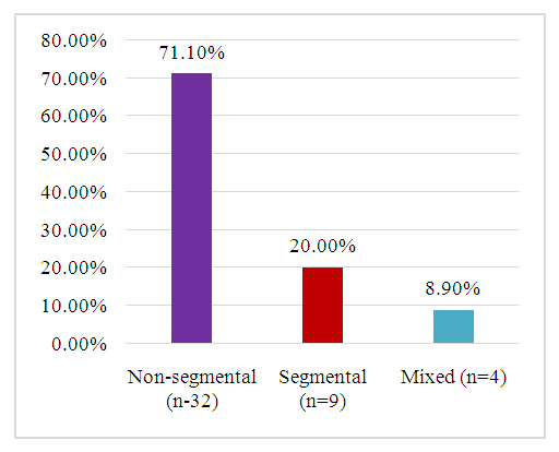 | Figure 1. Types of vitiligo among the study population |
|
|
|
|
4. Discussion
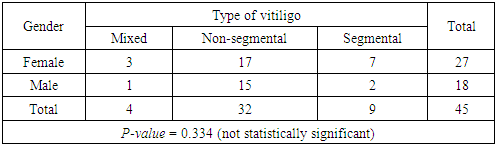
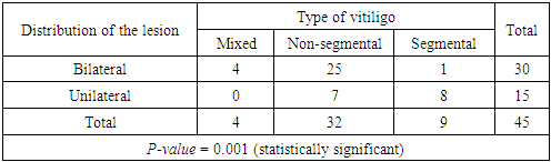
- This study provides highlights of vitiligo types, the distribution, affected sites, and associated symptoms. The findings contribute to a better understanding of the clinical presentation of vitiligo and its variations in patients with skin of color.This study delivers an in-depth description of the clinical and demographic characteristics of vitiligo in patients with skin of color, emphasising the distribution of disease subtypes, involved sits, and associated symptoms. The results will advance our knowledge of how vitiligo manifests clinically and how it varies, especially in individuals with skin of color. Forty-five vitiligo patients were included in the study; sixty percent of them were female. The female predominance is consistent with previous studies that found vitiligo to be more common in females, possibly as a result of hormonal factors or female's higher tendency to seek medical care [9]. Although the patients' ages ranged widely, the majority (51.1%) were under 35, which is in line with the fact that vitiligo usually appears in early adulthood [10]. However, the presence of patients aged over 65 years, seen in 6.7% of patients, highlights the fact that vitiligo can affect people of any age. The most common type was non-segmental vitiligo (71.1%), followed by segmental vitiligo (20%) and mixed vitiligo (8.9%). Global epidemiological data show that non-segmental vitiligo is the most common type of the disease, which is in line with the current study [11]. The different clinical characteristics of segmental and non-segmental vitiligo are further supported by the substantial correlation (p-value = 0.001) between the kind of vitiligo and lesion distribution. Segmental vitiligo was identified by the typical unilateral, localised patches, whereas non-segmental vitiligo manifested as bilateral, symmetrical lesions. Presence of associated symptoms such as joint pain and swelling (13.3%) and night sweats (8.9%) suggests the possibility of comorbidities or systemic involvement.Concerning the involved areas, palms/hands (91.1%) were the most affected by vitiliginous lesions, followed by face (57.8%) and fingers/toes (51.1%). These results are in line with the established tendency of vitiligo to develop in areas that are sun-exposed and friction-prone [12]. A noteworthy observation is the minimal percentage of individuals with genital (8.9%) and oral (11.1%) mucous membrane involvement. Although less frequent, mucosal involvement has been documented in other researches, i.e. Kovacevic et al., for who pointed out that vitiligo can manifest in a variety of ways [13]. The involvement of mucous membranes will give note of the systemic character of the disease and possible impact on the patient’s health.Regarding vitiligo distribution, in the current study, two-thirds of the patients (66.7%) had a bilateral distribution of vitiligo, whereas the remaining patients (33.3%) had a unilateral distribution. The results of this study support the classification of vitiligo into types according to its distribution and clinical appearance. The use of lesion distribution and vitiligo type as diagnostic criteria is supported by the significant association between the two. Furthermore, the absence of a significant correlation between the type of vitiligo and either gender or the onset of the disease implies that these factors might not be essential in identifying the clinical subtype of vitiligo.These results highlight the value of an extensive examination of lesion distribution and affected sites in the diagnosis and classification of vitiligo. The high presence of non-segmental vitiligo and its bilateral distribution suggest that systemic therapies targeting immune dysregulation may be more effective for this subtype. In contrast, segmental vitiligo, with its unilateral distribution, may respond better to localized treatments such as topical corticosteroids.
5. Conclusions
- In conclusion, this study highlights the clinical characteristics of vitiligo, which shows that non-segmental vitiligo is more common and that sun-exposed and friction-prone areas are frequently affected. Lesion distribution and vitiligo type are significantly correlated, which emphasizes the value of clinical evaluation in vitiligo diagnosis and classification. These findings contribute to a better understanding of vitiligo in skin of color and provide a foundation for future research and improved patient care.