American Journal of Medicine and Medical Sciences

p-ISSN: 2165-901X    e-ISSN: 2165-9036

2025;  15(1): 264-268

doi:10.5923/j.ajmms.20251501.52

Received: Jan. 2, 2025; Accepted: Jan. 25, 2025; Published: Feb. 3, 2025


Postnatal Development of the Thyroid Gland in Offspring under the Influence of Pyrethroid Pesticides During Pregnancy

Sh. A. Islomova

Assistant, Tashkent Medical Academy, Tashkent, Uzbekistan

Correspondence to: Sh. A. Islomova, Assistant, Tashkent Medical Academy, Tashkent, Uzbekistan.

Copyright © 2025 The Author(s). Published by Scientific & Academic Publishing.

This work is licensed under the Creative Commons Attribution International License (CC BY).
http://creativecommons.org/licenses/by/4.0/

Abstract

This article describes the negative complications observed in the endocrine system of offspring born under chronic exposure to pesticides in the mother's body. It is well known that, to date, protecting the environmental health of the population has become one of the pressing global issues. The widespread use of various chemical products in industry, households, and agriculture leads to environmental pollution. A significant portion of the toxic substances in the environment consists of pesticides, yet it is difficult to imagine the development and future prospects of the agricultural industry without them. However, in some cases, small doses of pesticides cause unpredictable effects that do not occur with higher doses. One such effect is the disruption of the endocrine system's function, leading to various negative complications.

Keywords: Endocrine system, Organophosphates, Organochlorines, Pyrethroids

Cite this paper: Sh. A. Islomova, Postnatal Development of the Thyroid Gland in Offspring under the Influence of Pyrethroid Pesticides During Pregnancy, American Journal of Medicine and Medical Sciences, Vol. 15 No. 1, 2025, pp. 264-268. doi: 10.5923/j.ajmms.20251501.52.

1. Introduction

In recent years, the use of highly toxic organophosphates and organochlorine pesticides has been banned or restricted in many countries [1,3,12,16]. These have gradually been replaced by new generations of pesticides, primarily pyrethroids or other classes of substances [2,6,4,8,9]. They are less toxic to humans and animals and generally exhibit high efficacy even in small doses. In Uzbekistan, more than 250 new generations of pesticides are currently used, most of which belong to the pyrethroid and pyrazole groups [4,5,8,9]. Despite the relatively low toxicity of these substances, their widespread use in Uzbekistan's agriculture poses a risk of contamination of water, air, and food, even in small amounts [7,10,11,12,16,17].
The aim of the study was to identify the structural and functional features of the early postnatal development of the thyroid system in offspring born under chronic pesticide exposure to the mother’s body [2,4,7,9].

2. Research Objectives

To study the morphological, morphometric, and ultrastructural features of the thyroid gland in offspring born under chronic pesticide exposure to the mother’s body, in dynamics;
To assess the proliferative activity of cells and the level of apoptosis in the thyroid gland of offspring born under chronic pesticide exposure to the mother’s body, using immunohistochemical methods;
To determine the concentration of marker hormones from the pituitary (TSH) and thyroid (T4, T3) in the mother and offspring over time under chronic pesticide exposure to the mother’s body.
The research subjects were adult, non-pregnant female Wistar rats weighing 150-180 g, as well as their offspring. The experimental groups of female rats received pesticides daily until the end of the experiments: fastokin at a dose of 8 mg/kg, or fipronil (Vigor) at a dose of 3.6 mg/kg. Material for the studies on offspring from all groups was collected on days 3, 7, 14, 21, and 30 post-birth. The pituitary, thyroid, adrenal glands, and serum from both the mothers and the offspring were studied over time.
The research subject was a comprehensive analysis of the early postnatal development of the thyroid system in offspring under the influence of small doses of pesticides through the maternal body; structural-functional mechanisms of the toxic effects of intrauterine and early postnatal pesticide exposure.

3. Research Methods

To address the tasks, the study used light microscopy, morphometry, transmission electron microscopy, immunohistochemistry, enzyme-linked immunosorbent assays (ELISA), and methods of variation statistics.
Morphometric Studies
Morphometric studies were conducted on paraffin and semi-thin sections using the Autandilova grid (Autandilov G.G., 1990). In some cases, the "Morphology-4" computer program by "Carl Zeiss Jena" (Germany) was used. In the thyroid gland, the outer and inner diameters of the follicles, areas of colloid, epithelium, height of thyrocytes, the number of thyrocytes in one follicle, areas of interfollicular epithelium, and connective tissue stroma were determined.
Assessment of Apoptosis and Proliferation Levels:
To evaluate the level of apoptosis and cell proliferation in the endocrine glands, immunohistochemical methods were used. Proliferating cell markers were determined using monoclonal rabbit antibodies to Ki-67, and apoptotic cells were detected with monoclonal rabbit antibodies to cas-3 and p-53 (Thermo Scientific, USA). Proliferating and apoptotic cells were identified on paraffin sections of the thyroid and adrenal glands using the UltraVision kit (Thermo Scientific, USA). Sections were counterstained with methylene blue or neutral red. The proliferation and apoptosis indices were calculated for 1000 thyroid cells (thyrocytes), expressed in per mille.
Hormonal Marker Determination:
Marker hormones from the pituitary and thyroid were determined in the serum of the animals. After euthanasia, blood was collected into dry sterile tubes, and the obtained serum was used to measure hormone concentrations. Thyroxine (T4), triiodothyronine (T3), and thyroid-stimulating hormone (TSH) in the serum were determined using enzyme-linked immunosorbent assays (ELISA) with special kits from "Human" (Germany) and a "Singl" spectrophotometer (Germany). Thyroxine (T4) and triiodothyronine (T3) were expressed in ng/ml, and thyroid-stimulating hormone (TSH) in mIU/L.
All numerical data were processed using the method of variation statistics. The calculations and statistical analysis were carried out with the use of a statistical package for Windows. All data were presented as mean ± standard deviation (SD). The statistical significance of differences between the control and experimental groups was compared using the Student's t-test, with values of P<0.05 considered statistically significant.
The "Effect of Chronic Pesticide Exposure on the Growth and Development of the Thyroid System in Offspring" describes the results of morphological, ultrastructural, morphometric, immunohistochemical, and functional studies of the pituitary-thyroid system of offspring under intrauterine and early postnatal pesticide exposure. The results showed that the thyroid system in offspring was sufficiently developed by the time of birth.
The thyroid gland in newborns is fairly well-formed and consists of two lobes connected by a thin isthmus. On days 1-3 after birth, the gland is surrounded by a thin connective tissue capsule. Occasionally, the parathyroid gland can be seen on the sections, surrounded by a common capsule for both glands. The parenchyma of the thyroid gland consists of formed follicles of various sizes. On the periphery of the gland, the follicles are relatively large and contain varying amounts of colloid. In the center of the gland, mostly small, developing follicles and interfollicular epithelial islands are observed. This morphological picture of the thyroid gland was characteristic of both control and experimental groups of newborn rats (Figure 1).
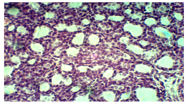
Figure 1. Characteristic of both control and experimental groups of newborn rats
The further dynamics of the growth and formation of the thyroid gland were uniform across all groups of the studied animals. This manifested as an increase in the size of the gland overall, a rise in the number of medium and large follicles, and a decrease in the number and size of interfollicular epithelial islands. By days 21 and 30 after birth, the thyroid gland was almost fully formed. The parenchyma of the gland mainly contained follicular complexes of small, medium, and large follicles. At the same time, large and medium follicles were primarily located on the periphery of the thyroid gland, while in the center of the organ, mainly small follicles and interfollicular islands were found.
Thus, the dynamics of growth and development of the thyroid gland after birth is of a similar character in both the control and experimental groups of offspring. However, morphometric studies revealed data indicating certain disruptions in the dynamics of thyroid growth and development in the offspring under pesticide exposure. The rates of increase in the total area of follicular epithelium under fastokin exposure were 10-17%, and under fipronil (FP) exposure, they were 15-30% lower than the control group (P1, P2<0.05). Correspondingly, the area occupied by colloid decreased by 15-25% and 20-40%, respectively. In contrast, the total area of interfollicular epithelial islands increased by 15-45% under fastokin exposure and 20-60% under FP exposure, compared to the control (P1, P2<0.05). It is known that interfollicular epithelial islands represent the cambial zone of the organ, where the formation of new follicles occurs. The increase in the area of this zone in the experimental rats is likely the result of a slowing down of the formation of new follicles. The delay in the growth and development of the structural-functional unit of the thyroid gland, the follicle, was most pronounced under fipronil exposure compared to fastokin exposure. However, the negative effects of pesticides were not limited to the inhibition of follicle formation alone. It was found that pesticide exposure led to a decrease in the overall follicle area, primarily due to reductions in the areas of the follicular epithelium and thyrocytes. The rates of increase in the height and average area of thyrocytes under fastokin exposure were 10-20%, and under FP exposure, 15-30% lower than in the control group (P1, P2<0.05).
Thus, morphometric studies clearly demonstrated that pesticide exposure via the maternal organism significantly reduces the rates of thyroid growth and development in postnatal ontogenesis. Notably, the low specific values of thyrocytes at all research time points indicate a decrease in the secretory activity of these cells. The morphometric data were fully corroborated by the results of electron microscopy studies.
Electron Microscopy:
Electron microscopy revealed that the follicles of the thyroid glands in control animals at all time points had a typical structure. Thyrocytes in the control animals were in various stages of the secretory cycle and generally exhibited ultrastructural signs of high activity of the synthetic and lysosomal machinery. The cytoplasm of individual thyrocytes contained a significant amount of cisterns of the granular endoplasmic reticulum (GER) (Figure 2). In some cells, the profiles of GER were expanded. The nuclei of the cells were located centrally or slightly shifted toward the basal part of the thyrocyte. The apical surface of the cells contained a few short microvilli. Lysosomes, 0.2 - 0.4 μm in size, were located under the apical plasma membrane, with their number varying depending on the phase of the secretory cycle of the thyrocyte.
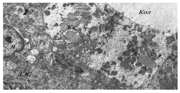
Figure 2. Significant amount of cisterns of the granular endoplasmic reticulum(GER)
Electron microscopy of the thyroid gland in the offspring of experimental animals showed certain submicroscopic changes indicative of disturbances in various phases of the thyrocyte secretory cycle. The walls of the follicles often contained cells with pronounced signs of disorganization in the GER profiles, such as excessive expansion and a decrease in the density of their internal contents. The cytoplasm of individual thyrocytes resembled a giant vacuole with light content, and the nucleus and few organelles were displaced to the periphery of the cell. Thyrocytes with swollen mitochondria, whose matrix appeared lightened and whose cristae were disorganized, were frequently observed. In some thyrocytes, a few lipid droplets of various sizes were noted, which were virtually absent in the control group. Finally, compared to the control, the offspring of the experimental animals showed a significantly higher frequency of thyrocytes exhibiting clear signs of apoptosis and destruction of cytoplasmic organelles.
In normal physiological conditions, thyrocytes are characterized by the presence of well-developed organelles responsible for the synthesis of thyroglobulin. This primarily refers to the structures of the granular endoplasmic reticulum (GER), which are generally well-developed in the thyrocytes of control animals. However, disorganization and total expansion of GER cisterns with a decrease in the density of their contents were observed only in the offspring of experimental animals. Similar ultrastructural changes in thyrocytes have been described by several researchers under toxic exposures and are considered signs of disrupted thyroglobulin synthesis and/or its transport into the follicular lumen (Yukina G.Yu. et al., 2004). It is evident that the submicroscopic changes we observed are also indicators of impaired synthetic and secretory activity in thyrocytes. Our morphometric data show that pesticide exposure via the maternal organism leads to a slowing down of the postnatal formation and growth of the main structural-functional unit of the thyroid gland the secretory follicles. Based on these data, it can be concluded that pesticide exposure causes significant disruption in the differentiation process of thyrocytes in the offspring's thyroid gland. Submicroscopic signs of organelle destruction responsible for the synthesis, transport, and cleavage of thyroglobulin support this conclusion. All of these factors ultimately lead to thyroid dysfunction and disturbances in metabolic processes in the developing offspring.
This raises the question what is the mechanism behind these disruptions, and what is the state of cell proliferation and apoptosis processes? To answer this question, we used immunohistochemistry with highly specific monoclonal antibodies to markers of proliferation and apoptosis. Quantitative analysis of the results showed that pesticide exposure leads to a decrease in the proliferation index compared to the control group (Table 1).
Table 1. Proliferation Index of Thyroid Cells (in per thousand) during Postnatal Development
     
The decrease in the proliferative activity of thyroid cells in both experimental groups is most pronounced on the 7th-21st postnatal day. During this observation period, the proliferation index is reduced by 40-45% when exposed to fipronil compared to the control, while exposure to fastokin reduces the proliferation index by 28-30% compared to the control (P1, P2<0.05). On the 30th day after birth, against the backdrop of a general decrease in cell proliferation activity, the reduction in the proliferation index with fastokin exposure is 13%, while with fipronil exposure, it is 18%, compared to the control.
Overall, this indicates a significant suppression of thyroid cell proliferation under in utero and early postnatal pesticide exposure. A more pronounced and statistically significant decrease in proliferation was observed with fipronil exposure compared to fastokin.
Somewhat different data were obtained when studying the apoptosis process (Table 2). As seen from the table, the apoptosis index in the experimental animals significantly exceeds the control values at all observation points. The most significant increase in the apoptosis index was observed on days 14-21 after birth, when the values in the experimental animals were 3.2-4.3 times higher than the control values. Furthermore, the induction of apoptosis in cells was more pronounced under the influence of fipronil compared to cyhalothrin exposure.
Table 2. Apoptosis Index of Thyroid Cells (in per thousand) during Postnatal Development
     
Thus, intrauterine and early postnatal exposure to even low doses of modern pesticides has an adverse effect on the thyroid system. This effect manifests as a slowdown in the growth and development of the thyroid gland, the formation and functioning of its secretory follicles. The basis of the slowed growth of the organ is an imbalance between the processes of proliferation and apoptosis, which is more pronounced under the influence of fipronil compared to fastokin. We believe that these changes form the morphological substrate for thyroid dysfunction, which arises in the postnatal ontogenesis of the organ under the influence of pesticides.

4. Conclusions

As a result of the conducted dissertation research on the topic: "Postnatal development of the thyroid gland under intrauterine exposure to low doses of pyrethroid pesticides," the following conclusions were made:
1. Chronic exposure to low doses of pesticides on the maternal organism during pregnancy and lactation leads to an "endocrine-disrupting" effect on the offspring in the early postnatal period.
2. Morphological, morphometric, and ultrastructural studies revealed structural mechanisms for the disturbances in the thyroid system of the offspring under pesticide exposure:
A slowdown in the growth and formation of thyroid follicles was identified.
Submicroscopic changes in the organelles of thyrocytes, indicating disruption in their secretory function, were observed.
3. Immunohistochemical research revealed an imbalance between the processes of proliferation and apoptosis of thyroid cells in the offspring under pesticide exposure:
A significant increase in the apoptosis index was observed, alongside a decrease in cell proliferation.
The enhancement of apoptosis in cells, along with reduced proliferation activity, is one of the mechanisms contributing to the slowdown in thyroid growth and formation.
Induction of apoptosis in thyroid cells is primarily due to hypothyroidism caused by pesticide exposure.
4. Chronic exposure to low doses of pesticides on the maternal organism during pregnancy and lactation leads to functional disorders in the thyroid system of both the mother and offspring:
The most pronounced hypothyroidism in experimental females was found on day 21 of pregnancy and on day 14 of lactation, with T4 and T3 concentrations decreasing by 1.3-1.5 times compared to the control group.
Pronounced neonatal hypothyroidism with a significant decrease in T4 and T3 concentrations was observed in the offspring at all time points, reaching its maximum on day 21 after birth.
An important factor in the development of thyroid dysfunction in the offspring is the imbalance between the processes of proliferation and apoptosis of thyrocytes, contributing to the slowdown in thyroid follicle formation, as well as submicroscopic changes in thyrocytes, indicating disruption in their secretory function.
5. A comparative analysis of the pesticides showed significantly stronger "endocrine-disrupting" toxicity of the pyrazole-based pesticide fipronil compared to the pyrethroid pesticide fastokin.

References

[1]  Sh.A. Islomova, N.S. Tukhtaev, "Growth and formation of the thyroid gland in offspring under maternal hypothyroidism conditions," XXI International Medical-Biological Conference of Young Researchers "Fundamental Science and Clinical Medicine – Man and His Health," April 14, 2018, St. Petersburg State University.
[2]  N.B. Zokirova, Sh.A. Islomova, "Impact of prolonged exposure of pyrethroid pesticides on the thyroid function of pregnant rats and their offspring," "Medicine of the Silk Road of the 21st Century: Modern Development Vector," Tashkent – 2020, April 20.
[3]  N.B. Zokirova, Sh.A. Islomova, N.B. Utepaova, N.K. Tukhtaev, "Use of modern innovative methods for studying postnatal morphogenesis of the endocrine and immune systems under pesticide exposure," International Scientific-Practical Conference, Samarkand, May 6, 2021.
[4]  N.B. Utepaova, S.N. Tukhtaev, Sh.A. Islomova, "Postnatal development of the thyroid gland and immune organs in offspring under intrauterine pesticide exposure," XXIV International Medical-Biological Conference of Young Researchers "Fundamental Science and Clinical Medicine – Man and His Health," April 24, 2021, St. Petersburg State University.
[5]  Sh.A. Islomova, N.B. Utepaova, S.N. Tukhtaev, "Postnatal development of the endocrine and immune organs of offspring under intrauterine pesticide exposure," "New Problems in Medical Science and Perspectives for Their Solutions," dedicated to the 30th Anniversary of the State Independence of the Republic of Tajikistan and the Years of Rural Development, Tourism, and Folk Crafts (2019-2021), with international participation, Dushanbe, April 30, 2021.
[6]  Sh.A. Islomova, N.B. Zokirova, "Postnatal development of the thyroid gland in offspring under intrauterine exposure to pesticides," "Innovative Approaches to the Urgent Problems of Medicine," 2022, Republic and International Young Scientists Scientific-Practical Conference, Andijan, May 12, 2022.
[7]  Sh.A. Islomova, N.B. Zokirova, M.B. Saifutdinova, "Toxic effects of pesticides on the endocrine system of offspring exposed through the maternal body during pregnancy and lactation," "New Day in Medicine" Scientific-Referative, Spiritual-Educational Journal.
[8]  Zokirova N.B., Islomova Sh., "Growth and formation of the thyroid gland in offspring obtained under conditions of maternal hypothyroidism," "Modern Medicine Through the Eyes of Young Scientists," 50th Republican Scientific-Practical Online Video Conference of the Student Scientific Society, dedicated to "The Year of Ensuring Human Interests and Strengthening Mahalla."
[9]  Islomova Sh.A., "Destructive effects of pesticides on the endocrine system of offspring under the conditions of their exposure through the mother's body during pregnancy and lactation," Iqro Junali, 2023.
[10]  Islomova Sh.A., Zokirova N.B., "Growth and formation of the thyroid gland in offspring obtained under conditions of maternal hypothyroidism," Bulletin of Fundamental and Clinical Medicine, 2022.
[11]  Khalikov P.Kh., Daminov A.O., Islomova Sh.A., Khusainova H.Zh., "Influence of Pesticides on Cytogenetic Effects," International Scientific Conference on "New Scientific Trends and Challenges," February 20, Rome, Italy.
[12]  Islomova Sh.A., Khamzaeva N.T., Olimjonova G.O., Khusainova Kh.J., "Growth and formation of the thyroid gland in offspring obtained under conditions of maternal hypothyroidism," E-Global Congress Hosted Online from Dubai, U.A.E., E-Conference, April 30, 2023.
[13]  Islomova Sh.A., Olimjonova G.O., "Toxic effects of pesticides on the endocrine system of offspring exposed through the maternal body during pregnancy and lactation," TMA Bulletin, No. 4, 2024.
[14]  Islomova Sh.A., "Toxic effects of pesticides on the endocrine system of offspring exposed through the maternal organism during pregnancy and lactation," Fundamental and Clinical Medicine Information, 2022.
[15]  Islomova Sh.A., Olimjonova G.O., "Postnatal development of the thyroid gland in offspring under pyrethroid pesticide exposure," Iqro Junali, 2023.
[16]  Islomova Sh.A., Khusainova H.J.B., Olimjonova G.O., "The toxic effect of pesticides through the maternal organism on postnatal generation in the thyroid glands," International Journal of Medical Sciences and Clinical Research.
[17]  Islomova Sh.A., Khamzaeva N.T., Khusainova Kh.Zh., Olimjonova G.O., "Postnatal development of the thyroid gland under fetal exposure to low doses of pyrethroid pesticides," Monograph.