American Journal of Medicine and Medical Sciences

p-ISSN: 2165-901X    e-ISSN: 2165-9036

2025;  15(1): 249-254

doi:10.5923/j.ajmms.20251501.49

Received: Dec. 17, 2024; Accepted: Jan. 12, 2025; Published: Feb. 3, 2025


In Vitro Study of the Effectiveness of Preparations Used in Superficial and Deep Fluoridation of Permanent Teeth Using Scanning Electron Microscopy

Daminova Shakhnoza Badriddinovna1, Tashpulatova Xurshidabonu Akhmadjon Qizi2, Iskandarov Nurmukhammad Ergashovich3

1DSc, Professor, Head of the Department of Prevention of Dental Diseases, Tashkent State Dental Institute, Tashkent, Uzbekistan

2PhD Student, Department of Prevention of Dental Diseases, Tashkent State Dental Institute, Tashkent, Uzbekistan

3Junior Researcher, Laboratory of Physical Chemical Research Methods, Center for Advanced Technologies under Ministry of Higher Education, Science and Innovation of the Republic of Uzbekistan, Tashkent, Uzbekistan

Correspondence to: Tashpulatova Xurshidabonu Akhmadjon Qizi, PhD Student, Department of Prevention of Dental Diseases, Tashkent State Dental Institute, Tashkent, Uzbekistan.

Email:

Copyright © 2025 The Author(s). Published by Scientific & Academic Publishing.

This work is licensed under the Creative Commons Attribution International License (CC BY).
http://creativecommons.org/licenses/by/4.0/

Abstract

Using scanning electron microscopy, morphological changes in the surface layer of 20 enamel samples of permanent teeth that have just erupted were studied in vitro under the influence of fluoride varnish and deep fluoride. It was found that the enamel of teeth that have just erupted has visual signs of insufficient mineralization and, accordingly, has an insufficient level of caries resistance; during the experiment, under the influence of the studied exogenous prophylactic agents, morphological changes are observed on the enamel surface, indicating an increase in the degree of mineralization.

Keywords: Enamel, Permanent teeth, Mineralization, Fluoride varnish, Fluoride, Deep fluoridation, Caries prevention

Cite this paper: Daminova Shakhnoza Badriddinovna, Tashpulatova Xurshidabonu Akhmadjon Qizi, Iskandarov Nurmukhammad Ergashovich, In Vitro Study of the Effectiveness of Preparations Used in Superficial and Deep Fluoridation of Permanent Teeth Using Scanning Electron Microscopy, American Journal of Medicine and Medical Sciences, Vol. 15 No. 1, 2025, pp. 249-254. doi: 10.5923/j.ajmms.20251501.49.

1. Introduction

The relevance of the problem of dental caries, especially in children, remains high in many countries, including Uzbekistan, and requires not only medical intervention, but also a comprehensive approach to prevention and treatment. Caries is one of the most common diseases among children, which determines its importance for both health care and public well-being. The most significant local risk factors for caries development are low initial mineralization levels, lack of preventive measures, poor oral hygiene, presence of orthodontic pathology and excessive carbohydrate consumption [2,3].
The lowest level of mineralization of hard tissues of permanent teeth is observed in the first years after their eruption [4]. This is confirmed by a significant increase in the prevalence and intensity of caries in permanent teeth in the period from 6 to 15 years [5]. Caries resistance of enamel of permanent teeth remains insufficient even 18 months after eruption [6].
Caries in the "white spot" stage is a reversible form of the disease, in which it is possible to restore the enamel structure. In the case of progression of the pathological process, irreversible changes occur in the enamel structure, resulting in the formation of a cavity defect [8,11,25,30]. Along with this, there is a loss of the aesthetic and functional value of the tooth. The size of the carious spot and its color indicate the degree of demineralization of hard dental tissues. With an increase in its size and color intensity (from white to black), the loss of calcium and phosphorus ions increases [9,10,11,13].
Achieving optimal enamel mineralization rates is possible through the additional use of exogenous caries prevention agents [4,9]. For this purpose, agents containing calcium, phosphorus, fluorine, magnesium, etc. are used [2,5,6,7]. The problem of studying the effectiveness and selection of remineralizing agents with different mechanisms of action is extremely relevant for both researchers and practicing physicians [1,5,8,9,15]. Therefore, substantiating the choice of the most effective exogenous prevention agents, especially during the period of secondary mineralization, will contribute to increasing the caries resistance of tooth enamel.
The aim: of the study is to investigate in vitro morphological changes in the surface layer of enamel of permanent teeth that have just erupted, under the influence of exogenous dental caries prevention agents that contain calcium and fluoride compounds.

2. Materials and Methods of the Study

The study was carried out on 20 enamel samples of permanent teeth that erupted at the same time. They were removed for orthodontic reasons (premolars of 10 12-year-old children, no later than 6 months after eruption). Immediately after removal, the roots of the teeth were cut at the level of the enamel-cement junction and the remains of soft tissues were removed. The coronary segments were cleaned using ultrasound and polishing paste and a brush.
5 enamel samples were examined at the beginning of the experiment. The other 15 samples were randomly divided equally into three groups. They were placed in three separate sealed boxes (5 samples per box), which were filled with artificial saliva (T. Fusayama, 1975). Subsequently, the enamel samples of the first group were treated with the fluorine varnish “Omega Dent”, Russia with “high fluoride content", the second - with the “Denta-Fluo”, Dentals Pharma GmbH, Uzbekistan. Denta-Fluo dental material is available in a set: liquid №1/liquid №2. Denta-Fluo dental material contains: active fluoridating components: sodium fluoride, calcium fluoride.
Liquid № 1-Water-based liquid contains fluorine, copper, and magnesium ions.
Liquid № 2 - is a suspension of highly dispersed calcium hydroxide in distilled water.
The samples of the third group served as a control, they were not treated with anything.
The treatment of the samples of the first group was carried out at the beginning of the study, after 3, 6 and 9 months in courses of 30 days, twice a day for 3 minutes according to the manufacturer's recommendation; the second group - at the beginning of the study, after 3, 6 and 9 months in courses of 10 days, twice a day for 5 minutes according to the manufacturer's recommendation.
Enamel samples for further studies were cut from the vestibular and oral surfaces of the crown part of the teeth at the equator level using a 0.2 mm thick diamond disk under running water, cleaned using ultrasound, degreased and vacuumed. The enamel surface was examined at the beginning of the experiment, after 6 and 12 months.
The surface structure of the samples was studied at the equator level in a scanning electron microscope (ZEISS MA15, Germany) with magnification from 1000 to 5000. During the study, the surfaces of the samples were not sprayed for maximum reliability of the result. The studies were carried out in the “Laboratory of Physical-Chemical Research Methods” Center for Advanced Technologies under Ministry of Higher Education, Science and Innovation of the Republic of Uzbekistan. Special thanks for the junior researcher of the department Iskandarov N.E.

3. Results of the Study and Their Discussion

During the study, morphological changes were established that occur on the surface of immature enamel of permanent teeth under the influence of the studied means of exogenous prophylaxis (Fig. 1, 2).
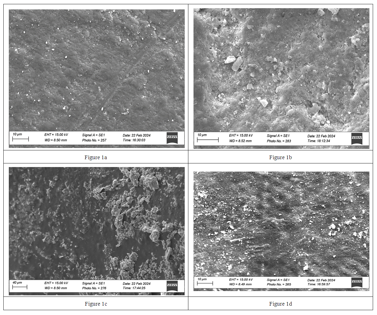
Figure 1. Surface of permanent teeth immature enamel: а – at the start of experiment, b – 12 months later in control group, c – treated with Fluorine varnish of fluoride ions 6 months later, d - treated with deep fluoridation – Denta-Fluo deep fluoride 6 months later (SEM, ×1000)
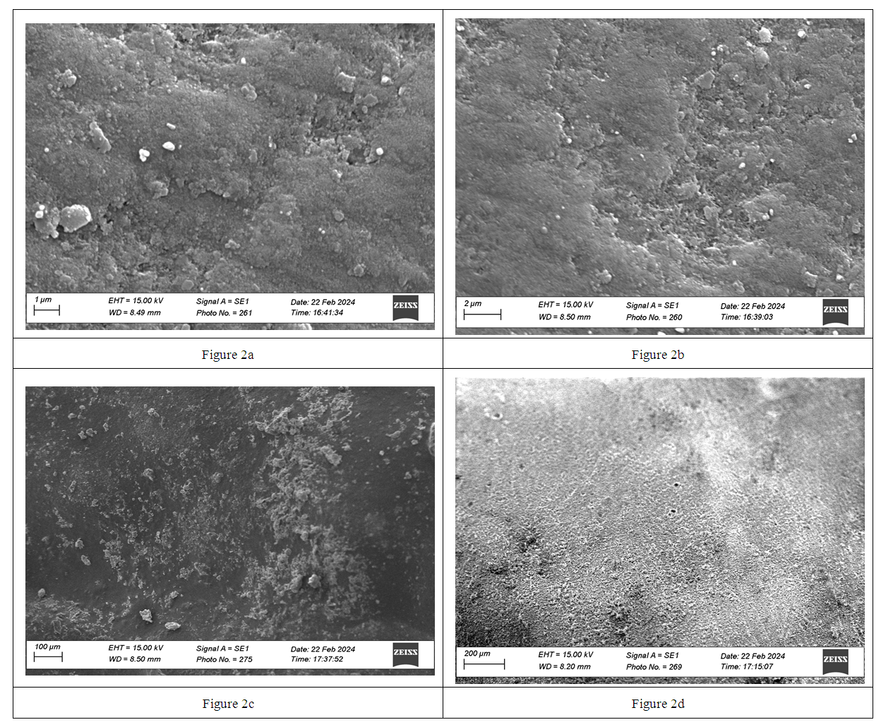
Figure 2. Surface of permanent teeth immature enamel: а – at the start of experiment, b – 12 months later in control group, c – treated with Omega dent of fluoride ions 12 months later, d - treated with “Denta- fluo” deep fluoride 12 months later (SEM, ×2000, 4000, 5000)
At the beginning of the experiment, perikymata (Retzius lines) are clearly visible on the enamel surface of the control group samples at 1000x magnification, at the border of which the exit of enamel arcade-shaped prisms is noted, the enamel surface is dull and rough (Fig. 1a). This indicates a low level of mineralization of the surface layer of enamel, the absence of a protective layer on the surface, high permeability of immature enamel, the presence of conditions for additional retention of microorganisms. The above factors predetermine low caries resistance of immature enamel and a high risk of enamel vulnerability during the period of secondary mineralization.
After 12 months of the experiment, perikymata are smoothed out on the surface of the control group samples (Fig.1b), the enamel remains matte and rough, a significant number of depressions are observed, which indicate the place of exit of enamel prisms. The observed changes may indicate that mineral substances are deposited on the surface of immature enamel, the number of retention points decreases and, accordingly, the permeability of the enamel decreases.
Under the influence of fluorine varnish “Omega Dent” after 6 months, at a magnification of 1000 times (Fig. 1c), the perikymata are pronounced, but there is a tendency for them to smooth out. The surface of the perikymata is smoothed and uniform. The places of enamel prisms both within the perikymata and at their border are almost not determined. This may be due to the deposition of fluorine compounds on the surface of the studied samples.
After 6 months of using the deep fluoridation method by using Denta -Fluo, at a magnification of 1000 times (Fig. 1d), the enamel surface looks lighter and shinier, and also less relief due to the smoothing of the perikymata. This confirms the hypothesis that teeth after treatment with mineralizing agents become clinically lighter (whitening effect) [12]. At the site of the enamel prisms coming out to the surface, depressions are observed, which may indicate the deposition of an additional layer of mineral substances on the enamel surface.
The obtained morphological changes in the enamel surface in both experimental groups indicate that under the influence of exogenous prophylaxis agents, which contain calcium and fluorine compounds, faster, but visually different processes occur on the enamel surface, which equally lead to a change in the surface relief, a decrease in the number of retention points and, accordingly, a decrease in the permeability of the enamel.
A more detailed difference in the site of the enamel prisms coming out to the surface during the 12 months of the experiment was established at a magnification of 5000 times (Fig. 2).
At the beginning of the study, the surface of immature enamel is dull, relief due to the emergence of a significant number of enamel prism craters on the surface (Fig. 2a).
After 12 months of the study, the surface of the enamel of permanent teeth in the control group becomes more uniform and smooth, the number of both open and closed craters of enamel prisms on the surface decreases (Fig. 2b).
Grooves and scratches, which are clearly visible on the surface of the enamel samples of the control group after 12 months of the study at different magnifications (Fig. 1b, 2b), may indicate that the layer of mineral substances that is deposited on the enamel surface during the experiment is less mineralized, and, accordingly, insufficient to perform the protective function. Under the influence of Omega Dent “fluoride varnish” after 12 months, the enamel surface becomes shiny, with both open and closed craters of enamel prisms emerging on the surface. Craters on the surface of the samples of the study group in comparison with a similar area in the control group (Fig. 2b, 2c) are visually deep and smaller in area.
After 12 months of treatment of immature enamel samples with deep fluoride “Denta Fluo”, the enamel surface is distinguished by its greatest homogeneity and shine due to a significant reduction in the number of unclosed craters and their area in comparison with the initial level and the results of other study groups in a similar period (Fig. 2). The obtained morphological data confirm the hypothesis about the deposition and long-term preservation of a layer of additional chemical compounds on the enamel surface after the use of the studied exogenous caries prevention agents containing calcium and fluorine compounds, even after mechanical and ultrasonic cleaning of the surface during sample preparation. Such changes on the enamel surface create conditions for its full mineralization and, accordingly, an increase in the level of caries resistance.
Therefore, in the period up to 18 months after the eruption of permanent teeth, it is necessary to additionally use products that promote increased mineralization of hard dental tissues. This period is the most effective and convenient for the treatment and prevention of caries in the initial stages. For clinical use immediately after the eruption of permanent teeth, exogenous prophylaxis of dental caries can be recommended, which are used for deep fluoridation and contain active fluoridating components: sodium fluoride, calcium fluoride. (1-2 times or 3-4 times procedures per year or after the achieved effect after 2 weeks) and products that surface fluoride in the form of fluoride varnish (2-4 courses per year for 2-4 weeks) for a period of at least twelve months.

4. Conclusions

The results of the study confirm that the enamel surface of newly erupted teeth shows signs of insufficient mineralization, which leads to a decrease in the level of caries resistance. Even 12-18 months after the eruption of teeth, without the use of preventive agents, the level of caries resistance of enamel remains insufficient.
At the end of the scientific study, the composition and effectiveness of the domestic drug “Denta-Fluo” for remineralizing therapy in the prevention and treatment of enamel caries at the "white spot" stage were clinically and laboratory substantiated. It was proven that this agent helps to increase resistance, reduce the acid solubility of enamel, and restore the enamel crystal lattice.
Based on the results of electron microscopy, it was established that the minimum penetration depth is possessed by the Omega Dent fluoride varnish - 95 ± 10 μm (p < 0.05), and the pronounced penetrating ability is possessed by “Denta-Fluo” - 163 ± 11 μm (p < 0.05) which exceeds the depth of demineralization in enamel caries 118 ± 10 μm (p < 0.05). When using the deep fluoridation method with the “Denta-Fluo” product in patients with a good and satisfactory level of oral hygiene, the effectiveness of enamel caries treatment is 96.7% and 93.5%, respectively, with an unsatisfactory and poor level of oral hygiene 85.3% and 81.8%, respectively.

References

[1]  Abou Neel, EA. Demineralization–remineralization dynamics in teeth and bone / EA. Abou Neel, A. Aljabo, A. Strange [et al.] // International Journal of Nanomedicine. – 2016. – № 11. – P. 4743–4763.
[2]  Ahrari, F. Enamel resistance to demineralization following Er: YAG laser etching for bonding orthodontic brackets / F. Ahrari, M. Poosti, P. Motahari // Dent Res J (Isfahan). – 2012. – № 9. – P. 472–477.
[3]  Aida M. The use of tiefenfluorid for desensitization of dentinal hyperesthesia / M. Aida, M. Agron, T. Emiljano, L. Monika, B.Christian // Balk J Dent Med. – 2014. – № 18. – P. 85–88.
[4]  Alavi, S. The effect of fluoride varnish and chlorhexidine gel on white spots and gingival and plaque indices in fixed orthodontic patients: A placebo controlled study / S. Alavi, N. Yaraghi // Dent Res J (Isfahan). – 2018. – № 15(4). – P. 276–282.
[5]  Duverger, O. Keratins as components of the enamel organic matrix / O. Duverger, E. Beniash, MI. Morasso // Matrix biology: journal of the International Society for Matrix Biology. – 2016. – № 52(54). – P. 260–265.
[6]  Fontana, M. Minimal intervention dentistry: part 2. Caries risk assessment in adults // M. Fontana, C. Gonzalez-Cabezas // BRITISH DENTAL JOURNAL. – 2012. – № 213. – P. 447–451.
[7]  Guo, J. Amelogenins as Potential Buffers during Secretory-stage Amelogenesis / J. Guo, DM. Lyaruu, Y. Takano, CW. Gibson, PK. DenBesten, ALJJ. Bronckers // Journal of Dental Research. – 2015. – № 94(33). – P. 412–420.
[8]  Hannig, M. Nanomaterials in preventive dentistry / M. Hannig, C. Hannig // Nat. Nanotechnol. – 2010. – № 5(8). – P. 565–569.
[9]  Lynch, R.J. Remineralization agents – new and effective or just marketing hype? / R.J. Lynch, S.R. Smith // Adv Dent Res. – 2012. – № 24(2). – P. 63– 67.
[10]  Moiseeva, N.S. Effciency of dental caries prevention with use of polymer-based toothpastes modified by the electromagnetic field / N.S. Moiseeva, A.A. Kunin, C.M. Haytac // The EPMA Journal. – 2018. – Vol. 9. – № 3. – P. 319–329.
[11]  Morrier, JJ. White spot lesions and orthodontic treatment. Prevention and treatment / JJ. Morrier // Orthod. – 2014. – № 85. – P. 235–244.
[12]  Najibfard, K. Remineralization of early caries by a nano-hydroxyapatite dentifrice / K. Najibfard, K. Ramalingam, I. Chedjieu, B.T. Amaechi // J. Clin. Dent. – 2011. – № 22(5). – P. 139–143.
[13]  Patil, N. Comparative evaluation of remineralizing potential of three agents on artificially demineralized human enamel: An in vitro study / N. Patil, S. Choudhari, S. Kulkarni, SR. Joshi // J Conserv Dent. – 2013. – № 16(2). – P. 116–120.
[14]  Ramos-Gomez, F.J. Minimal intervention dentistry: part 3. Paediatric dental care– prevention and management protocols using caries risk assessment for infants and young children // F.J. Ramos-Gomez, Y.O. Crystal, S. Domejean, J.D.B. Featherstone // BRITISH DENTAL JOURNAL. – 2012. – № 213. – P. 501–508.
[15]  Richard, J.W. Effects of self-assembling peptide p11-4, fluorides, and caries infiltration on artificial enamel caries lesions in vitro / J.W. Richards, K. Julian, E-O. Marcella, M-L. Hendrik // Caries Res – 2017. – № 51. – P. 451–459.
[16]  Senneby, A. Diagnostic accuracy of different caries risk assessment methods. A systematic review / A. Senneby, I. Mejàre, N-E. Sahlin, G. Svensӓter, M. Rohlin // J Dent. – 2015. – № 43. – P. 1385–1393.
[17]  Simmer, JP. A post-classical theory of enamel biomineralization… and why we need one / JP. Simmer, AS. Richardson, Y-Y. Hu, CE. Smith, Hu J. Ching-Chun // International Journal of Oral Science. – 2012. – № 4(3). – P. 129–134.
[18]  Simon-Soro, A. Solving the etiology of dental caries / A. Simon-Soro, A. Mira // Trends in Microbiol. – 2015. – № 23. – P. 76–82.
[19]  Somani, R. Remineralization potential of various agents on tooth erosion / R. Somani, S. Jaidka, J. Singh, V. Arora // Journal of oral biology and craniofacial research. – 2014. – № 4(2). – P. 104–108.
[20]  Uskokovic, V. Amelogenin in Enamel Tissue Engineering / V. Uskokovic // Advances in experimental medicine and biology. – 2015. – № 881. – P. 237–254.
[21]  Vahid, G.M. Remineralization effect of topical NovaMin versus sodium fluoride (1.1 %) on caries-like lesions in permanent teeth / G.M. Vahid, A. Sohrabi, M. Biria, G. Ansari // J. Dent (Tehran). – 2012. – № 1(9). – P. 68–75.
[22]  Vyavhare, S. Effect of three different pastes on remineralization of initial enamel lesion: an in vitro study / S. Vyavhare, DS. Sharma, VK. Kulkarni // J Clin Pediatr Dent. – 2015. – № 39(2). – P. 149–160.
[23]  Zohoori, FV. Fluoride retention in infants living in fluoridated and non fluoridated areas: effects of weaning / FV. Zohoori, N. Omid, RA. Sanderson, RA. Valentine, A. Maguire // Br J Nutr. – 2018. – № 5. – P. 1–8.
[24]  Modern means of exogenous prevention of oral diseases / [Khomenko L. A., Bidenko N. V., Ostapko E. I., Shmatko V. I.] – K.: Book plus, 2001. – 208 p.
[25]  Sorochenko G.V. Clinical and laboratory assessment of clinical effectiveness of preventive toothpastes in caries prevention: Dis… Cand. honey. Sciences: 01/14/22 / Sorochenko Grigory Valeriyovich – Kiev, 2011. – 173 p.
[26]  Grigorenko G.M. In vitro study of the surface enamel ball of permanent teeth in period of secondary mineralization / G.M. Grigorenko, L.O. Khomenko, G.V. Sorochenko, L.M. Kapitanchuk // Ukrainian Dental Almanac. – 2018. – No. 1. – P. 11–15.
[27]  Leontiev V.K. Prevention of dental diseases / V.K. Leontiev, G.N. Pakhomov - M., 2007. – 430 p. 8. Fedorov Yu.A. The role of fluorine in the vitality of the body: arguments, facts, assessments / Yu.A. Fedorov // Preventive dentistry for children. – 2017. - No. 2(11). – pp. 13-16.
[28]  Kobiasova I.V. Selection of methods, schemes and features of remineralization therapy in children / I.V. Kobiasova, S.K. Matelo // Preventive and children's dentistry. - 2014. - №1 (10). - P. 10-12.
[29]  Gjorgievska E. A preliminary study of enamel remineralization by dentifrices based on recaldent™ (cpp-acp) and novamin® (calcium-sodium-phosphosilicate) / E.Gjorgievska, J.W. Nicholson // Acta Odontol. Latinoam. – 2040. – Vol. 23. - №3. - P. 234-239.
[30]  Fattal R.K. Comparative assessment of clinical the effectiveness of modern drugs for remineralizing therapy / R.K. Fattal, J.V. Solovyova // Modern problems of science and education. – 2018. – No. 4. http://www.scienceeducation.ru/pdf/2014/4/525.pdf.