Sultonova N. A.1, Negmatullayeva M. N.2
1Department of Retraining and Advanced Training of Family Doctors, Bukhara State Medical Institute, Bukhara, Uzbekistan
2Department of Obstetrics and Gynecology in Family Medicine, Bukhara State Medical Institute, Bukhara, Uzbekistan
Copyright © 2025 The Author(s). Published by Scientific & Academic Publishing.
This work is licensed under the Creative Commons Attribution International License (CC BY).
http://creativecommons.org/licenses/by/4.0/

Abstract
The problem of spontaneous miscarriage in early pregnancy is considered an urgent today, both medical and social problem. In order to reduce this condition, it is important to identify its main causes, timely treatment and preventive measures. Research task: to determine the main cause by conducting a histological analysis of the placenta and endometrium in cases where spontaneous miscarriage is observed in early pregnancy. Research materials and methods: the first group was control group made up of 53 pregnant women with normal previous pregnancies (Group I), and the second group was made up of 51 pregnant women (Group II) with severe obstetric anamnesis. Anamnesis who had observed miscarriage 47 patients (Group III). In retrospective was included 98 women. The pregnant women included in the study were treated in the women's consultancies of Bukhara city, in the Regional perinatal Center and in the gynecology department of the Bukhara branch of the Republican urgent Medicine Scientific Center. Results of the study: this type of histological incidence was found in 15 out of 51 patients (29.4%) in Group 3, while in Group 2, 4 out of 47 (8.5%), spontaneous miscarriage or undeveloped pregnancies occurred between 6-8 weeks of gestation. Conclusion: taking into account all clinical and laboratory indicators, self-induced miscarriage in Anamnesis provides an opportunity to achieve a clear reduction in the risk of miscarriage as well as the incidence of miscarriage when taking complex treatment measures in women with observed miscarriage.
Keywords:
Spontaneous miscarriage, Morphological changes, Trophoblast, Stroma
Cite this paper: Sultonova N. A., Negmatullayeva M. N., Histological Analysis of the Placenta and Endometrium in Cases Where Spontaneous Miscarriage is Observed in Early Pregnancy, American Journal of Medicine and Medical Sciences, Vol. 15 No. 1, 2025, pp. 242-245. doi: 10.5923/j.ajmms.20251501.47.
1. Introduction
One of the most basic accurate methods of diagnosing the problem of spontaneous miscarriage is considered to be the study of the endometrium and the fetal placenta in a histological way [1,6,7,11]. A habitual miscarriage is the term given when a woman has had more than three miscarriages and it affects approximately 1-2% of women [4,9,10]. To do this, reproductive complications in Anamnesis, that is, in cases where women who have had multiple miscarriages experience spontaneous miscarriage in the 1st trimester of pregnancy, help to identify the main causes of the condition in which the fetus, its spleen and placenta are first microscopically studied after macroscopic [3,5,8].
2. Research Materials
The first group was control group made up of 53 pregnant women with normal previous pregnancies (Group I), and the second group was made up of 51 pregnant women (Group II) with severe obstetric anamnesis. Anamnesis who had observed miscarriage 47 patients (Group III). In retrospective was included 98 women. The pregnant women included in the study were treated in the women's consultancies of Bukhara city, in the Regional perinatal Center and in the gynecology department of the Bukhara branch of the Republican urgent Medicine Scientific Center. When histological study of macropreparate, attention is paid to the shape and size of the fetus and placenta, in which it can be reduced, deformed or misshapen. Its color may be pale, yellowish, or with blood clots. The consistency may be soft or frosted. In our patients, too, a biopsy was carried out on 19-21 days of the menstrual cycle. The main criteria for inclusion in the groups: in Anamnesis, women were included who were observed to have two or more fetuses by the 14th week of pregnancy. Criteria for exclusion from the group: women with genetic abnormalities or antiphospholipid syndrome (AFS) in the fetus, there is a genital infection or pregnant women with endocrine system pathology observed. Statistical processing was performed using Fischer-Student, the Statistica package. The heterogeneity was assessed using χ2 and I2. The fixed effect model was used to calculate data related to minor heterogeneity (I2> 50%, P< 0.1).
3. Results of the Study
For the purpose of microscopic study, the scraping sample from the endometrium and the micropreparations made from the placenta were dyed using hemotoxilin-eosin dye and magnified 100 times in the microscope ocular. This practice was performed in the 2nd and 3rd main groups. Due to the fact that patients who were part of Group 1 formed a control group and did not observe a complicated obstetric Anamnesis, they did not need histological scraping. Group 3 patients (51) were composed of women who had self-induced recurrent miscarriage in their Anamnesis who were not prepared for pregnancy, i.e. did not receive special preventive treatment measures, 32 had a risk of miscarriage (62.70%), 10 had no detection of fetal heart rate by 6-7 weeks of gestation (19.60%), 21 had spontaneous miscarriage by 8-9 weeks (41.17%), and 9 (17.6%), 10-12 it was observed to fall in the week. In patients of Group 3, during the 1st trimester, when the fetus was observed to fall during different periods of pregnancy, the umbilical cord from the endometrium was scraped by biopsy and histological examinations were carried out, taking a sample from the fetal placenta. Group 2 included women who had self-induced recurrent miscarriage in their Anamnesis, in order to determine the cause of the reproductive losses observed in their previous pregnancies, paypel was examined with removal before treatment and after the use of special complex treatment measures. In this, 6 out of 47 women in Group 3 (12.8%) had miscarriage. Of this, 3 had spontaneous miscarriage (6.38%) up to 8-9 weeks of gestation, and 4 (8.5%), falling at 10-12 weeks. 20 of 47 women in this group 2 (42.5%) were found to be at risk of miscarriage. The scraping was taken by the PayPal biopsy method. So, as for the results identified in our study, first of all, analyzing the results of histological analysis identified in patients of Group 2, in some cases, the insufficient thickness of the uterine endometrial floor is cited as the cause of miscarriage, that is, hypoplasia as the cause. This type of histological sample was found in 13 out of 30 Group 2 patients (27.6%), with this result occurring most of the time at 8-9 weeks of gestation when fetal miscarriage was observed, with a lower incidence of about one in 5 out of 10-12 weeks (10.63%). An example is the example in Figure 1.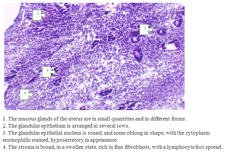 | Figure 1. Microscopic appearance of the lining of the uterus. Pregnancy 8-9hafta. Paint G-E. EU 10x10 OK |
As can be seen from this picture, the lining of the uterus has a small amount of glands compared to the norm, which are chaotically located and appear in different forms. The glandular epithelium is arranged in this in several rows, the core of which has a round or oblong shape, the nuclear structures are painted basophilically. Hyposecretory staining of the cytoplasm of the glands, as well as pronounced hydropic dystrophy, indicates endometrial hypoplasia. In the stroma, the accumulation of lymphocytes in the pervascular area is detected. Trophoblast cells are polygonal in shape and are arranged layer by layer. Blood vessels, fibrous structures and leukocytes are not found in this layer. In this case, it is determined that the intermediate cells are not sufficiently differentiated, the capillary mesh membranes are not sufficiently developed. As a result of the above histological changes, too, in the early term, a violation of the nutrient supply of the embryo occurs, in addition, vascular failure occurs miscarriage. This type of histological incidence was found in 15 out of 51 patients (29.4%) in Group 3, while in Group 2, 4 out of 47 (8.5%), spontaneous miscarriage or undeveloped pregnancies were found between 6-8 weeks of gestation. In patients of Group 2 and Part 3, however, it is possible to identify cells in which the Cystic altered glands are not well developed. They were found in Group 3 in 5 out of 51 (9.8%), while Group 2 was found in 2 out of 47 (4.2%). This revealed the case in Figure 2 below.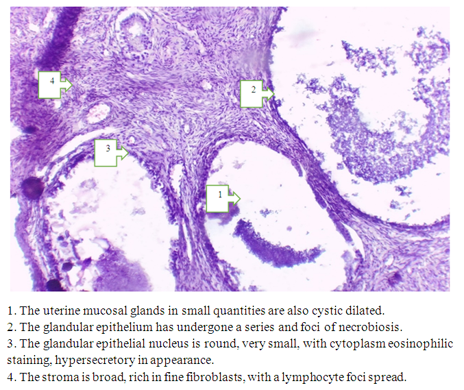 | Figure 2. Microscopic appearance of the lining of the uterus. Pregnancy is 10-12 weeks. Paint G-E. EU 10x10 OK |
In the histological analysis of the lining of the uterus, it can be recognized that the mucous membrane glands have expanded cysts, the amount of which is low. The size of its glands has increased sharply compared to the norm, and at the expense of its cystic expansion, the functional floor of the endometrium has thickened. When we examined the microscope closely (OK 10x10 ob); the epithelia appear cylindrical and cuboid, the glandular form was found to be glandular hyperplasia, expanding like an ordinary cyst. The nucleus is round, very small in size, the cytoplasm is eosinophilic in color, hypersecretory in appearance. In certain glands, the epithelium is located in a series, in which areas with foci of necrobiosis are identified. In addition, stroma hyperplasia is also found in histological analysis. Normal mitoses were found to be moderate in both epithelium and stroma. But in our preparations, a thickening of the stroma in relation to the norm, fullness of blood vessels and vasospasm were detected. Stroma enlarged, fine fibroblast-rich, rough, lymphocytes were found to have a focussed distribution in specific areas.At 10-12 weeks of gestation, when endometrial scraping was obtained at the time of fetal miscarriage, it was observed in 7 out of 7 (14%) of Group 3 patients. In this case, a sample of the tissue obtained contains blood vessels of the sinusoid type, in which it is determined that fullness and stasis have developed. In addition, just like in previous samples, the lining of the uterus is found to be low in the amount of glands, and some of them, cystic expansion. The scraping pattern is shown in Figure 3.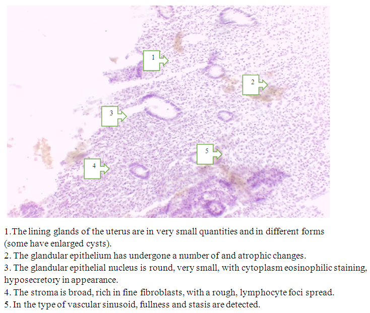 | Figure 3. Microscopic appearance of the lining of the uterus. Pregnancy 10-12 weeks. Paint G-E. EU 10x10 OK |
The basis of chorionic suckers stroma is connective tissue. At 6-8 weeks of pregnancy, chorionic suckers will have fibroblasts, macrophages, reticulin, and collagen fibers in the esophagus. The intercellular substance of the connective tissue has been found to contain glycosoaminoglycans (mucopolysaccharides) that capture hyaluronic and chondroitin sulfuric acids. From the chorial plate, branches of the umbilical artery grow into the stroma of the suckers and branch, dividing into capillaries. Capillaries transport blood saturated with oxygen and nutrients to the fetus through the umbilical vessels.In the biomaterials we take, when the following pathological condition occurs in early pregnancy, in particular at the beginning of the 1st trimester, histological analysis is carried out in incisions; in some of our preparations, a young chorionic sucker is detected and studied; thickening of external trophoblasts, hyperplasia and hypertrophy of syncytiotrophoblasts are observed. In addition to him, women with somatic diseases in the Anamnesis are observed that vorsins do not develop well or its hypoplasia. Chorionic vorsins are found in more areas of non-vascular, mesenchymal protein dystrophy in the underlying connective tissue of chorionic stroma, with connective tissueamucoid and fibrinoid staining observed. The same-looking fullness of the capillaries and sinusoids of certain chorionic vorsinas and blood clots in the stroma were detected. It can be seen from this that at the moment of miscarriage in the 6-7th week of pregnancy, chorionic vorsinas have not yet fully developed, and when its miscarriage is observed, it occurs due to the fact that the supply of blood vessels is not completely complete. Mucoid and fibrinoid staining of connective tissue fibrinoid necrosis is considered to be a typical condition for fetal miscarriage in early pregnancy. The detected changes in the endometrial Wrinkle sample taken at the time the fetus was observed to fall in this process are shown in Figure 4. By the end of the 1st trimester of pregnancy, there is remodeling and enlargement of the spiral arteries of the uterus. If inflammation and edema develop in the endometrium, this process is disrupted, and as a result, the proliferation of the cytotrophoblast of the embryo and its implantation into the endometrium are disrupted.In addition to it, the imperfection of the desidualization process may develop. As a result of all the above pathological conditions, premature displacement of the fetal bladder and miscarriage are observed.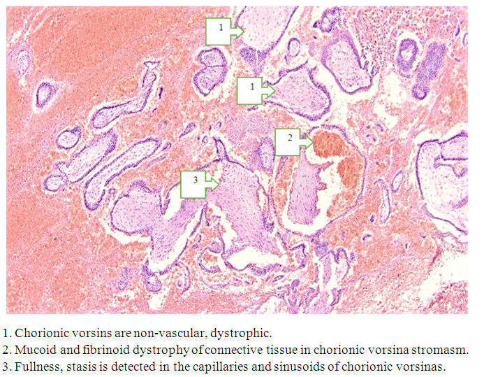 | Figure 4. Microscopic appearance of the lining of the uterus. Pregnancy 6-7 weeks. Bougg-E. EU 10x10 OK |
A study of a cross-sectional sample from the cilia of a fetus that had fallen in this process revealed the condition shown in Figure 5 below.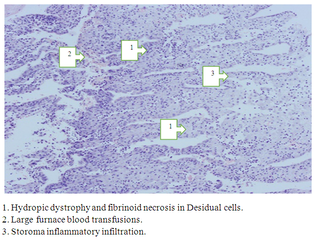 | Figure 5. Microscopic view of the caudal fin. Pregnancy 6-7 weeks. Paint G-E. Ob. 10x10 OK |
When a biomaterial was taken from a fetal cartilage sample for histological analysis, it was found that there was little desidual tissue compared to the norm. This process can develop due to several factors, among which inflammation (nospecific) is considered the most important. In our biomaterials, all signs of inflammation were detected, in particular, hydropic dystrophy in desidual cells, edema and leukocyte infiltration in the intermediate tissue, and destructive changes in the tissue furnace. As a result of the development of hydroponic protein dystrophy due to various causes (in nospesific inflammation) in Desidual cells, a displacement of the nucleus to the edge and a destructive change were detected following the appearance of vacuoles in the cytoplasm. In the cytoplasm, the interposition of vacuolar inclusions occurred and turned into a giant incision, and the color is not eosinophilic blurred. Common inflammatory infiltration of Stroma, fibrinoid necrosis, large blood spills are detected. In conclusion, it should be noted that all of the above causes can cause miscarriage in the first trimester of pregnancy, being considered important in determining the tactics of its prevention and treatment in the next period by analyzing specific causes. Taking into account all the clinical and laboratory indicators in addition to it, it provides an opportunity to achieve a clear reduction in the risk of miscarriage as well as the incidence of miscarriage when taking complex treatment measures in women who have had spontaneous miscarriage in their history.
References
| [1] | Akhmedov, F.K., Negmatullaeva, M.N., Tuksanova, D.I. Features of renal function in women with complicated preeclampsia. International Journal of Current Research and Review, 2021, 13(1), Р. 70–74. |
| [2] | Ashurova N.G., Bobokulova S.B. Prognozirovanie narushenij menstrual'no-ovarial'nogo cikla u devochek-podrostkov, osnovannoe na izuchenii geneticheskih markerov. Rossijskij vestnik akushera-ginekologa. 2024; 24(5): 12‑18. |
| [3] | Pasquali R., Gambineri A. Metabolic effects of obesity on reproduction [Journal] // Reprod Biomed Online. - 2006. - Vol. 12. - p. 542. |
| [4] | Regan L., Rai R. Epidemiology and the medical causes of miscarriage [Journal] // Baillieres Best Pract Res Clin Obstet Gynaecol. - 2000. - Vol. 14. - p. 839. |
| [5] | Rumbold A., Middleton P., Crowther C.A. Vitamin supplementation for preventing miscarriage [CD004073]. - [s.l.]: Cochrane Database Syst Rev, 2005. Sawaya G.F., Grady D., Kerlikowske K., Grimes D.A. Antibiotics at the time of induced abortion: the case for universal prophylaxis based on a meta-analysis [Journal] // Obstet Gynecol. - 1996. - Vol. 87. - p. 884. |
| [6] | Schleussner E., Kamin G., Seliger G., Rogenhofer N., Ebner S., Toth B., Schenk M., Henes M., Bohlmann MK., Fischer T., Brosteanu O., Bauersachs R., Petroff D., ETHIG II group. Low-molecular-weight heparin for women with unexplained reccurent pregnancy loss: a multicentertrial with a minimization randomization scheme // Ann Inter Med. 2015 May 5; 162(9): 601-9. |
| [7] | Sultonova N.A. Rannaya diagnostika nedostatochnosti placenty u zhenshchin s reproduktivnymi poteriyami v respublike Uzbekistana. Novyj den' mediciny // 2020. - 4 (34). - str. -366-368. |
| [8] | Sultonova N.A. Rol' patologii endometriya pri reproduktivnyh poteryah v rannih srokah beremennosti. Tibbiyotda yangi kun №4 (34) 2020 392-395 str. |
| [9] | Zaripova D.Ya. Diagnosticheskie kriterii vyyavleniya osteoporoza v perimenopauzal'nom periode. Reproduktivnoe zdorov'e vo stochnaya Evropa. 2024; 14 (5). S. 590-598. https://doi.org/10.34883/PI.2024.14.5.004. |
| [10] | Zaripova D.Ya., Abdullaeva M.A., Sultonova N.A., Ahmedov F.K., Nasirova Z.S., Umurov E.U., Shukrullaeva G.Zh. Optimizaciya mer diagnostiki rannej menopauzy i prezhdevremennoj menopauzy. Zhuranl Reproduktivnoe zdorov'e vo stochnaya Evropa. 2024; 14 (5). S. 617-628. |
| [11] | Negmatullaeva MN, Tuksanova DI, Zaripova DYa. Structural-optical properties of blood serum and their role in predicting the development of osteoporosis in perimenopause. Russian Bulletin of Obstetrician-Gynecologist. 2024; 24(3): 71‑76. (In Russ.) https://doi.org/10.17116/rosakush20242403171. |

 Abstract
 Reference
 Full-Text PDF
 Full-text HTML