American Journal of Medicine and Medical Sciences

p-ISSN: 2165-901X    e-ISSN: 2165-9036

2025;  15(1): 47-55

doi:10.5923/j.ajmms.20251501.09

Received: Dec. 20, 2024; Accepted: Jan. 12, 2025; Published: Jan. 16, 2025


Improvement of Methods of Prevention of Infection Generalization in Long-Term Non-Healing Wounds

Umarov Bakhtiyor Yatgarovich, Khamdamov Bakhtiyor Zarifovich

Bukhara State Medical Institute named after Abu Ali Ibn Sina, Uzbekistan

Copyright © 2025 The Author(s). Published by Scientific & Academic Publishing.

This work is licensed under the Creative Commons Attribution International License (CC BY).
http://creativecommons.org/licenses/by/4.0/

Abstract

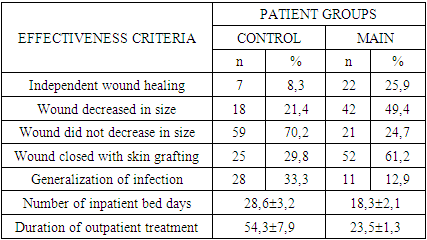
The use of our developed methods for predicting and preventing infection generalization in patients with NON-HEALING WOUNDS allowed us to reduce the incidence of this formidable complication by 2.6 times. This, in turn, resulted in a 1.6-fold reduction in the length of stay of patients in the clinic, and a 2.3-fold reduction in the duration of outpatient treatment.

Keywords: Wound, Infection, Prevention

Cite this paper: Umarov Bakhtiyor Yatgarovich, Khamdamov Bakhtiyor Zarifovich, Improvement of Methods of Prevention of Infection Generalization in Long-Term Non-Healing Wounds, American Journal of Medicine and Medical Sciences, Vol. 15 No. 1, 2025, pp. 47-55. doi: 10.5923/j.ajmms.20251501.09.

1. Introduction

Long-term wound healing contributes to the cumulation of the number of such patients, which reached more than 40 million people worldwide 10 years ago. This type of spread of DNI was noted by P. Driscoll [1,3,18,19] as a "silent epidemic". However, after 5 years, there were reports of the number of patients with DNI reaching 500 million [2,4,20]. Such an impressive figure leads to an increase in financial costs in the healthcare system [5,7,21,26,27]. In particular, M. Olsson et al. conducted a calculation and showed that in developed countries such costs make up to 3% of total healthcare costs. The physiological process of wound healing includes four stages: hemostasis, inflammation, proliferation and scarring, the coordinated work of which ensures proper healing [6,8,28,29]. However, when wounds do not undergo this organized process, soft tissue healing slows down, and this ultimately leads to the development of DNI with such common signs as exudation, re-infection, tissue necrosis, defective re-epithelialization, and decreased angiogenesis. All this served as a prerequisite for more in-depth studies of the mechanisms of the body's immune response as a guarantee of the possibility of improving the results of treatment of patients with DNI.
To date, the immunological aspects of DNI have been studied in certain nosological forms, in particular, in diabetic foot syndrome. Along with this, there is information regarding DNI in patients with concomitant diseases in the form of vascular diseases (both with arterial and venous lesions), obesity, and HIV infection [9,11,13,15,22,30].
Conclusions were made about the direct influence of the above-mentioned concomitant diseases on all stages of wound regeneration. Thus, according to M. Bagheri et al., NWRD in patients with diabetes mellitus are associated with a highly proinflammatory profile caused by excessive expression of inflammatory cytokines, such as TNF-α, and decreased production of mediators that promote healing, including IL-10 and TGF-β. As stated by P.M. Seraphim et al., this leads to polarization of macrophages towards the M1 phenotype, activation and degranulation of CD8+ T cells, which leads to tissue necrosis.
In non-healing wounds, certain factors are disrupted, which is partially responsible for the pathogenesis of injury. Mice deficient in IL-36 receptor antagonists exhibited impaired wound healing due to overproduction of IL-36γ, TGF-β, and CXCL1, excessive neutrophil and macrophage infiltration, and excessive granulation tissue formation [2,4,6,8,23,25]. Furthermore, the chemokine receptor CCR4 negatively affects chronic wounds induced by diabetes. Diabetic mice depleted of CCR4 exhibited decreased expression of wound healing-promoting cytokines such as IL-6, IL-12, IL-1β, TNF-α, and IL-10 [10,12,14,16,17,24]. During normal wound healing, cells in the injured area such as fibroblasts, keratinocytes and immune cells are induced by local mediators to secrete matrix metalloproteinase. Such mediators include various cytokines and growth factors involved in wound healing such as TGF-β, VEGF, EGF, interleukins and interferons. Matrix metalloproteinase is normally required in small amounts and is responsible for proper epithelialization and proliferation. However, their dysregulation leads to impaired epithelialization and is closely associated with DWRD. The process of complete DWRD regeneration does not occur when the immune system fails to continue the normal repair process, resulting in the prolonged presence of neutrophils and pro-inflammatory macrophages in the injured skin, which contributes to inflammation, tissue fibrosis and poor vascularization. Research in this area is ongoing, however, today it is necessary to clarify the causes of the development of generalization of the inflammatory process when using well-known methods of treating NCDs and to determine the role of changes in the immune status. This would allow us to develop effective methods of immunodiagnostics, as well as prediction and prevention of generalization of infection, which ultimately, in our opinion, can improve the treatment results for patients with NCDs.
Purpose of the study. Development and comparative evaluation of the effectiveness of methods for preventing generalization of infection in long-term non-healing wounds.

2. Material and Methods

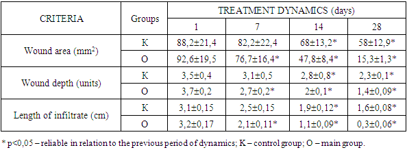
The development and evaluation of the effectiveness of methods for preventing generalization of infection was carried out in patients with NCDs of the main group. At the same time, the use of the method we developed for predicting the probability of generalization of infection showed that out of 85 patients in the main group, 32 (37.6%) patients had a pronounced probability of developing generalization of infection. Among them, in 15.3% of cases (13 patients), such patients were represented by neutrophic ulcers of diabetic foot syndrome, in 11.8% of cases (10 patients) - bedsores and in 10.6% of cases (9 patients) - trophic ulcers against the background of varicose veins of the lower extremities.
In 29 (34.1%) patients with DFU, the probability of developing generalization of the infection was insignificant. Among them, mainly (17.6% of cases - 15 patients) they were represented by bedsores. In the remaining 10.6% and 5.9% of cases, these were patients with trophic ulcers against the background of diabetic foot syndrome (9 patients) and varicose veins of the lower extremities (5 patients).
Only in 24 (28.2%) patients of the main group, the probability of developing generalization of the infection was absent. Among them, 11 patients (12.9%) had bedsores, 5 patients (5.9%) had trophic ulcers of diabetic foot syndrome, and 8 patients (9.4%) had varicose veins of the legs.
The complex of treatment and preventive measures in patients of the main group was based on the pathogenetic approach to influencing the course of the inflammatory process in the wound and preventing infection generalization.
The entire complex of developed measures was used against the background of regular wound care, the use of medications (platelet antiplatelet agents, phlebotonic drugs, peripheral vasodilators, metabolic drugs, immunotropic and antibacterial agents).
Surgical treatment of the wound (or debridement) was performed in the presence of a large amount of necrotic tissue and fibrin. Further tactics of local treatment of DNP depended on the degree of prognostic probability of infection generalization. In the absence of a prognostic probability of infection generalization, the method of choice for local wound treatment was the use of controlled negative pressure dressings (vacuum therapy).
To conduct sessions of controlled negative pressure exposure to the wound, we used polyurethane foam dressings with 400-600 micron micropores. This ensured uniform distribution of controlled negative pressure over the entire surface of the DNP. This treatment option is known to be optimal in terms of generating both physical and biological reactions in the wound.
Each session of controlled negative pressure was conducted in a vacuum mode of 0.1-0.15 atm. (76-115 mm Hg) for 9-10 minutes. Among the side effects of using this mode of controlled negative pressure exposure to the wound, a feeling of moderate pain in the wound area was noted by patients, which did not require the use of any analgesic medications.
We conducted this technique in patients with no prognostic probability of infection generalization in DNP for an average of 5.5 ± 0.5 days, which allowed us to subsequently use surgical methods of wound closure. This regime and duration of sessions of exposure to controlled negative pressure on the wound allowed to control the humidity and amount of exudate in the wound.
In case of patients with an insignificant prognostic probability of infection generalization, the regime of sessions using controlled negative pressure on the wound was extended on average to 8.4±0.8 days with the duration of each session up to 13-15 minutes. This allowed to minimize the moist environment of the wound and, accordingly, the amount of exudation, and thereby reduce the likelihood of its entry into the systemic bloodstream.
However, in case of detection of a pronounced prognostic probability of infection generalization in patients with DNP, sessions using controlled negative pressure on the wound were insufficient. Despite the extension of sessions of vacuum therapy on average to 13.6±2.1 days, such patients, after each session of vacuum therapy, also received laser photodynamic therapy (LPDT) according to the method of B.Z. Khamdamov.
For this purpose, after completion of the next vacuum therapy session, a photosensitizer was applied - 0.05% solution of mytilene blue belonging to the phenothiazine group with maximum absorption λ max (nm) - 668 nm with an exposure of 5 minutes. Then, after washing off the photosensitizer from the wound surface, the wound surface was illuminated with laser radiation using the ALT-Vostok device, model 03, corresponding to the technical conditions TSh 64-15302652-002:2010. The distance from the end of the light guide to the wound surface was 0.5-5.0 cm in the absence of thermal discomfort in the patient. The average wound irradiation time was 5.3 ± 0.5 minutes. For large wound areas, polypositional irradiation of wound surfaces was used by smoothly moving the terminal over the entire wound surface. The use of LFDT after each vacuum therapy session allowed to enhance the therapeutic effect on the wound not only by stimulating the growth of granulation tissue, but also by accelerating epithelialization due to maximum demarcation of the pathological focus.
The general preventive effect on the possible probability of infection generalization in patients with DNR was carried out by using Neupogen® and Infliximab according to the scheme we developed.
Neupogen® was used depending on the degree of prognostic probability of infection generalization in patients with DNR. Thus, patients with an insignificant probability of infection generalization were given Neupogen® by subcutaneous administration at a dose of 0.1-0.4 million U (1-4 μg) / kg per day once. In the presence of positive dynamics of changes in the prognostic probability of infection generalization on the 7th day of treatment, a repeated injection was used at the same dose. In patients with a high prognostic probability of infection generalization, Neupogen® was administered at a dose of 1.0 million IU (10 μg)/kg/day (maximum daily dose), with subsequent repeated administration on days 3 and 7 of the treatment. When the prognostic probability of infection generalization decreased to insignificant, the dose of Neupogen® was reduced to 0.1-0.4 million IU (1-4 μg)/kg to two injections every 72 hours. In the absence of a prognostic probability of infection generalization, Neupogen® injections were discontinued.
Neupogen® is known to be a hematopoietic growth factor and a highly purified non-glycosylated protein consisting of 175 amino acids. It regulates the formation of functionally active neutrophils and their release into the blood from the bone marrow. The effectiveness of its use is due to a significant increase in the number of active neutrophils in the peripheral blood already in the first 24 hours after administration with a slight increase in the number of monocytes. Patients receiving Neupogen® require lower doses of antibiotics.
Given the increased expression of TNF-α in patients with generalized infection, which in the long term can lead to endothelial dysfunction and progression of multiple organ failure, we also used Infliximab, known as a specific antibody against TNF-α, in patients with a high probability of sepsis. The drug was prescribed at a dose of 5 mg / kg on the 1st, 7th and 14th day of treatment. With an insignificant probability of developing generalized infection in patients with DNR, Infliximab was used at the same dose on the 1st and 7th days of treatment.
Thus, prevention of generalized infection in patients with DNR includes a differentiated approach to local (vacuum therapy and LFDT) as well as general effects on the course of both the wound and inflammatory process, provides for the use of pathogenetically substantiated methods for correcting local and general immunity disorders. Results and discussion. The development and evaluation of the effectiveness of methods for preventing infection generalization were carried out in patients with DUPID of the main group. At the same time, the application of the method developed by us for predicting the probability of infection generalization showed that out of 85 patients in the main group, 32 (37.6%) patients had a pronounced probability of developing infection generalization. Among them, in 15.3% of cases (13 patients), such patients were represented by neutrophic ulcers of diabetic foot syndrome, in 11.8% of cases (10 patients) - bedsores and in 10.6% of cases (9 patients) - trophic ulcers against the background of varicose veins of the lower extremities. In 29 (34.1%) patients with DUPID, the probability of developing infection generalization was insignificant. Among them, they were mainly (17.6% of cases - 15 patients) represented by bedsores. In the remaining 10.6% and 5.9% of cases, these were patients with trophic ulcers against the background of diabetic foot syndrome (9 patients) and varicose veins of the lower extremities (5 patients).
Only 24 (28.2%) patients of the main group had no probability of developing generalized infection. Among them, 11 (12.9%) patients had bedsores, 5 (5.9%) patients with trophic ulcers of diabetic foot syndrome and 8 (9.4%) varicose veins of the legs.
The complex of therapeutic and preventive measures in patients of the main group was based on the pathogenetic approach in influencing the course of the inflammatory process in the wound and preventing generalization of infection.
The entire complex of developed measures was used against the background of regular wound care, the use of medications (platelet disaggregants, phlebotonic drugs, peripheral vasodilators, metabolic drugs, immunotropic and antibacterial agents). Surgical treatment of the wound (or debridement) was performed in the presence of a large amount of necrotic tissue and fibrin. Further tactics of local treatment of DNP depended on the degree of prognostic probability of infection generalization.
In the absence of prognostic probability of infection generalization, the method of choice in local treatment of wounds was the use of dressings with the effect of controlled negative pressure (vacuum therapy). To conduct sessions of exposure to controlled negative pressure on the wound, we used polyurethane foam dressings with micropores of 400-600 microns. This ensured uniform distribution of controlled negative pressure over the entire surface of the DNP. This treatment option is known to be optimal in terms of generating both physical and biological reactions in the wound.
Each session of controlled negative pressure was carried out in a vacuum mode of 0.1-0.15 atm. (76-115 mm Hg) for 9-10 minutes. Among the side effects of using this mode of exposure to the wound of controlled negative pressure, a feeling of moderate pain in the wound area was noted by patients, which did not require the use of any analgesic medications. This technique in patients with no prognostic probability of infection generalization in DNP was carried out by us for an average of 5.5 ± 0.5 days, which allowed us to subsequently use surgical methods of wound closure. This mode and duration of sessions of exposure to DNP of controlled negative pressure allowed us to ensure control over the humidity and amount of exudate in the wound. In the case of an insignificant prognostic probability of infection generalization in patients, the mode of application of sessions using controlled negative pressure on the wound was extended to an average of 8.4 ± 0.8 days with the duration of each session up to 13-15 minutes. This allowed us to minimize the moist environment of the wound and, accordingly, the amount of exudation, and thereby reduce the likelihood of it entering the systemic bloodstream. However, in the case of detection of a pronounced prognostic probability of infection generalization in patients with DNP, sessions using controlled negative pressure on the wound were not sufficient. Despite the extension of vacuum therapy sessions to an average of 13.6±2.1 days, such patients were also given laser photodynamic therapy (LPDT) after each vacuum therapy session using the B.Z. Khamdamov method.
For this purpose, after the completion of the next vacuum therapy session, a photosensitizer was applied - a 0.05% solution of mytilene blue belonging to the phenothiazine group with an absorption maximum λ max (nm) - 668 nm with an exposure of 5 minutes. Then, after washing off the photosensitizer from the wound surface, the wound surface was illuminated with laser radiation using the ALT-Vostok model 03 device, which complies with the technical specifications TSh 64-15302652-002:2010 (Figure 1). The distance from the end of the light guide to the wound surface was 0.5-5.0 cm in the absence of thermal discomfort in the patient.
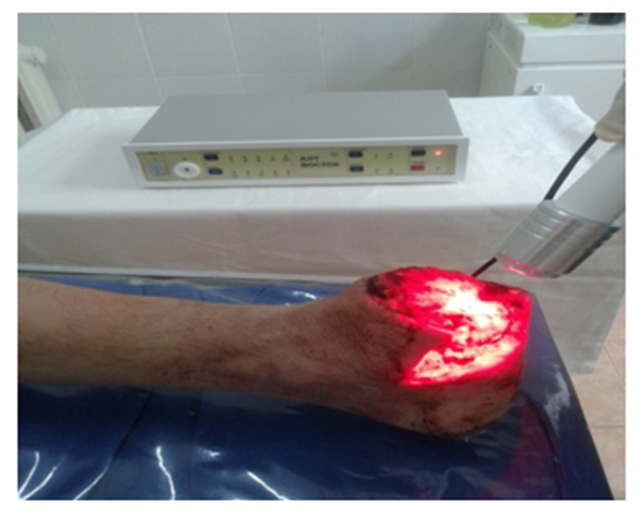
Figure 1. General view of the application of laser photodynamic therapy in the treatment of long-term non-healing wounds
The average wound irradiation time was 5.3±0.5 minutes. For large wound areas, polypositional irradiation of wound surfaces was used by smoothly moving the terminal over the entire wound surface. The use of LFDT after each vacuum therapy session made it possible to enhance the therapeutic effect on the wound by not only stimulating the growth of granulation tissue, but also accelerating epithelialization due to maximum demarcation of the pathological focus.
General preventive action on the possible probability of infection generalization in patients with DNR was carried out by using Neupogen® and Infliximab according to the scheme we developed. Neupogen® was used depending on the degree of prognostic probability of infection generalization in patients with DNR. Thus, patients with an insignificant probability of infection generalization were given Neupogen® by subcutaneous administration at a dose of 0.1-0.4 million U (1-4 μg)/kg per day once. In the presence of positive dynamics of changes in the prognostic probability of infection generalization on the 7th day of treatment, a repeat injection was used at the same dose.
In patients with a pronounced prognostic probability of infection generalization, Neupogen® was administered at a dose of 1.0 million U (10 μg) / kg / day (maximum daily dose), followed by repeated administration on the 3rd and 7th days of treatment. If the prognostic probability of infection generalization decreased to insignificant, the dose of Neupogen® was reduced to 0.1-0.4 million U (1-4 μg) / kg up to two injections every 72 hours. In the absence of a prognostic probability of infection generalization, Neupogen® injections were discontinued.
It is known that Neupogen® is a hematopoietic growth factor and a highly purified non-glycosylated protein consisting of 175 amino acids. It regulates the formation of functionally active neutrophils and their release into the blood from the bone marrow. Its effectiveness is due to a significant increase in the number of active neutrophils in the peripheral blood already in the first 24 hours after administration with a slight increase in the number of monocytes. Patients receiving Neupogen® require smaller doses of antibiotics.
Considering the increased expression of TNF-α in patients with generalized infection, which in the long term can lead to endothelial dysfunction and progression of multiple organ failure, we also used Infliximab, known as a specific antibody against TNF-α, in patients with a high probability of sepsis. The drug was prescribed at a dose of 5 mg / kg on the 1st, 7th and 14th day of treatment. With an insignificant probability of developing generalized infection in patients with DNR, Infliximab was used at the same dose on the 1st and 7th days of treatment.
Thus, the prevention of generalized infection in patients with DNR includes a differentiated approach to local (vacuum therapy and LFDT) as well as general effects on the course of both the wound and inflammatory process, provides for the use of pathogenetically substantiated methods for correcting local and general immunity disorders. In this case, the initial values of the cell count were almost identical in both the control (4.52±0.3%) and main (4.48±0.3%) groups.
A significant increase in neutrophils in the wound imprints of patients in the main group begins already on the 7-14th day of treatment compared to the control group by 1.34 (p < 0.05) and 1.41 times (p < 0.05).
A comparative assessment of the nature of changes in the main parameters of neutrophils in wound imprints revealed a predominant increase in the number of band neutrophils starting from the 7-14th day of treatment in patients in the main group by 2.0-2.1 times (up to 9.2±1.8% and up to 9.8±2.1%, respectively; p < 0.05) with a stable ratio of them on the 28th day of treatment (10.1±2.15%). In contrast, in patients of the main group, we found a decrease in segmented neutrophils compared to the data of patients of the control group from 29.7±2.8% to 20.3±3.1% on the 14th day of treatment and from 31.9±3.1% to 24.5±3.5% on the 28th day of treatment (p < 0.05).
However, we noted more reliable changes in the comparative dynamics of changes in degenerative forms of neutrophils. Their number significantly decreased on the comparative difference starting from the 14th day of treatment from 15.7±2.3% in the control group of patients and to 7.2±0.5% in the main group of patients, as well as on the 28th day of treatment from 15.2±2.45% in patients of the control group to 2.3±0.1% in patients of the main group (p < 0.001). Thus, starting from the 14th day of using the treatment and prophylactic algorithm developed by us, the difference in the progressive decrease in the number of degenerative forms of neutrophils was significant in favor of the indicators of patients in the main group. Thus, if on the 14th day of treatment the number of degenerative forms of neutrophils in the wound in patients in the main group was 2.2 times less than in patients in the control group (p < 0.05), then on the 28th day of treatment this difference had already increased to 6.6 times (p < 0.001). All this in turn led to a significant improvement in the cytomorphometric picture of the wound, which was manifested by an increase in the regenerative-degenerative index by 1.91 times (p < 0.05) on the 14th day and by 6.16 times (p < 0.001) on the 28th day of treatment.
More visual comparative changes can be noted in relation to leukocytes and lymphocytes. In the main group of patients, we noted an increase in the intensity of T-lymphocytes and their regulatory subpopulations, in particular CD3+, CD4+ and CD8+ cells. Unlike the control group of patients, the values of the main group ultimately reached reference values. The use of the treatment and diagnostic algorithm developed by us for the prevention of generalization of infection in patients with DND had a relatively pronounced immunocorrective effect due to an increase in the functional state of the immune system.
The content of immunocompetent cells increased, in particular T-lymphocytes in the form of an increase in their absolute number, which ultimately reached reference values (p < 0.001).
Ultimately, we noted a stable level of cellular immunity indicators over the course of 14-28 days of treatment, which may indicate the high efficiency of the treatment and diagnostic algorithm we developed for preventing generalization of infection in patients with DND.
The issue of progressive decrease in CD38+ cell subpopulations, both among absolute and relative values, remains interesting, which was apparently associated with the use of granulocyte neutrophil growth factor.
An identical nature of the positive dynamics of change can be noted in relation to the indicators of the B-lymphocyte system and humoral immunity in general in the examined patients. The increase in the number of CD20+ and CD23+ cells noted among the patients in the control group was associated with the ongoing inflammatory process (p < 0.001), which was not stopped by traditional treatment methods. In contrast, among the patients in the main group, due to the use of the treatment and diagnostic algorithm developed by us, all B-lymphocyte indices reached reference values on the 28th day of treatment (p < 0.001), which may indicate the suppression of the inflammatory process and the disappearance of the likelihood of infection generalization, which ultimately made it possible to use the final options for closing the surface of the DNZ. Thus, the results of studying the parameters of the B-lymphocyte system in patients with DNZ before and after complex treatment showed that all parameters were restored after 7 days and remained at a stably high level after the complex treatment. Both the parameters of the T-lymphocyte system and the B-lymphocyte system indicators were distinguished by a high intensity of changes after treatment, stability of a high content of immunocompetent cells, and normalization of all indicators of these links of the immune system in the examined patients with DND. High intensity of changes after 7 days and stability of results after 28 days after complex treatment were also observed for the level of lymphocytes with a marker of activation and apoptosis, as well as NK cells in patients with DND.
Reduced parameters of CD25+ and CD95+ lymphocytes detected before treatment increased sharply after the complex treatment - respectively, after 7 days (p < 0.001). It is evident that both parameters reached the values of the control group, not significantly different from them (p > 0.05), which indicates normalization of the parameters after treatment. Such changes were not established after traditional treatment. Only complex treatment leads to normalization of the above-mentioned cells of the patients' immune system.
The relative level of CD16+ cells after the complex treatment, on the 7th day of treatment remained reliably high, not only in relation to the data before treatment (P < 0.001), but also in relation to the reference values (p < 0.001). However, in the subsequent periods, the number of CD16+ cells in patients of the main group progressively decreased, reaching the reference values, which indicates a stable abatement of the inflammatory process. Thus, the content of lymphocytes with activation and apoptosis markers (CD25+ and CD95+ cells), as well as NK cells in the peripheral blood of patients with DNZD after the complex treatment were significantly increased, reaching the control values (CD25+ and CD95+ cells) and remained high in relation to normal values (p < 0.001). The intensity of changes in the dynamics of immunoglobulins in the blood of patients in the main group was completely different from that of patients in the control group. Initial high values of IgM, IgA and IgG indicated the intensity of the inflammatory process at the initial level of examination of patients. However, in the dynamics of the application of the treatment and diagnostic algorithm developed by us, the intensity of the inflammatory process decreased more significantly compared to the control group of patients (p < 0.05). Such changes can be noted in relation to IgM and IgA.
Ultimately, on the 28th day of treatment, these indicators reached reference values (p < 0.05), indicating the elimination of the active inflammatory process and a decrease in the prognostic probability of the development of generalization of the infection. Along with the above, the dynamics of IgG changes indicated pronounced immunological responses of the body, corresponding to their intense manifestation and higher production of this type of immunoglobulin (p < 0.05). The dynamics of changes in the concentration of the proinflammatory cytokine IL-1β in the blood of patients in the main group at the time of the initial examination was higher than in patients in the control group by more than 10% with a low reliable difference (p > 0.05). The use of the treatment and diagnostic algorithm developed by us in the main group of patients led to a decrease in the concentration of IL-1β to 117.9 ± 18.5 pg / ml (p < 0.05), which was already 30% less than among patients in the control group at this time of using traditional methods of treatment. The effectiveness of the treatment and diagnostic measures developed by us aimed at preventing generalization of infection in patients with DNP in the main group was proven by a reliable decrease in the concentration of IL-1β on days 14-28 by 2.1 times (p < 0.05) and 16.9 times (p < 0.05) with the achievement of reference values, which could not be obtained among patients in the control group. The intensity of changes in the dynamics of TNF-α in patients of the main group, in contrast to the control group, was more pronounced, characterized by a progressive decrease in the concentration of this cytokine already on the 7th day of using the treatment and diagnostic algorithm developed by us from 42.8 ± 9.3 pg / ml to 18.9 ± 3.7 pg / ml, that is, almost 2.3 times (p < 0.05). As can be seen from the values of this indicator in the blood of patients of the main group, its excess of reference values was at a reliable level (p < 0.05) only before the start of treatment. We deliberately focus on these data, since on the 14th-28th day of treatment, the concentration of TNF-α in patients of the main group reached reference values (p < 0.05), which was not noted among patients of the control group.
The opposite, but positive, comparative dynamics of the concentration of TGF-β in the blood was noted by us. This indicator, as is known from the previously presented data, increased in the blood of patients in the control group throughout the study. The maximum peak of TGF-β occurred on the 28th day of using traditional methods of treatment (p < 0.05). An interesting fact is the increase in the concentration of TGF-β in the blood of patients with DND of the main group, too, despite lower values than in patients in the control group before the start of treatment. Already on the 7th day of using the treatment and diagnostic algorithm developed by us, the concentration of TGF-β in the blood increased to 13.7 ± 0.4 pg / ml (p < 0.05), and on the 14th day - already to 19.4 ± 1.3 pg / ml (p < 0.05). A more reliable value was found by us on the 28th day of the treatment, when the concentration of TGF-β reached reference values and exceeded the indicators of the control group of patients at this time by 1.7 times (p < 0.05). In general, it can be noted that the use of the treatment and diagnostic algorithm developed by us allowed us to achieve not only regression of the inflammatory process in patients of the main group with DNR, but also to raise the level of the indicator of strengthening the regenerative properties of the wound process.
Thus, the intensity of changes in the concentration of proinflammatory cytokines in the blood of patients of the main group may indicate the high efficiency of the treatment and diagnostic measures developed by us, manifested by a decrease in inflammatory processes that could cause generalization of infection in patients with DNR. A decrease in the intensity of the general inflammatory reaction and its transition to a local form allows us to achieve positive treatment results, which was manifested by an increase in tissue growth indicators. The dynamics of changes in such indicators as MIP-1α, MIP-2β and PDGF, indicating the activity of cellular factors at the beginning of wound regeneration was ambiguous. Unlike the patients in the control group, the changes among the patients in the main group were not pronounced, which was apparently due to the direction of the vector of the treatment and diagnostic algorithm developed by us in the direction of preventing the generalization of infection. Meanwhile, the process associated with changes in the concentration of MIP-1α and MIP-2β in the blood was identical, in particular, the concentration of MIP-1α in the blood of patients in the main group increased from 9.2 ± 1.4 pg / ml on the 1st day of treatment to 14.1 ± 2.6 pg / ml on the 28th day of treatment (p < 0.05). At the same time, the concentration level of this indicator corresponded to the reference values. Against this background, we found a reverse dynamics of MIP-2β in the blood from 22.7±3.8 pg/ml on the 1st day of treatment to 16±1.8 pg/ml on the 28th day of treatment (p < 0.05).
Platelet growth factor increased among both control and main group patients. It should be noted that in the control group of patients, the increase in PDGF in the blood was less intense (from 16.1±2.7 pg/ml to 19.8±3.1 pg/ml; p<0.05), whereas among patients in the main group, the increase in PDGF in the blood was more intense (from 8.9±1.3 pg/ml to 22.1±4.5 pg/ml; p < 0.05). All this led to the achievement of reference values in patients of the main group on the 28th day of treatment (p < 0.05), which indicates an increase in the intensity of regenerative processes in the wound.
Thus, the study of the indices of cellular and humoral immunity showed significant changes with the achievement of reference values under the conditions of application of the treatment and diagnostic algorithms developed by us, which indicates their high efficiency.
When assessing the immediate results of treatment, local and general changes in the course of the DNZR were taken into account. Among 85 patients of the main group, such changes were ambiguous. In this regard, the obtained data were assessed depending on the prognostic probability of the development of generalization of the infection, which we subdivided, as indicated above, into absent, insignificant and pronounced. At the same time, the description of the signs of the pathological process was based on the point gradation improved by us, described in the second chapter of the dissertation.
In the majority of patients (54.1%) of the main group, the active inflammatory process in the DNZR was either absent (17 patients) or accompanied by no tissue necrosis (29 patients), which was apparently associated with an increase in the skills of independent dressing change. The remaining 39 patients had a necrobiotic process in the wound in the DNPZ. Among them, a mixed type of necrosis (24.7%) and the presence of dry necrosis (12.9%) were predominantly noted. Among patients with no probability of infection generalization, cases of the absence of an inflammatory process in the wound were predominantly recorded (62.5%), among patients with an insignificant probability of infection generalization, cases of the presence of an inflammatory process in the wound, but without necrobiosis, were a priority (72.4%), and among patients with a pronounced probability of developing infection generalization, cases with mixed necrosis in the DNPZ were predominantly recorded.
According to the type of tissue in the DNR bed, the advantage was (34.1%) for patients with fragile and pale granulation tissue. Among those with no probability of infection generalization, there were 37.5% of such patients, among those with a slight probability - 58.6%, and with a high probability - 9.4%. In second place (21.2%) were patients with the frequency of dense and red granulation tissue. Moreover, among patients with no probability of infection generalization, they were in predominant quantity (54.2%), among patients with a slight probability - in 13.8% of cases, and among patients with a high probability - in 21.2% of cases. The nature of the DNR wound exudate was varied. Predominantly (43 patients; 50.6%) it was purulent (27.1%) or serous-purulent (23.5%). Among the remaining 49.4% of cases, hemorrhagic (17 patients; 20%), serous-hemorrhagic (14 patients; 16.5%) and serous (11 patients; 12.9%) nature were established.
Among patients with a low prognostic probability of infection generalization, we diagnosed the presence of only serous (45.8%) and serous-hemorrhagic (54.2%) wound exudate. In patients with an insignificant prognostic probability of infection generalization, the presence of hemorrhagic (55.2%) and serous-hemorrhagic (37.9%) wound exudate was predominantly noted. At the same time, among patients with a pronounced prognostic probability of infection generalization in the DNPZ, purulent (68.8%) and serous-purulent wound exudate prevailed. The color of the exudate of the DNP in patients of the main group was predominantly red (28.2%) or yellow (21.2%). This type of exudate was typical for patients with no prognostic probability of infection generalization (58.3%) and for patients with a low prognostic probability (62%). Among patients with a pronounced prognostic probability of infection generalization, this type of exudate was noted only in 31.3% of patients. It should be noted that for patients of this subgroup, DNP had a characteristic exudate of green or dirty gray color (50%).
The nature of the consistency of the wound exudate was distributed evenly among patients with DNP in the main group. The predominant concentration of indicators accounted for the bloody and thick watery nature of the consistency of the wound exudate (54.1%). This type of exudate consistency accounted for patients with a low prognostic probability of infection generalization. Almost in the same proportion were watery (18.8%) and thick character of consistency of wound exudate (17.6%). At the same time, among patients with a pronounced prognostic probability of infection generalization, thick consistency of wound exudate (65.7%).
Microbial contamination of DNR in patients of the main group was represented by mixed flora in all studies. Staphylococcus aureus was predominant, which was sown in almost half of the patients (49.4%). Relatively less frequent was the presence of Pseudomonas aeruginosa in DNR, which was noted by us in 42.8% of cases. Staphylococcus haemolyticus (32.9%) and Acinetobakter (29.7%) also played a major role in the organization of combined symbiosis of DNR microbes in patients of the main group.
Among other pathogens identified were Proteus vulgaris, Proteus mirabilis, St. Epidermidis, Candida albicans and E. coli. The average level of the microbial flora of the wound in patients of the main group fluctuated between 107 and 109 CFU/cm2 of the wound surface, which significantly exceeded the permissible limit (x102 CFU/cm2) of microbial contamination of the wound. Comparative nature of changes in local clinical manifestations of the wound showed the degree of increase in the severity of the change after the use of the treatment and diagnostic algorithm developed by us. Thus, the area of the wound surface in patients of the control group decreased dynamically from 1.1 to 1.2 times, while in the main group of patients after 1 week of treatment the decrease in the wound area was 1.2 times, and in subsequent periods of treatment this trend only increased. In contrast to the control group of patients, the difference increased by 1.6 times on the 14th day of treatment (p < 0.05) and by 3.1 times on the 28th day of treatment (p < 0.05) (Table 1). The initial depth of the lesion of the DNPZ was almost identical between patients in both the control and main groups. After 7 days of treatment, this indicator decreased, by 1.1 times among patients in the control group and by 1.3 times among patients in the main group (p < 0.05). In subsequent periods of treatment, the dynamics of changes in the wound depth in patients in the control group was stable, still by 1.1 times, but among patients in the main group, due to the use of the treatment and diagnostic algorithm developed by us, the difference in filling the wound with tissues only increased and was already 1.4 times (p < 0.05).
Table 1. Comparative nature of the dynamics of changes in local clinical signs of DNZR
     
A similar nature of changes can be noted in relation to the reduction in the extent of the infiltrate around the wound. In patients of the control group, it did not have such pronounced changes as among patients of the main group. Thus, after 7 days of the studies, the extent of the infiltrate among patients of the control group decreased by 1.2 times, and in the subsequent periods of the study, that is, on the 14th and 28th day, by 1.3 and 1.2 times, respectively. However, in patients of the main group, as a result of applying the treatment and diagnostic algorithm developed by us, the intensity of the reduction of the infiltrate zone around the wound was significant and was expressed in 1.6 (p < 0.05), 1.8 (p < 0.05) and 4.2 times (p < 0.05) on the 7th, 14th and 28th day of the treatment.
It should be noted that the comparative nature of the intensity of the changes occurring was noted by us between patients of the control and main groups also in terms of the probability of infection generalization. In general, complete spontaneous wound healing among patients with DNI was achieved in 17.2% of cases (29 patients). Among them, patients of the main group prevailed by 3.1 times, which confirms the effectiveness of the treatment and diagnostic algorithm developed by us (Table 2). The use of differentiated approaches to wound treatment depending on the prognostic probability of infection generalization allowed us to achieve 2.3 times more cases of wound size reduction. In general, we noted such a treatment result in 35.5% (60 patients) of cases.
Table 2. Comparative evaluation of the effectiveness of the developed methods for predicting and preventing generalization of infection in patients with DNR
     
Despite the use of a complex of therapeutic measures, we failed to achieve the desired results of wound healing in 80 patients (47.3%). In such patients, wound cleansing, reduction in depth and changes in the nature of necrobiotic processes in tissues were achieved. However, the use of the treatment and diagnostic algorithm developed by us among patients of the main group allowed us to reduce the number of such cases by 2.8 times.
In 77 (45.6%) patients, wounds were closed using skin grafting, which turned out to be very effective among patients of the main group. The use of the differentiated approach developed by us in the treatment of DNPZ; skin grafting was performed 2.1 times more often among patients of the main group.
The use of the methods developed by us for predicting and preventing generalization of infection in patients with DNPZ allowed us to reduce the incidence of this formidable complication by 2.6 times. This, in turn, was reflected in the reduction in the length of stay of patients in the clinic by 1.6 times, and the duration of outpatient treatment - by 2.3 times. All this may indicate the effectiveness of the methods we have developed for predicting and preventing infection in patients with long-term non-healing wounds.

3. Conclusions

1. The use of differentiated approaches to wound treatment depending on the prognostic probability of infection generalization allowed us to achieve 2.3 times more cases of wound size reduction. In general, we noted such a treatment result in 35.5% (60 patients) of cases.
2. The use of the methods we developed for predicting and preventing infection generalization in patients with DNR allowed us to reduce the incidence of this formidable complication by 2.6 times. This, in turn, was reflected in a reduction in the length of stay of patients in the clinic by 1.6 times, and the duration of outpatient treatment - by 2.3 times.

References

[1]  Trophic ulcers of the lower extremities - a review of the problem. / V.N. Obolensky, G.V. Rodoman, V.G. Nikitin, M.A. Karev. // RMJ. 2019; 17: 25(364): 1647-1662.
[2]  Vasculitic and autoimmune wounds. / V.K. Shanmugam, D. Angra, H. Rahimi, S. McNish. // J. Vasc. Surg. Venous Lymphat Disord. 2017; 5(2): 280-292.
[3]  Tamrazova O.B. Long-term non-healing ulcers of the lower extremities: pathogenetic substantiation of the tactics of choosing therapy: Diss.... Doctor of Medical Sciences. Moscow, 2013.
[4]  Melnik V.R. Development of technologies and components of the drug Neupogen // Pharmacology and pharmaceutical industry. -2012; 3(1): 218-225.
[5]  Podgorny P.R., Sibiryakova R.N., Martynenko V.V. Hematopoietic effect of Neupogen in patients with blood diseases // Oncohematology. - 2010; 2: 34-39.
[6]  Smorodina G.V. To what extent is the use of Neupogen required in conditions of enhanced antibiotic therapy for sepsis? (literature review) // Bulletin of Pharmacology. -2013; 3: 23-29.
[7]  Khamdamov B.Z. Method of laser photodynamic therapy in the treatment of wound infection in diabetic foot syndrome. // Biology of diseases. Samarkand, 2020. No. 1 (116) - P. 142-148. (14.00.00; No. 19).
[8]  Khramilin V.N. Modern aspects of local treatment of chronic wounds of the lower extremities in patients with diabetes mellitus. // Scientific and practical medical journal of the Endocrinology Research Center of the Russian Academy of Medical Sciences. 2015; 4: 26-30.
[9]  Stone R.C. A bioengineered living cell construct activates an acute wound healing response in venous leg ulcers. // Sci. Transl. Med. 2017; 9(371): eaaf8611.
[10]  Burden of venous leg ulcers in the United States. / J.B. Rice, U. Desai, A.K. Cummings, et al. // J. Med. Econ. 2014; 17: 347-356.
[11]  Driscoll P. Wound prevalence and wound management, 2012–2020. 2013. // Accessed on 14 September 2015. Available online: http://blog.mediligence.com/2013/01/29/wound-prevalence-and-wound- management-2012-2020/.
[12]  Garraud O., Hozzein W.N., Badr G. Wound healing: Time to look for intelligent, “natural” immunological approaches? // BMC Immunol. 2020; (18): 23.
[13]  An economic evaluation of the impact, cost, and medicare policy implications of chronic nonhealing wounds. / S.R. Nussbaum, M.J. Carter, C.E. Fife, et al. // Value Health. 2018; 21: 27–32.
[14]  The humanistic and economic burden of chronic wounds: A systematic review. / M. Olsson, K. Jarbrink, U. Divakar, et al. // Wound Repair. Regen. 2019; 27: 114–125.
[15]  Mesenchymal stem cells for chronic wound healing: Current status of preclinical and clinical studies. / Y.Z. Huang, M. Gou, L.C. Da, et al. // Tissue Eng. Part B. Rev. 2020; 26: 555–570.
[16]  Goldberg S.R., Diegelmann R.F. What makes wounds chronic. // Surg. Clin. New Am. 2020; 100: 681–693.
[17]  Khamdamov B.Z. Indicators of immunocitocine status in purulent-necrotic lesions of the lover extremities in patients with diabetes mellitus. // American Journal of Medicine and Medical Sciences, 2020, 10 (7). -Р 473-478. DOI: 10.5923/j.ajmm.20201007.08.
[18]  Khamdamov B.Z., Sayfiddinov S.I., Khamdamov I.B., Teshaev U.Sh. The role and place laser photodynamic therapy in prevention postoperative complications at treatment of diabetic foot syndrome. // 5th International scientific conference “European Applied Sciences: challenges and solutions” December 10th 2015. Stuttgart, Germany. -Р. 27-31.
[19]  Khamdamov B.Z., Khamdamov I.B., Teshaev U.SH. Laser photodynamic therapy in prevention postoperative complication at treatment of diabetic foot syndrome. // 3rd International Congress Wounds and Wound Infections. Collection of Abstracts. Moscow, 2016. – С. 325-327.
[20]  Khamdamova M.T., Akramova D. E. Genetic aspects of genital prolapse in women of reproductive age // New day in medicine. Bukhara, 2023. - No. 5 (55). - Р. 638-643.
[21]  Khamdamova M.T., Teshaev Sh.Zh., Hikmatova M.F. Morphological changes of the thymus and spleen in renal failure in rats and correction with pomegranate seed oil // New day in medicine. Bukhara, 2024. - N. 3(65). - Р. 167-187.
[22]  Khamdamova M. T. The state of local immunity in background diseases of the cervix // Eurasian journal of medical and natural sciences Innovative Academy Research Support Center. Volume 3 Issue 1, January 2023 ISSN 2181-287X R. 171-175.
[23]  Khamdamova M.T., Khasanova M.T. Various mechanisms of pathogenesis of endometrial hyperplasia in postmenopausal women (literature review) // New day in medicine. Bukhara. 2023. - No. 8 (58). - Р. 103-107.
[24]  Khamdamova M.T. Reproductive Health of Women Using Copper-Containing Intrauterine Contraception // Eurasian Medical Research Periodical Volume 28 January 2024, ISSN: 2795-7624. www.geniusjournals.org P. 39-45.
[25]  Khamdamov I.B. Advantages Of Laparoscopic Hernioplasty in Obesity Women of Fertile Age // Eurasian Medical Research Periodical Volume 28 January 2024, ISSN: 2795-7624. www.geniusjournals.org P. 33-38.
[26]  Khamdamova M.T., Zhaloldinova M.M., Khamdamov I.B. The state of nitric oxide in blood serum in patients with cutaneous leishmaniasis // New day in medicine. Bukhara, 2023. - No. 5 (55). - Р. 638-643.
[27]  Khamdamova M.T., Zhaloldinova M.M., Khamdamov I.B. The value of ceruloplasmin and copper in blood serum in women wearing copper-containing intrauterine device // New day in medicine. Bukhara, 2023. - No. 6 (56). - Р. 2-7.
[28]  Khamdamova M. T. Bleeding when wearing intrauterine contraceptives and their relationship with the nitric oxide system // American journal of pediatric medicine and health sciences Volume 01, Issue 07, 2023 ISSN (E): Р. 2993-2149. R. 58-62.
[29]  High glucose activates ERK1/2 to stabilize AP1 and increase MMP9 expression in diabetic foot ulcers. / J. Lang, C. Yang, L. Liu, et al. // Exp. Cell Res. 2021; 112550.
[30]  Efficacy of Neupogen in the complex of cytotoxic therapy / R. Ching, F. Bore, Y. Yangu, et al. // Bulletin of Oncohematology. -2021; 8(11): 1428-1433.