Jamolova Aziza Urinbek Qizi 1, Akhmedova Sayyora Mukhamadovna 2
1Kimyo International University in Tashkent, Tashkent, Uzbekistan
2Tashkent Medical Academy, Tashkent, Uzbekistan
Copyright © 2024 The Author(s). Published by Scientific & Academic Publishing.
This work is licensed under the Creative Commons Attribution International License (CC BY).
http://creativecommons.org/licenses/by/4.0/

Abstract
In the available literature, both in foreign and domestic, there is no information about the unified terminology and systematization of anatomical variants of the portal vein, there is a lack of unified approaches and principles for studying it in variant anatomy. The presented data on the morphometric characteristics of the portal vein vary significantly, the data on its extreme forms and the range of anatomical differences are very contradictory, discussions continue about the boundaries of the norm beyond which pathology begins. A prospective study was conducted among practically healthy children aged 0 to 18 years in the period 2023 to 2024. Descriptive statistics (mean, standard deviation, frequency, and percentages) and Pearson product-moment correlation were used for the analysis. Statistical significance was considered at P<0.05, P<0.01, P<0.001. The results of the study showed that the average diameter of the portal vein in boys was 7,57±0,78 mm, and in girls 7,04±0,95 mm. The average diameter of left and right branches of the portal vein in boys was 5,36±0,88 and 5,42±0,87 mm , and in girls 5,10±0,70 and 5,05±0,72 mm. The diameter of left and right branches of the portal vein positively correlates with some anthropometric indicators in gender dependence.
Keywords:
Portal vein, Left and right branches, Ultrasound diagnostics, Anthropometric parameters, Morphometry and children
Cite this paper: Jamolova Aziza Urinbek Qizi , Akhmedova Sayyora Mukhamadovna , The Study of Morphometry of the Trunk of the Portal Vein, Its Roots and Branches in Gender and Age Differences, American Journal of Medicine and Medical Sciences, Vol. 14 No. 12, 2024, pp. 3445-3450. doi: 10.5923/j.ajmms.20241412.79.
1. Introduction
The liver plays an important role in the inter-organ and intersystem interactions of the body, since the metabolism of other organs and systems, as well as the adaptive activity of the whole organism under the influence of various extreme factors of exogenous and endogenous origin, largely depends on the activity of the transformation processes and functional synthesis of energy and plastic substances carried out in the liver. The portal blood vessel is formed by the fusion of the splenic, inferior and superior mesenteric veins. Pancreatic and gastric veins (right and left) flow directly into the portal vascular trunk. The portal blood vessel of the liver ensures the normal functioning of the digestive tract and is an important node of the circulatory system, the pathologies of which affect the functioning of the entire body. However, few studies have clarified how variable portal vein anatomy in children varies by sex, age, and constitution, and the available data are often contradictory. The size of the normal portal vein in adults has been widely studied, but little is known about the size of the portal vein in the growing child. At the same time, the rapid development and introduction into clinical practice of new highly informative diagnostic methods has led to a change in the existing ideas about the variant anatomy of internal organs, including the quantitative parameters of their structure. The use of intravital research methods has become one of the most promising areas in morphology. The results obtained can be used to identify and interpret topographic-anatomical variations at the preoperative stage and have important applied significance in surgery of the upper floor of the peritoneal cavity. The morphometry of the hepatic portal vein is of clinical importance, particularly in pre-operative assessments, surgical management, and diagnoses of liver conditions. This systematic review and meta-analysis aimed to characterize the morphometry of the normal portal vein in both pediatric and adult patients.Purpose of the study: determination of the linear dimensions of the portal vein its roots and branches in children based on age, gender and anthropometric indicators.
2. Materials and Research Methods
The basis of the study is the analysis of the results of ultrasound diagnostics, which was carried out on 138 patients aged 0 to 18 years. An ultrasonic device Canon (Toshiba) Aplio 500 (Japan) with a curvilinear transducer with a frequency of 3.75 MHz was used. Quality control and maintenance of equipment before measurements were carried out by the medical physicist of the department as planned. Measurements were taken using electronic calipers of an ultrasound machine after freezing the image. Anthropometric parameters such as height, weight and body mass index were measured for each participant. A total of 138 children took part in the study. Of the children included in the study, there were 82 boys (59.42%) and 56 girls (40.58%).
3. Discussion of the Results
As can be seen from the table, in the general population, depending on gender, the average diameter of the portal vein trunk in boys under the age of 1 year was 4.85±0.70, from 1 to 3 years - 6.10±0.79; from 4 to 7 years - 7.46±1.00; from 8 to 12 years - 8.21±0.86; from 13 to 15 years - 8.8 ± 1.25; from 15 to 18 years - 10.0 ± 0.86. Accordingly, for girls it is 4,27±0,74; 6,0±1,11; 6,82±0,79; 7,9±0,75; 8,91±1,16 and 8.34±1.13.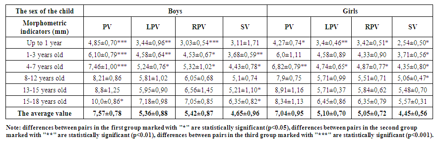 | Table 1. Variability of linear indicators depending on age and gender |
In our study, the average diameter of the portal vein trunk in the general sample of boys ranged from 3.6 to 11.1 mm, while 15.22% of the observations ranged from 6 to 9.6 mm. Accordingly, in girls - in the range from 3 to 10.1 mm, while 13.04% of observations were in the range from 5.4 to 8 mm. The average diameter of the portal vein in boys was 7.57±0.78 mm, which is significantly more by 7% than in girls - 7.04±0.95 mm. [1]It was found that the average diameter of the left branch of the portal vein in boys was 5.36±0.88 mm (range 2.5-8.4 mm) and was significantly smaller than the right branch of the portal vein (Table 1). In girls, this indicator was lower and amounted to 5.10±0.70 mm (range 2.7-7.6 mm), which is also larger than the diameter of the right branch. The difference in the diameter of the left branch of the portal vein in boys and girls is statistically significant (p <0.05). The diameter of the right branch of the portal vein in boys averaged 5.42±0.87 mm with a range of 2.3-8.1 mm. In girls, this indicator was 5.05±0.72 mm (range 2.5-7.4 mm). Differences in the average diameter of the right branch of the portal vein in men and women are statistically significant (p <0.05).For the left branches of the portal vein, the average diameter in boys was 5.36±0.88 mm and was significantly larger than the average diameter of the left branch in girls (5.10±0.70 mm) by 4.85%. The increase in the diameter of the left branch of the portal vein in boys with age is statistically significant. The change in the average diameter of the left branch of the portal vein in boys occurred incrementally with age, starting from 3.44±0.96 mm and reaching 7.18±0.98 mm, as well as in girls ranging from 3.4±0.46 mm to 6.45±0.86 mm. For the right branches, the average diameter in boys was 5.42±0.87 mm and was significantly larger than the average diameter of the right branch in girls (5.05±0.72 mm) by 6.82%. With age, the diameter of the right branch of the portal vein in boys changed incrementally from 3.03±0.54 mm to 7.05±0.85 mm, in the same way as in girls in the range from 3.42±0.51 mm to 6.35±0.79 mm. [2]The average diameter of the splenic vein, taking into account gender differences in boys in the study, was 4.65±0.96 mm with a range from 2.0 mm to 7.8 mm. In girls, the average diameter of the splenic vein was 4.45±0.56 mm with a range of 2.2 mm – 6.0 mm. The diameter of the splenic vein in boys is 4.3% significantly larger than the diameter of the splenic vein in girls (p <0.05).It was found from the table 2 that the average height, weight and BMI in boys was 119.35±11.69, 27.14±8.07 and 17.4±3.33, and in girls - 119.27±8.45, 28.2±5.78 and 17.2±2.58, respectively. The average body mass index in boys during the first and second childhood reached the lowest values of 16.06±3.05 and 16.06±1.91 and has a moderate inverse correlation with the average diameter of the trunk of the portal vein. [3]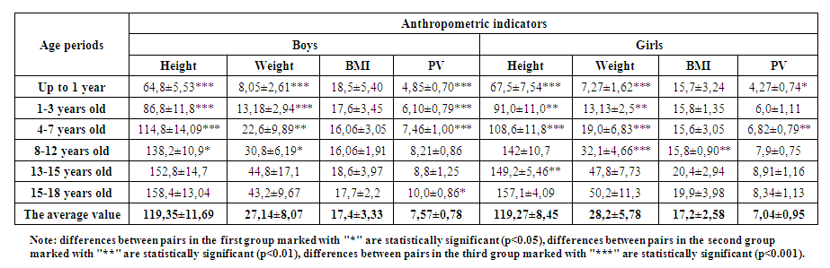 | Table 2. Variability of height, weight, BMI and portal vein in relation to age and gender |
In girls, BMI values were stable until adolescence and averaged 15.8±0.90, then there was a sharp increase in BMI values in adolescence and adolescence and reached up to 20.4±2.94. When studying the correlation of BMI and the diameter of the portal vein trunk with age, a direct weak connection was revealed.When studying the variability of liver size indicators depending on gender and age, the results presented in Table 3 were obtained.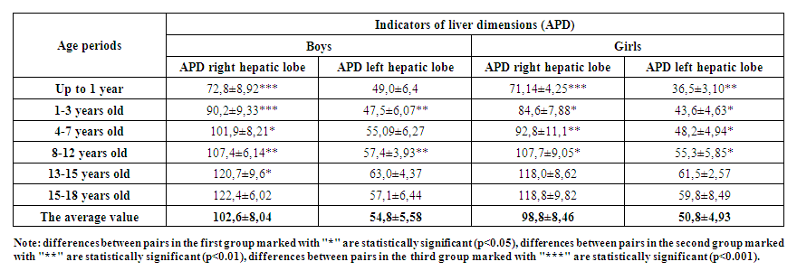 | Table 3. Variability of anterior-posterior dimensions of the liver in relation to age and gender |
As a result of statistical processing of data obtained during ultrasound examination of the linear dimensions of the liver lobes and the diameter of the portal vein (Table 3), it was found that all the studied parameters in boys were significantly (p <0.05) higher than in girls. The data presented in the above tables convincingly show that when interpreting the results of ultrasound examination of the liver and the diameter of the portal vein, it is necessary to use evaluation tables separately for boys and girls. [4]The average APD of the right lobe of the liver in boys was 102.6±8.04 mm, which is significantly higher than the right lobe of the liver in girls by 3.7% and is 98.8±8.46 mm. Also, the average values of the left lobe of the liver in boys is 54.8 ±5.58 mm and is significantly higher (50.8±4.93 mm) than the left lobe of the liver in girls by 7.3%. As can be seen from Table 3, the anterior-posterior dimension of the right lobe of the liver in boys and girls increased moderately with age, reaching the highest values in youth period - 122.4±6.02 mm and 118.8±9.82 mm. But the values of the left lobe in boys and girls differed in that the maximum size reached in adolescent period and is 63.0 ± 4.37 mm and 61.5±2.57 mm. A significant direct correlation was found between the anterior-posterior dimension of the right lobe and the left lobe in the sample in boys, while in girls a significant direct correlation was found between the anterior-posterior dimension of the right lobe and the left lobe of the liver. [5]Significant variability in the diameter of the right and left branches of the trunk of the portal vein allowed us to identify groups of their extreme variants (Table 4).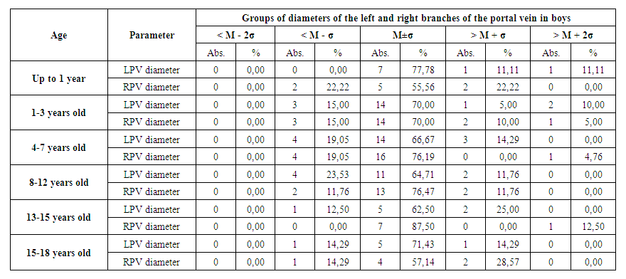 | Table 4. Variants of the diameter for the right and left branches of the trunk of the portal vein |
For the diameter of the left branch of the portal vein in boys in infants, the average value of M±σ was determined by an interval of values from 2.48 to 4.4 mm. 77.78% (7 out of 9) of the measurements corresponded to the specified interval. [6] For the following periods such as: early childhood, first childhood, second childhood, adolescent period and youth period, the average value of M±σ was determined by an interval of values from 3.94 to 5.22 mm, from 4.48 to 6.0 mm, from 4.79 to 6.83 mm, from 5.05 to 6.85 mm and from 6.2 to 8.16 mm, respectively. The indicated intervals corresponded to 70% (14 out of 20), 66.67% (14 out of 21), 64.71% (11 out of 17), 62.50% (5 out of 8) and 71.43% (5 out of 7) measurements. The average value of M±σ, the above intervals, corresponded to 55,56%, 70%, 76,19%, 76,47%, 87,50% and 57.14% of the measurements of the diameter of the right branch of the portal vein and they were in the range from 2.49 to 3.57 mm, from 3.86 to 5.2 mm, from 4.3 to 6.34 mm, from 5.37 to 6.73 mm, from 5.11 to 8.01 mm and from 6.2 to 7.9 mm. This range corresponded to 5 out of 9, 14 out of 20, 16 out of 21, 13 out of 17, 7 out of 8 and 4 out of 7 measurements. The extreme diameter variants of the right and left branches of the portal vein formed 2 groups in which the studied parameters had a value less than the average and greater than the average. Extreme variants of the diameter of the left branch of the portal vein, of the above periods, were revealed, respectively, in 22.22% (2 out of 9), 30% (6 out of 20), 33.33% (7 out of 21), 35.29% (6 out of 17), 37.5% (3 out of 8) and 28.57% (2 out of 7) observations. For the diameter of the right branch of the portal vein, the proportion of extreme variants was 44.44% (4 out of 9), 30% (6 out of 20), 23.81% (5 out of 21), 23.53% (4 out of 17), 12.5% (1 out of 8) and 42.86% (3 out of 7) (Figure 1).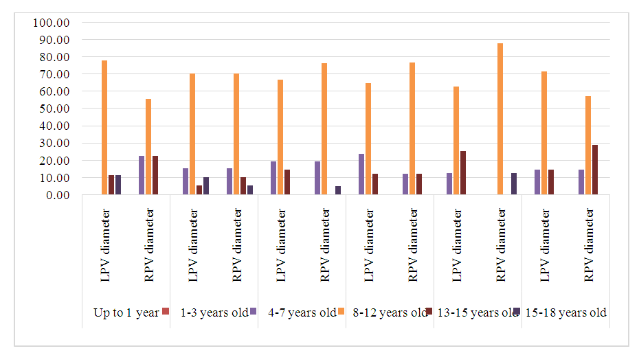 | Figure 1. Variants of the diameter of the left and right branches of the portal vein in boys |
The diameter values of the right and left branches of the portal vein are less than the average, and were divided into two variant subgroups: measurements with a small value (M-2σX>M+σ) and measurements with an extremely large value (>M+2σ). [7]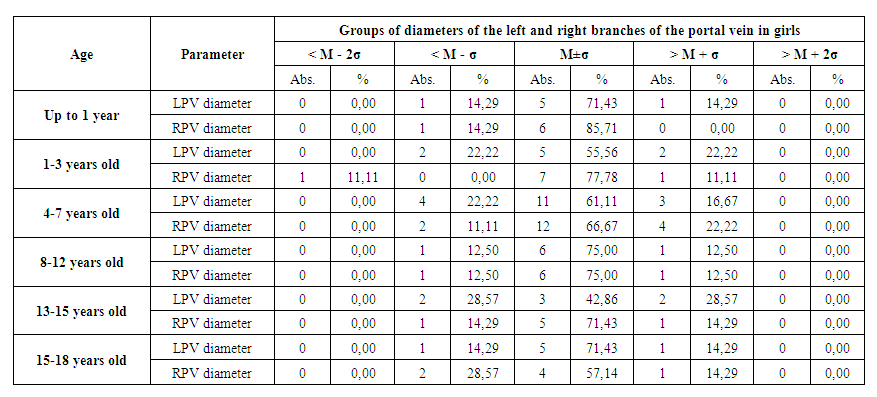 | Table 5. Variants of the diameter for the right and left branches of the trunk of the portal vein |
For the diameter of the left portal vein in girls in the above age groups, the average value M±σ was determined by the range of values from 2.94 to 3.86 mm, from 3.69 to 5.47 mm, from 4.09 to 5.39 mm, from 4.72 to 6.7 mm, from 5.34 to 6.08 mm, from 5.59 to 7.31 mm. The indicated intervals corresponded to 71.43% (5 out of 7), 55.56% (5 out of 9), 61.11% (11 out of 18), 75% (6 out of 8), 42.86% (3 out of 7) and 71.43% (5 out of 7) measurements. The average value of M ±σ, the above intervals, measurements of the diameter of the right branch of the portal vein corresponded to 85,71%, 77,78%, 66,67%, 75%, 71,43% and 57.14% and they were in the range from 2.91 to 3.93 mm, from 3.43 to 5.23 mm, from 4.1 to 5.64 mm, from 4.8 to 6.22 mm, from 5.22 to 6.46 mm and from 5.56 to 7.14 mm. This range corresponded to 6 out of 7, 7 out of 9, 12 out of 18, 6 out of 8, 5 out of 7 and 4 out of 7 measurements. The extreme variants of the length and diameter of the portal vein formed 2 groups in which the studied parameters had a value less than the average and greater than the average. Extreme variants of the diameter of the left branch of the portal vein in girls of the above periods were revealed, respectively, in 28.57% (2 out of 7), 44.44% (4 out of 9), 38.89% (7 out of 18), 25% (2 out of 8), 57.14% (4 out of 7) and 28.57% (2 of 7) observations. For the diameter of the right branch of the portal vein, the proportion of extreme variants was 14.29% (1 of 7), 22.22% (2 of 9), 33.33% (6 of 18), 25% (2 of 8), 28.57% (2 of 7) and 42.86% (3 of 7) (Figure 2). [8]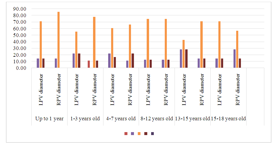 | Figure 2. Variants of the diameter of the left and right branches of the portal vein in girls |
4. Conclusions
138 children participated in the study, of which the largest group were children of the first childhood, accounting for 28.26%. While the smallest group consisted of youth period children – 10.14%. Individual differences in the lifetime linear dimensions and characteristics of the trunk, branches and roots of the portal vein make up the correct range, in which the minimum and maximum values are most rarely observed, and values corresponding to the range M±σ are most common. And the diameter of the branches of the portal and splenic veins has significant sex and age differences and are significantly larger in boys. Also, the linear dimensions of the left and right branches of the portal vein have significant sex and age differences.The conducted ultrasound examination in comparison with the literature data made it possible to summarize and supplement the available information on the anatomical variability of linear parameters of ultrasound examination of the liver, trunk of the portal vein and its branches in healthy children aged 0 to 18 years of both sexes. [9]The study obtained aim to improve the understanding of portal vein anatomy, especially with relevance to surgical interventions of the liver in both pediatric and adult patients. Measurements from ultrasound imaging closely approximates the generated pooled PVD mean for pediatric and adult patients. The results of the study significantly complement the available morphological data on the patterns of growth and development of the inhabitants of the Republic of Uzbekistan at certain stages of ontogenesis and can serve as a scientific basis for the development of biomedical programs to strengthen the health of the population in the studied region. [10]At the same time, the rapid development and introduction into clinical practice of new highly informative diagnostic methods has led to a change in the prevailing ideas about the variant anatomy of internal organs, including the quantitative parameters of their structure. The use of lifetime research methods has become one of the most promising areas in morphology. The results obtained can be used to identify and interpret topographic and anatomical variations at the preoperative stage and are of great applied importance in surgery of the upper floor of the peritoneal cavity.
References
| [1] | Adeyekun, A.A. Grey-scale sonographic evaluation of portal vein diameter in healthy nigerian adults / A.A. Adeyekun, H.B. Tsebi // JMBR. - 2014. - Vol 13, № 1. - P. 17-24. |
| [2] | Anakwue, A.C. Sonographic evaluation of normal portal vein diameter in Nigerians / A.C. Anakwue, R.C. Anakwue, A.C. Ugwu [et al.] // Euro J. Sci. Res. 2009. Vol. 36, № 1. P. 114– 117. |
| [3] | Arora, J Ramification pattern of portal vein in right lobe of liver - a corrosion cast study/ J. Arora, V. Kapur, A. Kakkar // J. Anat. Soc. India. - 2003. - Vol. 52, № 1. - P. 12-14. |
| [4] | Atri, M. Intrahepatic portal venous variations: prevalence with ultrasound / M. Atri, P.M. Bret, M.A. Fraser-Hill // Radiology. – 1992. – Vol. 184. – P. 157–158. |
| [5] | Baba, Y. Intrahepatic portal venous variations: Demonstration by helical CT during arterial portography / Y. Baba, H. Hokotate, H. Nishi [et al.] // J. Comput. Assist. Tomogr. - 2000. - Vol. 24, № 5. - P. 802–808. |
| [6] | Bhattacharya, J. Sonographical assessment of portal vein diameter in northern part of West Bengal, India / J. Bhattacharya, A. Dasb, A. Bhowmikc // Oct. Jour. Env. Res. - 2013. - Vol. 1, № 3. - P. 231-234. |
| [7] | Carneiro C, Brito J, Bilreiro C, Barros M, Bahia C, Santiago I, Caseiro-Alves F. All about portal vein: a pictorial display to anatomy, variants and physiopathology//Insights Imaging. 2019 Mar 21; 10(1): 38. |
| [8] | Guerra A, De Gaetano AM, Infante A, Mele C, Marini MG, Rinninella E, Inchingolo R, Bonomo L. Imaging assessment of portal venous system: pictorial essay of normal anatomy, anatomic variants and congenital anomalies // Eur Rev Med Pharmacol Sci. 2017 Oct; 21(20): 4477-4486. |
| [9] | Madhusudhan KS, Vyas S, Sharma S, Srivastava DN, Gupta AK. Portal vein abnormalities: an imaging review. // Clin Imaging. 2018 Nov-Dec; 52: 70-78. |
| [10] | Özbayrak M, Tatlı S. Cross-sectional imaging of congenital and acquired abnormalities of the portal venous system. // Diagn Interv Radiol. 2016 Nov-Dec; 22(6): 501-507. |

 Abstract
 Reference
 Full-Text PDF
 Full-text HTML