American Journal of Medicine and Medical Sciences

p-ISSN: 2165-901X    e-ISSN: 2165-9036

2024;  14(12): 3425-3429

doi:10.5923/j.ajmms.20241412.74

Received: Dec. 12, 2024; Accepted: Dec. 26, 2024; Published: Dec. 31, 2024


Clinical Manifestations of Papillomavirus Infection in Women with Secondary Infertility

L. R. Agababyan, Z. Sh. Israilova

Department Samarkand State Medical University, Samarkand, Uzbekistan

Copyright © 2024 The Author(s). Published by Scientific & Academic Publishing.

This work is licensed under the Creative Commons Attribution International License (CC BY).
http://creativecommons.org/licenses/by/4.0/

Abstract

This article analyzes the main factors of female infertility, including tubal-peritoneal, endocrine, immunological, infertility caused by genital diseases, and idiopathic infertility. The etiological aspects are considered, including the influence of infectious agents on female fertility. Particular attention is paid to the role of sexually transmitted infections - the human papillomavirus, in the development of infertility. Data from numerous studies on the prevalence of HPV and its impact on women's reproductive health are presented. The article emphasizes the importance of a comprehensive approach to the diagnosis and treatment of female infertility to improve treatment outcomes and increase the chances of successful conception.

Keywords: Secondary infertility, Papillomavirus infection, Condyloma acuminata, Diagnosis of "thin" endometrium

Cite this paper: L. R. Agababyan, Z. Sh. Israilova, Clinical Manifestations of Papillomavirus Infection in Women with Secondary Infertility, American Journal of Medicine and Medical Sciences, Vol. 14 No. 12, 2024, pp. 3425-3429. doi: 10.5923/j.ajmms.20241412.74.

1. Introduction

In the field of reproductive health today, infertility occupies a special place. According to modern sources, 8 to 12% of married couples have infertile marriages, in which the woman's share is about 50-80% [3,6,10,29,38].
There are five main groups of factors of female infertility:
Tubal-peritoneal
Endocrine
Infertility caused by genital diseases and anatomical abnormalities
Immunological
Idiopathic (infertility of unknown origin)
Analyzing these factors, it should be recognized that infectious agents can become a predictor of female infertility in all five groups. Undoubtedly, infection affects the condition of the fallopian tubes and endometrium, but the inflammatory autoimmune process contributes to the disruption of the structure and function of the ovaries. And finally, the disruption of the vaginal microbiota in connection with the presence of microorganisms with impaired immunoreactivity contributes to the development of antisperm antibodies and impaired sperm motility [7,9,8,11,12,23,14,31,24,34]. In this regard, the problem of sexually transmitted infections (chlamydia, ureaplasmosis, mycoplasmosis, gonorrhea), as predictors of infertility, has already been sufficiently studied. However, the etiological role of viral sexually transmitted infections (STIs) in fertility impairment, including human papillomavirus infection, has not yet been studied. The frequency of human papillomavirus infections (HPV) in the world, according to various researchers, has increased more than 10 times over the past decade [5,4,2,20,21,16,22,35,36,27]. Almost all of the reports cited are based on the detection and treatment of oncogenic HPV strains. However, in practice, clinical manifestations of the human papillomavirus in the form of condyloma acuminata are increasingly encountered, which is the main reason for women to seek medical attention, in whom, upon closer examination, other infections are also detected [19,17,37,40]. This state of affairs even led to the need to publish clinical guidelines for the management of women with condyloma acuminata [32]. Although most researchers agree that human papillomavirus infection itself does not lead to infertility in women, but can be the cause of male infertility in her partner [5,25,26,30,18,39]. the frequent referral of infertile patients with manifestations of condyloma acuminata served as the reason for conducting this study.
Objective of the study: To study the condition of the endometrium in infertile patients with condyloma acuminata.

2. Materials and Methods

Using ultrasound, the condition of the endometrium was studied in 78 patients with secondary infertility who applied to the private medical clinic MCHJ "Mama i Ya" in 2022-2023 (main group). The control group consisted of 50 patients with secondary infertility without condyloma acuminata.
The inclusion criteria for the study were: age not older than 35 years; absence of developmental anomalies of the pelvic organs; history of pregnancy; absence of pregnancy for 12 months without the use of contraceptives; signed informed consent to participate in the study; condyloma acuminata detected during genital examination.
Exclusion criteria: age over 35 years; absence of condyloma; congenital malformations of the pelvic organs; somatic diseases that are contraindications for pregnancy and childbirth; patient's refusal to participate in the study; alcohol abuse, drug or drug addiction; male infertility.
Standard clinical and gynecological research methods were used. In the middle of the menstrual cycle, an ultrasound examination of the pelvic organs with Dopplerography was performed on a Sonoscape 3D/4D device.
The obtained results were subjected to statistical processing using methods presented in the Statistica 6.0 program. To assess the differences between groups according to quantitatively measured indicators, the parametric Student's t-test was used. To compare the control and main groups according to qualitative characteristics, the Fisher's multifunction criterion was used. Using the χ2 criterion, the frequency of Dopplerometry changes in the groups was compared. Statistically significant differences were considered at p<0.05 (95% confidence level).

3. Results

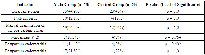
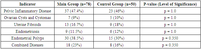
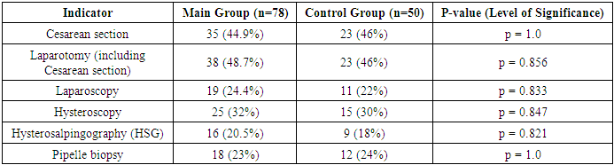
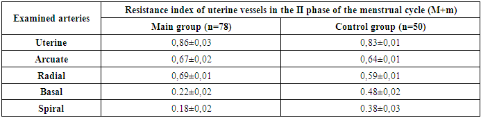
The patients included in the study were aged 25 to 35 years. We did not find statistically significant differences in age between the study groups: the average age of patients in the main group was 30.5±1.2 years, patients in the control group – 30.2±1.29. When comparing the main characteristics of the menstrual cycle according to anamnestic data (menarche, duration of the menstrual cycle, duration and nature of menstruation), it was shown that the groups are comparable in all these parameters. There were no significant differences in marital status and education level between the patients in both groups.
Of the somatic diseases, anemia, which was found with the same frequency in both groups of women – 50% and 52% respectively, is noteworthy. Mostly it was chronic iron deficiency anemia of moderate severity.
Particular attention was paid to the obstetric history and gynecological morbidity in the examined women, as well as to the types of surgical interventions performed on the pelvic organs (Tables 1, 2, 3).
Table 1. Obstetric history of observed patient
     
Table 2. Gynecological morbidity of observed patients (anamnestic data)
     
Table 3. Past surgical interventions on the pelvic organs of served patients
     
The presented data demonstrate that both in terms of frequency and structure of the incidence of gynecological pathology, the groups are comparable (p≥0.05). It is obvious that the high frequency of endometrial polyps in the structure of past gynecological diseases (38.5% and 30% respectively), as well as information from the obstetric history about complications of childbirth and a high frequency of cesarean section (44.9% and 46% respectively) are quite understandable, since almost all patients included in the study had a "thin" endometrium, which can be considered as a predictor of complications that led to infertility. Laparotomy and laparoscopy on the pelvic organs in the history were performed in 71% (n=128) of patients in both groups. As can be seen from Table 3, in general, statistically significant differences in the frequency of past surgical interventions between the groups were not revealed (p≥0.05).
The vaginal microflora was disrupted (according to anamnestic data) in 36 patients in both groups, which amounted to 28.1%. Bacterial vaginosis occurred in 20 (25.7%) patients in the main group and in 5 (10%) patients in the control group (p≥0.05). Vulvovaginal candidiasis was diagnosed in 8 (10.3%) women in the main group and in three (6%) in the control group. The presence of a sexually transmitted infection (STI) in the history was diagnosed in both groups of observed patients: chlamydial infection in the history was more than 3 times more common in patients in the main group – 7.7% (n=6) compared to patients in the control group – 2% (n=1).
CINI-II was diagnosed 5 times more often in the group of women with condyloma acuminata compared to the control group (20.5% and 4% respectively). It should be noted that all patients with condyloma acuminata and CIN were not vaccinated against HPV.
All patients in this group were prescribed appropriate therapy. Due to the presence of an inflammatory process of the genital organs, invasive diagnostic procedures were postponed until the end of treatment.
In the structure of infertility in the main group of patients, the predominant factor was the violation of the morphological and functional ability of the endometrium - "thin" endometrium.
The condition of the pelvic organs was assessed according to ultrasound data (Fig. 1) and
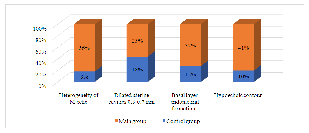
Figure 1. Dopplerometry
Table 4. Indicators of the median M-echo in the examined groups of patients.
In the conducted study, the echographic parameters of patients in the main and control groups were analyzed. The results showed the presence of significant differences in a number of indicators between the groups:
Heterogeneity of the M-echo was observed in 36% of patients in the main group and in 8% of patients in the control group, which indicates a significant difference between the groups (p<0.001).
Expanded uterine cavities (0.3-0.7 mm) were noted in 23% of patients in the main group and in 18% in the control group. Statistical analysis showed no significant differences in this indicator (p = 0.501), which suggests that this symptom may be common in both groups, regardless of the presence of condyloma acuminata.
Formations of the basal layer of the endometrium were detected in 32% of patients in the main group and in 12% of the control group, which also indicates a statistically significant difference between the groups (p = 0.015).
Hypoechoic contour was found in 41% of patients in the main group and in 10% of the control group, which also demonstrates significant differences (p<0.001).
These data emphasize that patients in the main group have more pronounced pathological changes in the endometrium, which requires further in-depth study of their clinical significance and the development of appropriate treatment methods.
Table 4 shows the resistance index of uterine vessels in patients with "thin" endometrium in the second phase of the menstrual cycle.
Table 4. Index of uterine vascular resistance in patients with “thin” endometrium
     
In patients in the main group, the resistance index in the uterine arteries was 0.86±0.03, which is slightly higher than in the control group, where this indicator is 0.83±0.01. This indicates a slight increase in resistance in the main group, although the differences are not significant (p≥0.05).
In the main group, the resistance index in the arcuate arteries is 0.67±0.02, while in the control group it is 0.64±0.01 (p≥0.05).
RI in the radial arteries in the main group is significantly higher (0.69±0.01) compared to the control group (0.59±0.01) (p<0.05).
In the main group, the resistance index in the basal arteries was 0.22±0.02, which is significantly lower than the control group (0.48±0.02) (p<0.05). All this indicates a lower resistance of blood vessels in patients with "thin" endometrium in the main group.
RI in the spiral arteries in the main group (0.18±0.02) is significantly lower compared to the control group (0.38±0.03) (p<0.05). These data indicate significant differences in vascular resistance between the groups.
Overall, the resistance index of uterine vessels in patients in the main group shows pronounced changes compared to the control group, especially in the radial, basal and spiral arteries. These data may indicate impaired blood supply to the uterus in patients with "thin" endometrium, which requires further study to understand the pathophysiological processes underlying these changes that led to secondary infertility.

4. Discussion

Analyzing the data we obtained, we can argue that HPV-positive patients have prerequisites for the development of infertility. This is also evidenced by studies conducted by Yu.N. Banashkaeva and co-authors (2022), which indicate that even in the treatment of infertility using assisted reproductive technologies, the lowest frequency of pregnancy after cryoprotocols of IVF was observed in the group of HPV-positive women (33.3%) compared to HPV-negative (73.9%) and previously vaccinated patients (60.7%); p<0.05 [1]. The work of Italian researchers showed that in HPV-positive patients, unsuccessful IVF attempts were almost 3 times more common compared to HPV-negative (40 and 13.5%) [28]. According to the data of the US university clinic, when conducting ART programs in women with HPV persistence, the frequency of pregnancy was significantly lower than in women without the virus (57 and 23.5%) [15].
The negative impact of HPV on reproductive function is also confirmed in a number of other works. Thus, a systematic review conducted in Germany demonstrates a reliable association between spontaneous abortion, spontaneous preterm birth and the presence of HPV both in the cervix and in the placenta [13].

5. Conclusions

The combination of condyloma acuminata with "thin" endometrium in patients with secondary infertility requires further research and determination of the tactics for managing these women.

References

[1]  Banashkaeva Yu.N., Zarochentseva N.V., Krasnopolskaya K.V., Isakova K.M. The effectiveness of embryo transfers in patients in cryoprotocols of in vitro fertilization // Obstetrics and Gynecology: news, opinions, training. 2022. Vol. 10, No. 2. P. 23–30. DOI: https://doi.org/10.33029.
[2]  Bebneva T.N., Dikke G.B. Recurrent violations of the vaginal biocenosis associated with bacterial vaginosis in pregnant women with HPV infection // Obstetrics and Gynecology. – 2018. – No. 9. – P. 55-60.
[3]  Golyshkin M.S. et al. Female infertility as a factor of emotional distress: the importance of psychotherapy in the treatment of infertility // Neurology, neuropsychiatry, psychosomatics. – 2021. – Vol. 13. – No. 2. – P. 97-103.
[4]  Zarochentseva N.V. et al. The effectiveness of the in vitro fertilization program for infertility in patients with human papillomavirus // Problems of Reproduction. – 2021. – Vol. 27. – No. 4. – P. 108-116.
[5]  Ibishev Kh.S., Ryabenchenko N.N., Magomedov G.A. Papillomavirus infection of the ejaculate associated with male infertility // Urology. – 2021. – No. 1. – P. 79-83.
[6]  Oganesyan MV et al. Tubal infertility: medico-social and psychological aspects of the problem. Obstetrics and Gynecology. 2020; (11): 213-24.
[7]  Prokopets V.I., Strizhak D.A. Female infertility of inflammatory origin // Young Scientist. – 2016. – No. 22-1. – P. 31-34.
[8]  Rakhmetova M.R. et al. Features of diagnosis and treatment of endocrine infertility // Academic research in educational sciences. – 2022. – Vol. 3. – No. 4. – P. 722-728.;
[9]  Semenova E.V., Kirillov N.A. Etiology of female infertility // Modern problems of medicine and natural sciences. – 2018. – P. 79-83.
[10]  Smoley N.A. Modern approaches to the diagnosis and treatment of female infertility // BBK 57.16+ 57.3 ya43 A437. – 2020. – P. 96.
[11]  Yuzko A.M. Female infertility of tubal origin (Literature review) // Women's Health. – 2017. – No. 2. – P. 126-131.
[12]  Anwar S., Anwar A. Infertility: A review on causes, treatment and management // Womens Health Gynecol. – 2016. – Vol. 5. – P. 2-5.
[13]  Ambühl L.M.M., Leonhard A.K., Widen Zakhary C., Jorgensen A., Blaakaer J., Dybkaer K. et al. Human papillomavirus infects placental trophoblast and Hofbauer cells, but appears not to play a causal role in miscarriage and preterm labor // Acta Obstet. Gynecol. Scand. 2017. Vol. 96, N 10. P. 1188–1196. DOI: https://doi.org/10.1111/aogs.13190 Epub 2017 Aug 14. PMID: 28699189.
[14]  Barbieri R.L. Female infertility // Yen and Jaffe's reproductive endocrinology. – Elsevier, 2019. – P. 556-581. e7.
[15]  Gomez L.M., Ma Y., Ho C., McGrath C.M., Nelson D.B., Parry S. Placental infection with human papillomavirus is associated with spontaneous preterm delivery // Hum. Reprod. 2008. Vol. 23, N 3. P. 709–715. DOI: https://doi.org/10.1093/humrep/dem404.
[16]  Hu J.P. et al. Prevalence and genotype distribution of human papillomavirus infection among 66000 women from 2014 to 2023 in the plateau region of Southwest China // Virology Journal. – 2024. – Vol. 21. – No. 1. – P. 176.
[17]  Halim H.M. Successful treatment of giant anogenital condyloma accuminata with combined cryotherapy and intralesional tuberculin purified protein derivative: a case report // The Journal of Sexual Medicine. – 2022. – Vol. 19. – No. 11. – P. S24.
[18]  Isaguliants M, Krasnyak S, Smirnova O, Colonna V, Apolikhin O, Buonaguro FM. Genetic instability and anti-HPV immune response as drivers of infertility associated with HPV infection. Infect Agent Cancer. (2021) 16: 29. doi: 10.1186/s13027-021-00368-1.
[19]  Jin G. et al. Efficacy and safety of Chinese patent medicine paiteling in condyloma acuminatum: A systematic review and meta-analysis // Journal of Ethnopharmacology. – 2024. – Vol. 318. – P. 116894.
[20]  Kesheh M.M., Keyvani H. The prevalence of HPV genotypes in Iranian population: an update // Iranian journal of pathology. – 2019. – Vol. 14. – No. 3. – P. 197.
[21]  Lewis R.M. et al. Prevalence of genital human papillomavirus among sexually experienced males and females aged 14–59 years, United States, 2013–2014 // The Journal of infectious diseases. – 2018. – Vol. 217. – No. 6. – P. 869-877.
[22]  Lin K. et al. Cervical HPV infection and related diseases among 149,559 women in Fujian: an epidemiological study from 2018 to 2023 // Frontiers in Microbiology. – 2024. – Vol. 15. – P. 1418218.
[23]  Mustafa M. et al. Male and female infertility: causes, and management // IOSR Journal of Dental and Medical Sciences. – 2019. – Vol. 18. – No. 9. – P. 27-32.
[24]  Mishra A. et al. Impacts and Causes of Female Infertility: An Observational Study // The Scientific Temper. – 2022. – Vol. 13. – No. 02. – P. 19-24.
[25]  Moreno-Sepulveda J, Rajmil O. Seminal human papillomavirus infection and reproduction: a systematic review and meta-analysis. Andrology. (2021) 9:478– 502. doi: 10.1111/andr.12948.
[26]  Muscianisi F, De Toni L, Giorato G, Carosso A, Foresta C, Garolla A. Is HPV the novel target in male idiopathic infertility A systematic review of the literature. Front Endocrinol. (2021) 12: 643539. doi: 10.3389/fendo.2021.643539.
[27]  Nikolic N. et al. Prevalence of Carcinogenic Genotypes of HPV-Infected Women in a Ten-Year Period (2014–2023) in Vojvodina, Serbia // Medicina. – 2024. – Vol. 60. – No. 6. – P. 922.
[28]  Perino A., Giovannelli L., Schillaci R. et al. Human papillomavirus infection in couples undergoing in vitro fertilization procedures: impact on reproductive outcomes // Fertil. Steril. 2011. Vol. 95, N 5. P. 1845–1848. DOI: https://doi.org/10.1016/j.fertnstert.2010.11.047.
[29]  Purewal S, Chapman SCE, van den Akker OBA. A systematic review and meta-analysis of psychological predictors of successful assisted reproductive technologies. BMC Res Notes. 2017 Dec 7; 10(1): 711. doi: 10.1186/s13104-017-3049-z.
[30]  Pellavio G, Todaro F, Alberizzi P, Scotti C, Gastaldi G, Lolicato M, et al. HPV infection affects human sperm functionality by inhibition of aquaporin-8. Cells. (2020) 9: 1241. doi: 10.3390/cells9051241.
[31]  Rakhimova M. Infertility in women classification, symptoms, causes and factors, recommendations for women // Science and innovation. – 2022. – Vol. 1. – No. D7. – P. 245-250.
[32]  Sindhuja T., Bhari N., Gupta S. Asian guidelines for condyloma acuminatum // Journal of Infection and Chemotherapy. – 2022. – Vol. 28. – No. 7. – P. 845-852.
[33]  Starc A, Trampus M, Pavan Jukic D, et al. Infertility and sexual dysfunctions: a systematic literature review. Acta Clin Croat. 2019 Sep; 58(3): 508-15. doi: 10.20471/acc.2019.58.03.15.
[34]  Sahana Devi K.J., Yendapalli V. Women infertility: a systematic review of effects and causes // SN Computer Science. – 2022. – Vol. 4. – No. 2. – P. 103.
[35]  Sharipova I.P. et al. Prevalence of high-risk human papillomavirus genotypes among women in Uzbekistan, 2021–2023 // Journal of Gynecologic Oncology. – 2024. – Vol. 36.
[36]  Sivars L. et al. Prevalence of human papillomavirus (HPV) types 16 and 18 in cervical cancer in Stockholm, Sweden during 2019–2023 compared to 2003–2008 // Acta Oncologica. – 2023. – Vol. 62. – No. 12. – P. 1649-1652.
[37]  Stuqui B. et al. Condyloma acuminata: An evaluation of the immune response at cellular and molecular levels // PloS one. – 2023. – Vol. 18. – No. 4. – P. e0284296.
[38]  Zegers-Hochschild, F. The International Glossary on Infertility and Fertility Care, 2017 / F. Zegers-Hochschild, G.D. Adamson, S. Dyer et al. // Fertility and Sterility. – 2017. – Vol. 108, No. 3. – P. 393-406.
[39]  Yuan H. et al. Epidemiology of human papillomavirus on condyloma acuminatum in Shandong Province, China // Human Vaccines & Immunotherapeutics. – 2023. – Vol. 19. – No. 1. – P. 2170662.
[40]  Yuan S. et al. Human papillomavirus infection and female infertility: a systematic review and meta-analysis // Reproductive BioMedicine Online. – 2020. – Vol. 40. – No. 2. – P. 229-237.