-
Paper Information
- Next Paper
- Previous Paper
- Paper Submission
-
Journal Information
- About This Journal
- Editorial Board
- Current Issue
- Archive
- Author Guidelines
- Contact Us
American Journal of Medicine and Medical Sciences
p-ISSN: 2165-901X e-ISSN: 2165-9036
2024; 14(12): 3414-3416
doi:10.5923/j.ajmms.20241412.71
Received: Dec. 12, 2024; Accepted: Dec. 26, 2024; Published: Dec. 31, 2024

Morphological Structure of the Stomach of White Outbred Rats in the Norm
 Abstract
 Reference
 Full-Text PDF
 Full-text HTML
Oltiyev E. D., Baymuradov R. R.
Bukhara State Medical Institute, Bukhara, Uzbekistan
Copyright © 2024 The Author(s). Published by Scientific & Academic Publishing.
This work is licensed under the Creative Commons Attribution International License (CC BY).
http://creativecommons.org/licenses/by/4.0/

This article provides an analysis of the morphological and morphometric parameters of the stomach of white outbred rats. The macro-anatomy of the stomach and its topographic anatomy in the abdominal cavity are considered. In addition, emphasis is placed on the layered structure of the stomach, the location and functioning of the main and parietal cells.
Keywords: Stomach, Morphology, Laboratory animals
Cite this paper: Oltiyev E. D., Baymuradov R. R., Morphological Structure of the Stomach of White Outbred Rats in the Norm, American Journal of Medicine and Medical Sciences, Vol. 14 No. 12, 2024, pp. 3414-3416. doi: 10.5923/j.ajmms.20241412.71.
Article Outline
1. Introduction
- Scientists have long been interested in digestion and the role of the stomach in maintaining human health [1]. The stomach is traditionally considered as a hollow muscular organ that initiates the second phase of digestion. However, this simplistic view ignores the fact that it is the most complex endocrine organ with a unique anatomy, physiology, biochemistry, immunology, and microbiology. Everything that we consume, including our food, must first pass through the stomach, and therefore it is perhaps the most important part in the gastrointestinal tract [2].The gastric mucosa performs its immune function through innate and adaptive immunity, attracting immune cells and releasing appropriate cytokines, which are inextricably linked to diseases of the digestive system. Whether it is infectious gastric diseases caused by Helicobacter pylori, Epstein-Barr virus or another microbe, non-infectious gastric diseases or stomach cancer, the immunity of the gastric mucosa plays an important role in the occurrence and development of pathology [3].With the deepening of research, it is now believed that the gastric mucosa can perform its immune function through immunity [4] and maintain the balance of microbes in the mechanism of immune homeostasis [5]. When pathogens such as bacteria and viruses enter the gastric mucosa, both epithelial cells and innate immune cells begin to protect them through physical, chemical, and biological processes [3].Based on the above, it is very important to study the structure of the stomach of laboratory animals for their further use in various experiments.Objective of the study. To study and evaluate the morphological features of the stomach structure of white outbred rats.
2. Materials and Methods
- To conduct morphological studies of the stomach tissues 20 white outbred rats weighing from 231 to 304 grams were selected. The animals were kept in normal vivarium conditions, where all ethical and methodological standards approved by the Ethics Committee were observed.The rats were kept in individual cages with room temperature, natural light and ventilation. At the initial stages of the scientific experiments, all sexually mature rats were placed in a seven-day quarantine, after which they were transferred to the usual vivarium regime. During the experiment, the physiological state and behavior of the rats under study were carefully monitored. In the process of using experiments on laboratory rats, our actions complied with the requirements of the document "Rules for conducting work with experimental animals" (No. 18 dated 16.01.2018) of the Ethics Committee of the Bukhara State Medical Institute named after Abu Ali ibn Sina, in addition, the International Medical Declaration adopted by the association in Helsinki in 1964 and completed in 1975, 1983, 1989, 1996, 2000, 2002, 2004, 2008, 2013 was strictly observed, methodological recommendations and ethical principles for working with laboratory animals were used.The animals were slaughtered at the appropriate time in the morning, on an empty stomach by instant decapitation under ether anesthesia. All animals in turn underwent routine sectional removal of the anterior wall of the chest and abdominal cavities and photographing their contents. After this, they resorted to traditional anatomical dissection, which consisted of removing the stomach from the abdominal cavity.The general morphological picture of changes in the studied sections of the stomach was studied using hematoxylin and eosin staining and Van Gieson staining.
3. Results of the Research and Their Discussion
- To study the features of the morphological structure of the stomach of white outbred rats, 20 individuals were examined that were not subjected to experimental intervention, which made it possible to establish the norms of the morphological parameters of the stomach.To identify the topographic and anatomical boundaries and areas of the rats' abdomen, two horizontal lines were drawn: the upper intercostal (linea costarum) and the lower interridge (linea spinarum), thus the abdominal wall of the rats was divided into three areas: the upper - epigastric, the middle - mesogastric and the lower - hypogastric, to identify the location of the stomach in these areas of the abdominal cavity.In the studied rats, body weight ranges from 231 to 304 grams, with the average value being 265 grams.The stomach of white rats is a saccular formation of the digestive tract. In white rats, the stomach has a more curved and relatively constant hook-shaped form, as well as a transverse position in relation to the horizontal and sagittal planes. The narrowed esophageal part of the stomach is located to the left of the esophagus and is devoid of digestive glands. From the inside, this part is separated from the glandular part by a tortuous folded edge. The distal end of the esophagus is shifted to the middle of the lesser curvature, is located with a large contiguous place with the outlet of the stomach. This allows easy evacuation of food into the duodenum. Depending on the age, the degree of their fullness and the functional state of the muscular membrane of the wall, the shape of the stomach is variable. The stomach of rats has two surfaces: the anterior lower contacts the diaphragm and most of the abdominal wall and a small part covered by the left lobe of the liver. The posterior upper surface is located under the liver at the level of the body of the organ, on the right is the spleen, on the left is the pancreas (near the porta hepatis) in the abdominal cavity. Relative to the left part of the abdominal cavity at the level of the XI and XII thoracic vertebrae, dorsal to the liver and with the long axis, the stomach is directed transversely to the sagittal plane.In rats, three sections are distinguished by the location of the stomach: the bottom - facing the diaphragm, the body - to the abdominal wall and the pyloric section facing the porta hepatis.The length of the stomach in white outbred rats is on average - 3.21 ± 0.05 cm, and the width of the stomach at the level of the bottom of the organ is on average - 2.11 ± 0.08 cm, at the level of the body - 2.83 ± 0.09 cm, at the level of the pyloric section - 2.14 ± 0.06 cm.The walls of the stomach of rats are divided into two parts: transparent and opaque. This part of the stomach, divided by low tissue, a vertically limiting ridge that passes just below the esophagus along the circumference of the greater and lesser curvature of the stomach. The transparent part is the vestibule of the rat stomach, which receives food and serves as a receptacle for it. The length of the transparent part of the stomach in rats averaged 2.88±0.06 cm. The opaque part of the organ is a relatively small part of the stomach, which is located on the right side in relation to the transparent part of the stomach; the pyloric sphincter that controls the movement of food from the body of the stomach into the duodenum is not expressed in it.The greater curvature of the rat stomach is located distally and below, it faces the anterior abdominal wall and is more mobile than the lesser curvature. The lesser curvature is formed by the upper edge of the organ. When the stomach is empty, its cardiac opening is located behind at the level of the oscillating thoracic vertebrae, and the pyloric opening is at the level of the upper lumbar vertebrae.In rats, the length of the greater curvature is on average 6.83±0.34 cm. The lesser curvature is located proximally under the liver and from above it faces the spinal column. The length of the lesser curvature in rats was on average - 1.14 ± 0.02 cm.In rats, the length of the abdominal part of the stomach is on average - 0.16 ± 0.003 cm. Along the lesser curvature on the right, at some distance from the esophageal opening, there is a second opening - the pyloric part of the stomach, where the pyloric sphincter of the rat stomach is located, passing into the initial part of the duodenum.The wall of the rat stomach has two morpho - functionally different sections. After the transition of the esophagus into the stomach, the non-glandular (cardiac) section of the organ begins, lined with stratified squamous epithelium. It is followed by the glandular (pyloric) section, lined with a single-layer cylindrical epithelium, containing the gastric glands. These two sections are clearly separated by a limiting ridge.The wall of the stomach consists of a mucous membrane, submucosa, muscular and serous membranes, as shown in Fig. 1.
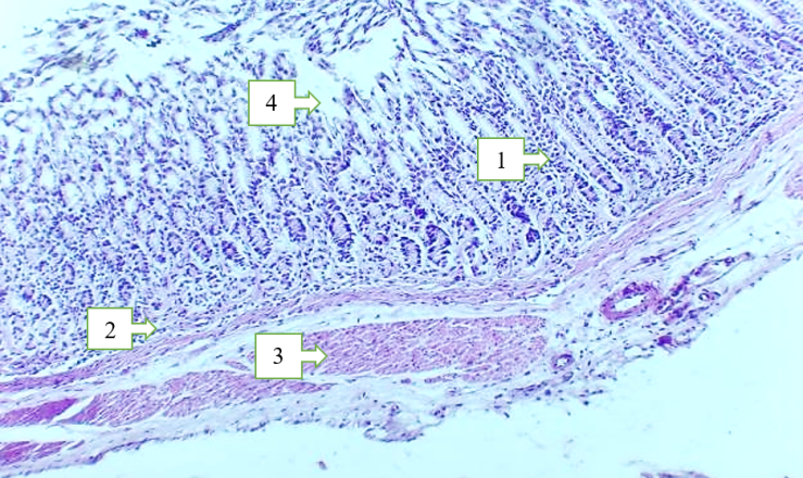 | Figure 1. Pyloric part of the stomach of rats. 1 - mucous membrane, 2 - submucosa, 3 - muscular membrane, 4 - depression between the folds. Hematoxylin and eosin staining. 10x20 |
4. Conclusions
- The obtained morphological indices and morphometric data of the stomach of white outbred rats did not reveal any pathological changes. Therefore, these parameters can be used in comparative analysis to study different pathological conditions.