Shodikulova Gulandom Zikriyayevna, Khasanov Oybek Gafurovich, Atoev Tulkin Tolmasovich
Samarkand State Medical University, Samarkand, Uzbekistan
Copyright © 2024 The Author(s). Published by Scientific & Academic Publishing.
This work is licensed under the Creative Commons Attribution International License (CC BY).
http://creativecommons.org/licenses/by/4.0/

Abstract
Introduction. Osteoarthritis is the most common joint disease. According to the World Health Organization 2023 data, OA affects more than half a billion people worldwide, an increase of 113% since the 1990s. Approximately 3/5 of people living with OA are older than 55 years of age, and 3/5 of them are represented by the female gender. Aim of the study was to investigate the clinical, immunological and genetic features of the development of osteoarthritis of hip joints in the Uzbek population who underwent COVID-19 in order to develop an algorithm for early diagnosis of the disease. Materials and methods of research: All clinical studies were conducted for the period 2021-2023 on the basis of Samarkand City Medical Association and Samarkand branch of the Republican Specialized Scientific and Practical Medical Centre of Traumatology and Orthopedics. A comprehensive approach including clinical, genetic, laboratory, ultrasound, radiological and statistical methods of research was used for the present work. Conclusions. Thus, in-depth study of clinical, immunological and genetic features of hip OA development opens new opportunities for early diagnosis and individualization of treatment, which increases the chances of slowing disease progression and improving the quality of life of patients.
Keywords:
Hip Osteoarthritis, COVID-19, COL1A1 and GDF5 gene polymorphisms, Clinical and immunological features
Cite this paper: Shodikulova Gulandom Zikriyayevna, Khasanov Oybek Gafurovich, Atoev Tulkin Tolmasovich, Clinical, Immunological and Genetic Peculiarities of Development of Ostearthritis of Hip Joints in COVID-19 Patients of Uzbek Population, American Journal of Medicine and Medical Sciences, Vol. 14 No. 12, 2024, pp. 3278-3282. doi: 10.5923/j.ajmms.20241412.42.
1. Introduction
Osteoarthritis is the most common joint disease [1,3]. According to the World Health Organization (WHO) 2023 data, OA affects more than half a billion people worldwide, an increase of 113% since the 1990s. Approximately 3/5 of people living with OA are older than 55 years of age, and 3/5 of them are represented by the female gender [11]. Coxarthrosis (deforming osteoarthritis of the hip joint) is a chronic degenerative, progressive and multifactorial disease of the hip joint characterized by pain, limping and limitation of daily activities. This disease leads to surgical intervention (total joint endoprosthesis) in 80% of cases, reduced work capacity in 60%, and disability in 11.5% [9].Modern scientific research demonstrates that in addition to mechanical factors of OA, such as increased stress and trauma, genetic predisposition and hereditary factors have a significant influence on the development of the disease. Genetic studies make it possible to identify a predisposition to osteoarthritis at preclinical stages, when symptoms have not yet manifested themselves and degenerative changes in the joint are insignificant. This opens up new possibilities for early diagnosis and prevention of the disease, which is especially important for patients with a history of osteoarthritis. Research in the field of osteoarthritis genetics is aimed at identifying specific genetic markers associated with disorders in articular cartilage metabolism, inflammatory processes, and tissue regeneration [10]. To date, many genetic variants (polymorphisms) that may be associated with the development of osteoarthritis have been identified. Among them, special attention is paid to genes regulating collagen synthesis, matrix metalloproteinases (MMPs), and genes responsible for the regulation of inflammatory processes. One of the key genes involved in the development of osteoarthritis is the COL1A1 gene, which encodes type II collagen, which is the main structural protein of articular cartilage. Mutations in this gene can lead to impaired collagen synthesis and, as a consequence, increased vulnerability of cartilage tissue to mechanical damage and degeneration [9].In recent years, almost ubiquitous pathology of COVID-19 has been adversely affecting various organs and systems of the human body. There are multiple reports in the available literature regarding the development of osteonecrosis after COVID-19. Data of M.A. Panin et al. (2022) demonstrated that osteonecrosis in COVID-19 patients developed in a shorter time frame compared to the patients without coronavirus infection in anamnesis [10].Meanwhile, the results of studies evaluating immunogenetic mechanisms of OA initiation, for early diagnosis, do not have unambiguous conclusions, so additional research in this direction is necessary and relevant.Therefore, the aim of the study was to investigate the clinical, immunological and genetic features of the development of osteoarthritis of hip joints in the Uzbek population who underwent COVID-19 in order to develop an algorithm for early diagnosis of the disease.
2. Materials and Methods of Research
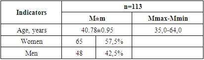
All clinical studies were conducted for the period 2021-2023 on the basis of Samarkand City Medical Association and Samarkand branch of the Republican Specialized Scientific and Practical Medical Centre of Traumatology and Orthopaedics. A comprehensive approach including clinical, genetic, laboratory, ultrasound, radiological and statistical methods of research was used for the present work.Inclusion and exclusion criteria for the study were drawn up according to the planned clinical and laboratory investigations and the developed algorithms. A total of 113 patients with a reliable diagnosis of hip osteoarthritis were examined. These patients underwent general clinical methods of investigation, biochemical methods of investigation (renal and hepatic indices, rheumatic tests, calcium and vitamin D levels, IL4, IL6, IL10, TNF levels), determination of COL1A1 and GDF5 gene polymorphism, familiarization with case histories, protocols of primary examination, protocols of MSCT, MRI, radiological and X-ray densitometric studies. The functional status of joints was assessed using the FIHOA (Functional Index of Hand OA), KOOS, HOOS (Knee/Hip injury and Osteoarthritis Outcome Score), WOMAC Knee, Hip (Western Ontario and McMaster Universities Arthritis Index) scales. The impact of OA on quality of life and pain level, general health status, coping strategies for coping with pain were assessed using SF-36 (short form of index quality of life) and VAS (visual analogue scale) scales.All patients were divided into two groups: Group I - 63 patients with hip OA and COVID-19 in anamnesis and Group II - 50 patients with idiopathic OA.The age range of the patients included in the study varied from 25 to 65 years. There were more women than men (7.32%). The mean age of the patients was 40.78±0.95 (table 1).Table 1. Sex and age indicators in the examined groups of patients
 |
| |
|
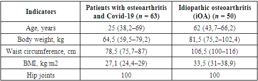
Evaluation of the occurrence of osteoarthritis by group, revealed that 52.9% of the subjects had a history of SARS-CoV-2 infection.In a general clinical study of osteoarthritis patients, we examined the frequencies of risk factors predisposing to the development of the disease, among which heredity (22.8% and 26.9%), trauma (39.6% and 54.1%), obesity (39.8% and 42.9%) were the most common in patients with the articular form of the disease.To clarify and verify the diagnosis of osteoarthritis, all patients underwent radiography of the hip joints, with subsequent assessment using the Kellgren-Lawrence and OARSI scales. To confirm comorbidities (obesity, hypertension and type 2 diabetes mellitus), all patients were examined according to the relevant criteria. Prior to inclusion in the study, patients filled out an informed consent form. Characteristics of the groups are presented in Table 2.Table 2. Characteristics of the examined groups of patients, median (interquartile intervals)
 |
| |
|
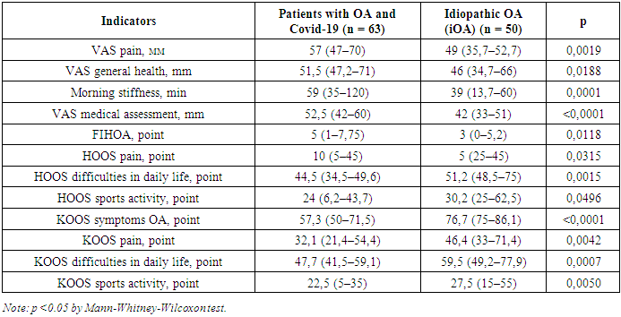
Clinical assessment of osteoarthritis activity depending on the form of the disease showed a correspondence of pain level on VAS 53.2 mm and 65.6 mm; the number of painful joints was 1.6 times more prevalent among hip osteoarthritis patients. A similar pattern was observed with regard to the number of swollen joints, as well as starting pains. Table 3. Distribution of patients by osteoarthritis groups
 |
| |
|
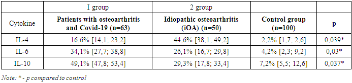
As it can be concluded from Table 3, COVID-19 patients with osteoarthritis are characterized by a high level of pain, which is most pronounced in the hip joints. The general health/wellness of this category of patients suffers, stiffness in the joints in the morning hours lasts longer. Indicators of functional activity are significantly lower, which affects the performance of various physical loads, daily activities and work. Patients with idiopathic osteoarthritis do less physical exercise, the doctor's assessment of the disease is significantly higher in post-occlusive osteoarthritis.In our study we found a statistically significant increase in IL10 concentrations (p=0.03) in group I patients and a significant increase in IL4, IL6 concentrations in group I patients compared to group II patients. Table 4. Levels of cytokines in patients in comparison groups, pg/ml
 |
| |
|
The following fact is noteworthy: our study revealed that blood IL6 is statistically significantly correlated with OA in COVID-19 patients.Thus, the study showed that IL4, IL6, and IL10 have the highest sensitivity and specificity in osteoarthritis.As a result of this study, a statistically significant decrease in the concentrations of Ca, vitamin D and an increase in the rate of erythrocyte sedimentation in the blood in idiopathic osteoarthritis was found.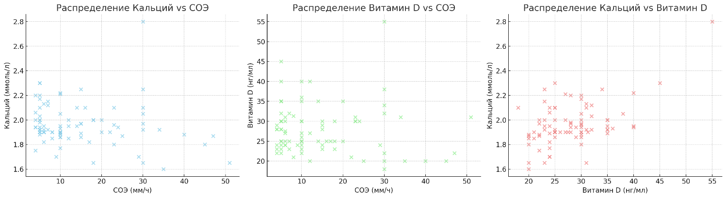 | Figure 1. Concentrations of calcium, vit D, and ESR levels |
Figure 1 demonstrates the percentages of reduced calcium, vitamin D levels and elevated ESR for patients with idiopathic and post osteoarthritis. We found that in idiopathic osteoarthritis, 82.98% had decreased calcium levels, while vitamin D levels were within normal limits. This group also showed an increase in COE to an average of18.6 mm/h. In the group with post-malignant osteoarthritis, these parameters were changed in the following order: 74.42% and 2.33% decrease in calcium and vitamin D levels, respectively, as well as an increased COE level up to 25.5 mm/hour.As a result of the X-ray examination, the de-centration of the head and its congruence to the acetabulum (III-IV stage according to J. Kellgren and J. Lawrence) was detected in group I patients compared to group II (p=0.03 and p=0.003, respectively). Group II patients showed narrowing of the articular gap, presence of subchondral sclerosis compared to Group I (p=0.06; p=0.8; p=0.5 and p=0.9, respectively).According to the results of our study, group I patients showed pronounced radiological changes (necrosis of the femoral head) compared to group II.The results of the study of GDF5 gene polymorphism in patients with osteoarthritis in the general group of patients (n=85) showed that the proportion of occurrence of unfavourable allele A slightly exceeds this index in the group with AA (20.5% vs. 19.4%). The greater registration of the A allele was observed due to the predominant share of its carriage among patients with osteoarthritis who underwent COVID-19 (n=45), which was 46.4%, whereas with GA (n=45) this indicator was 52.2%. At the same time, with regard to the frequency of G/G genotype, the carriage rate was found to be 27.2% in 27.2% of cases and 39.7% in controls. At the same time, the frequency of heterozygous G/A genotype among patients of the general group was 29.2%, while in the group with G/A this indicator was slightly less frequent (19.3%). Also, as for allele A, cases of carriage of unfavourable G/A and A/A genotypes in 1A (52.28% and 20.45%, respectively) and 1B (21.9% and 3.1%, respectively) subgroups of patients were registered more frequently compared to GG (19.3% and 1.8%, respectively). Collected data indicate a possible role of functionally unfavourable G/A and A/A genotypes of the studied polymorphism in increasing the risk of developing this pathology (Table 5).Table 5. Differences in the frequency of allelic and genotypic variants of the G/A polymorphism in the GDF5 gene in patient groups
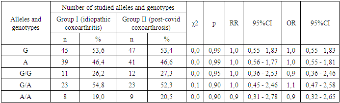 |
| |
|
Whereas an increase in the proportion of carriage of the G allele of the polymorphic variant of the GDF5 gene in the group of idiopathic osteoarthritis patients and in the group of COVID19 patients with osteoarthritis tended to increase the risk of developing the disease almost twofold (χ2=3. 344; P=0.07; OR=1.919; 95%CI:0.954-3.859) and 1.65-fold (χ2=1.57; P=0.211; OR=1.65; 95%CI:0.756-3.594), while in the group of patients with GG the risk of disease development was statistically significantly increased 2.58-fold (χ2=4.512; P=0.037; OR=2.58; 95%CI:1.076-6.188). The reduction in the frequency of wild-type A/A genotype among patients compared with GA was not statistically significant (in the combined osteoarthritis patient group, χ2=3. 073; P=0.084; OR=0.517; 95% CI: 0.247-1.081; in the COVID19-treated osteoarthritis patient group - χ2=1.713; P=0.193; OR=0.58; 95% CI: 0.257-1.311 and in the CC group - χ2=3.336; P=0.072; OR=0.406; 95% CI: 0.154-1.068).In our studies on the carrier patterns of COL1A1 gene polymorphism, the proportion of carriers of the unfavourable A allele (46.5% vs. 24.5%; χ2=5.349; p=0.021; OR=1.963; 95%CI: 1.108-3. 477) and heterozygous genotype C/A (53.94% vs. 19.3%; χ2=4.1 21; p=0.045; OR=2.011; 95%CI: 1.024-3.948) of the COL1A1 gene polymorphism in the group of patients with osteoarthritis were statistically significantly higher than those among conventionally healthy individuals. The obtained data may indicate a possible contribution of COL1A1 in the early diagnosis of osteoarthritis. The analysis of allele frequency distribution showed a greater proportion of C allele carriers among osteoarthritis patients compared to controls (60.8% vs. 71.0%). The increase in the frequency of this allele was observed due to the high shares in both groups of patients, osteoarthritis patients with COVID-19 reached 43.2%, and in the group and osteoarthritis patients with CC - 26.8% (table 6).Table 6. Differences in the frequency of allelic and genotypic variants C/A polymorphism in the COL1A1 gene in patient groups
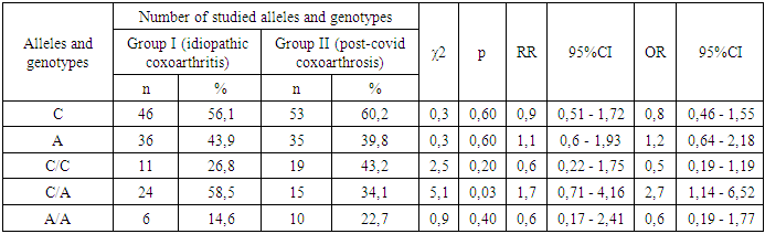 |
| |
|
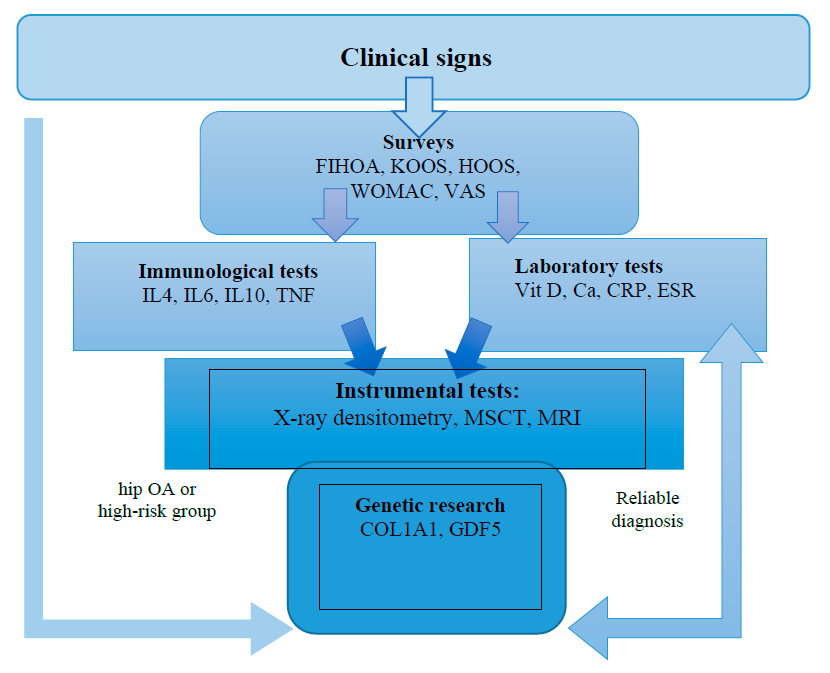 | Figure 2. Algorithm for early diagnosis of hip osteoarthritis |
Based on the presented scheme, the following scientific conclusion can be formulated: The scheme integrates a multidimensional approach to diagnosis, combining clinical data with biochemical, immunological, instrumental and genetic studies for a comprehensive assessment of the patient's condition. The use of a variety of questionnaires (FIHOA, KOOS, HOOS, WOMAC, VAS) allows detailing the subjective symptoms of the disease, while immunological studies (IL4, IL6, IL10, TNF) provide data on molecular and cellular mechanisms of inflammation and immune reactivity. Laboratory tests such as vitamin D, calcium, C-reactive protein and erythrocyte sedimentation rate levels further confirm the presence or absence of pathological processes. Instrumental diagnostic techniques including X-ray densitometry, multislice computed tomography (MSCT) and magnetic resonance imaging (MRI) provide visualization of anatomical and structural changes in tissues. Genetic studies (COL1A1, GDF5) can identify hereditary predisposition to certain diseases, allowing a personalized approach to prevention and treatment.Thus, in-depth study of clinical, immunological and genetic features of hip OA development opens new opportunities for early diagnosis and individualisation of treatment, which increases the chances of slowing disease progression and improving the quality of life of patients.Information about the source of support in the form of grants, equipment, and drugs. The authors did not receive financial support from manufacturers of medicines and medical equipment.Conflicts of interest. The authors have no conflicts of interest.
References
| [1] | Shodikulova G. Z. et al. The Correlation among Osteoporosis, Calcium-Phosphore Metabolism and Clinical Symptoms of Main Disease in Patients with Rheumatoid Arthritis //Annals of the Romanian Society for Cell Biology. – 2021. – С. 4185-4190. |
| [2] | Zikriyaevna S. G., Kamolidinovna T. Z. Stratification of Cardiovascular Risk in Patients with Rheumatoid Arthritis // Telematique. – 2023. – Т. 22. – №. 01. – С. 1114-1119. |
| [3] | Shodikulova G. Z., Pulatov U. S. Efficiency evaluation of treatments patients with rheumatoid arthritis by dependence of clinic course and genetic polymorphism of haptoglobins // Toshkent tibbiyot akademiyasi axborotnomasi. – 2020. – №. 1. – С. 175-178. |
| [4] | Ritchlin C.T. The pathogenesis of psoriatic arthritis. Сurr Opin Rheumatol 2005; 17: 406—12. |
| [5] | Avramescu C, Biciusca V, Daianu T et al. Cytokine panel and histopathological aspects in the systemic lupus erythematosus. Rom J Morphol Embryol. 2010; 51(4): 633–40. |
| [6] | Babamuradova Z. B., Shavazi N. N. Assessment of the efficacy and safety of biological agents in rheumatoid arthritis // Journal of Advanced Medical and Dental Sciences Research. – 2021. – Т. 9. – №. 6. – С. 26-31. |
| [7] | Clark KEN, Lopez H, Abdi BA. Multiplex cytokine analysis of dermal interstitial blister fluid defines local disease mechanisms in systemic sclerosis. Arthritis Res Ther. 2015; 17: 73. doi: 10.1186/s13075- 015-0575-8. |
| [8] | Hinchcliff M, Huang CC, Wood TA, et al. Molecular signatures in skin associated with clinical improvement during mycophenolate treatment in systemic sclerosis. J Invest Dermatol. 2013; 133: 1979–89. doi: 10.1038/jid.2013.130. |
| [9] | Ergashеvich I. M. et al. Avascular necrosis of the femoral head after covid-19: a case series // Journal of biomedicine and practice. – 2023. – Т. 8. – №. 2. |
| [10] | Kompantsev D. et al. Substantiation of the composition of a rectal dosage form containing ketoprofen and glucosamine sulfate for the treatment of rheumatoid arthritis //Publishing Director. – С. 44. |
| [11] | Furkatovich S. B., Mamatmuradovna M. G., Kobilovna G. N. Evaluation of the effectiveness of ultrasound diagnosis of hip displasiya in children under 6 months // Journal of biomedicine and practice. – 2023. – Т. 8. – №. 2. |

 Abstract
 Reference
 Full-Text PDF
 Full-text HTML