-
Paper Information
- Next Paper
- Previous Paper
- Paper Submission
-
Journal Information
- About This Journal
- Editorial Board
- Current Issue
- Archive
- Author Guidelines
- Contact Us
American Journal of Medicine and Medical Sciences
p-ISSN: 2165-901X e-ISSN: 2165-9036
2024; 14(11): 3031-3034
doi:10.5923/j.ajmms.20241411.78
Received: Nov. 3, 2024; Accepted: Nov. 26, 2024; Published: Nov. 28, 2024

Determination of Immunohistochemical Changes in the Testes of Purebred Rats After Experimental Pulmonary Fibrosis Using Pomegranate Seed Oil
 Abstract
 Reference
 Full-Text PDF
 Full-text HTML
Asadov Baxriddin Sayfiddinovich
Bukhara Medical Institute named after Abu Ali ibn Sino, Bukhara, Uzbekistan
Correspondence to: Asadov Baxriddin Sayfiddinovich, Bukhara Medical Institute named after Abu Ali ibn Sino, Bukhara, Uzbekistan.
Copyright © 2024 The Author(s). Published by Scientific & Academic Publishing.
This work is licensed under the Creative Commons Attribution International License (CC BY).
http://creativecommons.org/licenses/by/4.0/

The expression of the Ki-67 marker makes it possible to distinguish cells that are in the active phase during the entire duration of the cell cycle (G1-, S-, G2- and M-phases). Proliferative activity refers to the growth rate of cells that are becoming malignant and of cells that have already become tumor cells. This is one of the most prominent features of the tumor. It mainly helps to evaluate the growth rate of the tumor, the risk of metastases, the response to treatment and the outcome of the diseases. Evaluation of pathogenetic activity of tumor cells is important not only for evaluation of biological properties of tumor cells, but also for choosing treatment tactics.
Keywords: Rats, Experiment, Immunohistochemical examination, Pomegranate seed oil, Correction
Cite this paper: Asadov Baxriddin Sayfiddinovich, Determination of Immunohistochemical Changes in the Testes of Purebred Rats After Experimental Pulmonary Fibrosis Using Pomegranate Seed Oil, American Journal of Medicine and Medical Sciences, Vol. 14 No. 11, 2024, pp. 3031-3034. doi: 10.5923/j.ajmms.20241411.78.
Article Outline
1. Introduction
- Changes in the environment (pollution), various chemical and physical factors affect the normal functioning of the body. In this regard, the reproductive system is a "mirror" (highly sensitive) of the body, where the nature of reactions to various factors is not unique [1,2,3]. The male reproductive system is a separate topic for long discussions, and now, as an equal participant in the reproductive process, a man is distinguished by a higher morbidity and mortality rate than a woman, and, accordingly, the average life expectancy [4,5]. Of all the organs of the male reproductive system, the testis and epididymis are called universal experimental organs, where the effects of factors at different levels can be studied quickly and accurately for preliminary reports and long-term results. In addition, according to modern concepts, the epididymis is an additional gonad in which the final maturation and accumulation of mature spermatozoa takes place [6].Baimurodov R.R. in his research (2021) studied the morphological changes in the testis and post-testis during radiation. In the era of scientific and technical progress, radioactive pollution, whose sources are not only natural factors, but also anthropogenic activity to a greater extent, is particularly dangerous among all pollutions [5]. The effect of ionizing radiation significantly disrupts the integrity of structures and the metabolic balance that maintains homeostasis in the cells of various tissues of the body. The development of radiation-biochemical effects is more evident in radiosensitive tissues of the body, including spermatogenic epithelial tissues. The morphometry of the testes showed uneven changes in their weight, length and thickness during postnatal ontogeny. Comparing the rate of growth of body weight and length with the weight and size of the testicles shows that with the increase in their size, the body weight increases more than the length. Testis weight increases 1.16 times faster than body weight, and at 90 days of age, the highest rate of testis growth is observed. In the experiment, all anatomical parameters of the testicles lag behind the control parameters. Irradiation, which negatively affects the morphological structure of the testicles, causes a delay in all morphometric parameters, including the thickness of the white layer and spermatogenic epithelium. The delay is more pronounced in white-bred rats at 180 days of age and less pronounced at 360 days of age. This is due to the activation of the body's protective and compensatory mechanisms, which is more clearly manifested in the groups that received ASD-2f. Puberty is delayed with chronic radiation sickness. This is manifested by disturbances in the stages of spermatogenesis, changes in the cells of the spermatogenic line, and the delay of the sperm system in the cavity of the tubes. In white rats of the control group, at the age of sexual maturity (90 days), quantitative changes are observed in Leydig cells, characterized by a sharp increase in their number by 11.2 times, and at adulthood (360 days), this indicator is 1, increases by 4 times. The number of Leydig cells, as well as their diameter, is lower in all experimental groups than in the control group. Chronic radiation sickness adversely affects the vasculature of the testes and seminiferous tubules, where the diameter of arterioles and venules, as well as the diameter of the curvature of the seminiferous tubule, are significantly higher in the radiation-exposed group than in the ASD-2f exposure and compared to the control group. 'more decreases [5,6,7].Up to 58% of the pathology that causes infertility in adult men begins in childhood and adolescence. In some cases, significant disturbances occur in the early stages of sexual formation and fetal development [1]. Hypoxia is one of the main factors that cause the decay processes of adaptation to extrauterine life. Most complications of pregnancy (preeclampsia), somatic and infectious diseases of a pregnant woman, adverse factors are accompanied by a lack of oxygen in the organs and tissues of the fetus [12]. It is known that hypoxia has a negative effect on all systems of the growing body, especially the central nervous and cardiovascular systems. Antenatal damage to the reproductive system is also important, because the consequences of this damage can be irreversible and can lead to testicular dysfunction in the postpartum period and the development of primary infertility [8,9].
2. Research Aim
- Analysis of age-related immunohistochemical changes in the testes of white outbred rats after correction with pomegranate seed oil after experimental pulmonary fibrosis.
3. Materials and Methods
- Immunohistochemical studies were conducted on 36 samples of the obtained materials. Serial sections 4–6 μm thick were deparaffinized, dehydrated, unmasked, and stained with antigens using a specialized automated Ventana Benchmark XT system, Roche, Switzerland. The study was conducted with Ki-67 antigens. Counting was done as a percentage for the Ki-67 marker (proliferative index) and the expression level was evaluated as mild, moderate, and high expression. White outbred rats were selected for the study and 4 months, 5 months and 6 months of age were selected. From the selected rats, they were divided into three groups: the control group, the experimental group with pulmonary fibrosis, and the group with pulmonary fibrosis.
4. Results and Discussion
- In order to determine the level of expression in testicular tissue of white rats after experimental pulmonary fibrosis, determination of the immunohistochemical proliferation index is an important test. It is widely used in the diagnosis of various pathological processes and diseases. This is one of the most prominent features of the tumor. It mainly helps to evaluate the growth rate of the tumor, the risk of metastases, the response to treatment and the outcome of the diseases. Evaluation of the pathogenetic activity of tumor cells is important not only for evaluating the biological properties of tumor cells, but also for choosing treatment tactics. The index of proliferative activity in various tumors is important for predicting the clinical course and consequences of diseases. The proliferation index is determined based on the expression level of the Ki-67 marker, in which 500 cells in the field of view are counted, and how many of the nuclei of the counted cells are positively expressed cells are counted, and the counted cells are divided by the total number of positively expressed cells, and the resulting result is calculated as %, the expression index was determined. The expression level of Ki-67 was evaluated as a percentage. Expression staining of Ki-67 markers was quantified by relative percentages and graded as light, moderate, and strong expression, and these values were: 0 (no staining); 1+(<20% cells, weakly stained); 2+(20-60% cells, moderately stained); 3+ (>60% cells, strongly stained).Immunohistochemical studies were conducted on 36 samples of the obtained materials. Serial 3-μm-thick sections were deparaffinized, dehydrated, unmasked, and stained with antigens using a specialized automated Ventana Benchmark XT system, Roche, Switzerland. The study was conducted with Ki-67 antigens. Counting was done as a percentage for the Ki-67 marker (proliferative index) and the expression level was evaluated as mild, moderate, and high expression. White outbred rats were selected for the study and 4 months, 5 months and 6 months of age were selected. From the selected rats, they were divided into three groups. In the experiment, the expression of the Ki-67 marker in the micropreparations isolated from the group treated with pomegranate seed oil was studied and compared.The expression of the Ki-67 marker was studied in the tissues isolated from the testicular tissue of white outbred rats aged 7, 9, and 12 months in the age dynamics group, and the following results were obtained. The following changes were found when the histological tissues isolated from the testicular tissue of white outbred rats in this group were studied for the expression of Ki-67 markers.
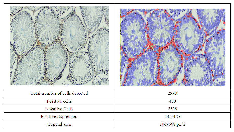 | Figure 1. Low expression of Ki-67 marker in the testicular tissues of 4 month old white outbred rats in the experimental lung fibrosis then corrected with pomegranate seed oil group.QuPath-0.4.0.ink. was scanned in the program and the level of expression was determined. Expressed cells are in red |
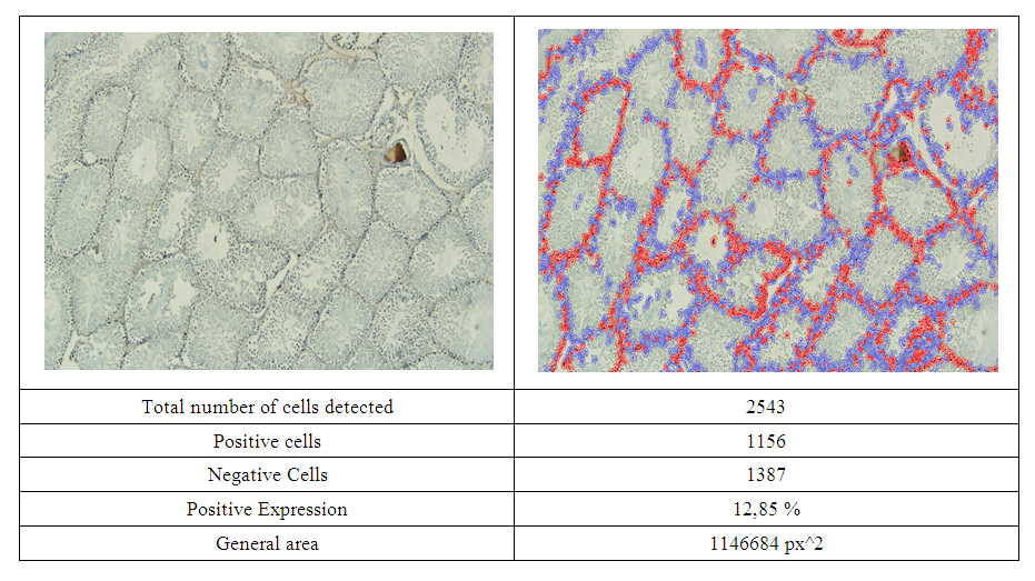 | Figure 2. Low expression of Ki67 marker in the testicular tissues of 6 month old white outbred rats in the experimental lung fibrosis then corrected with pomegranate seed oil group.. QuPath-0.4.0.ink. was scanned in the program and the level of expression was determined. Expressed cells are in red |
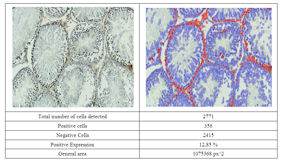 | Figure 3. A low level of Ki67 marker was expressed in testicular tissues of 9-month-old white outbred rats in the experimental pulmonary fibrosis followed by pomegranate seed oil correction. Dab is painted by the chromogenic method. Image magnified 400 times. QuPath-0.4.0. was scanned in the program and the level of expression was determined. Expressed cells are in red |
5. Conclusions
- Quantitative indicators of Ki-67 expression level from immunohistochemical markers in testicular tissue in experimental pulmonary fibrosis and when pomegranate seed oil was used for its correction. the average index of expression of the Ki 67 marker was 14.34%, and at 6 and 9 months it was 12.85%. Proliferative activity at 4, 6, and 9 months was mainly due to spermatogonial and myoid cells in the spermatogenic epithelial layer of many tubules and a small amount of spermatocyte 1 cells.