-
Paper Information
- Next Paper
- Previous Paper
- Paper Submission
-
Journal Information
- About This Journal
- Editorial Board
- Current Issue
- Archive
- Author Guidelines
- Contact Us
American Journal of Medicine and Medical Sciences
p-ISSN: 2165-901X e-ISSN: 2165-9036
2024; 14(11): 3028-3030
doi:10.5923/j.ajmms.20241411.77
Received: Oct. 25, 2024; Accepted: Nov. 20, 2024; Published: Nov. 28, 2024

Results of Immunohistochemical Study of Morphological Changes in the Liver of White Rats with Craniocerebral Injury
 Abstract
 Reference
 Full-Text PDF
 Full-text HTML
Olimova Aziza Zokirovna, Teshaev Shukhrat Jumaevich
Bukhara State Medical Institute, Uzbekistan
Correspondence to: Olimova Aziza Zokirovna, Bukhara State Medical Institute, Uzbekistan.
| Email: |  |
Copyright © 2024 The Author(s). Published by Scientific & Academic Publishing.
This work is licensed under the Creative Commons Attribution International License (CC BY).
http://creativecommons.org/licenses/by/4.0/

The expression level of the immunohistochemical marker Bcl-2 was low in 80% of the materials and 28.4% in 20% of the materials, it was moderately expressed. Thus, it was proven that the expression level of the marker Bcl-2 has a low level of apoptosis (as a result of aging or exposure to various damaging factors) in liver tissue cells in brain injury, i.e. programmed cell death.
Keywords: Liver, Morphology, Traumatic brain injury, Immunohistochemistry
Cite this paper: Olimova Aziza Zokirovna, Teshaev Shukhrat Jumaevich, Results of Immunohistochemical Study of Morphological Changes in the Liver of White Rats with Craniocerebral Injury, American Journal of Medicine and Medical Sciences, Vol. 14 No. 11, 2024, pp. 3028-3030. doi: 10.5923/j.ajmms.20241411.77.
Article Outline
1. Introduction
- The high rate of death and disability of patients with brain injury determines the undoubted socio-medical importance of the problem of brain injury (TBI). Frequency and severity of traumatic brain injury, high mortality rate (from 26.8-81.5%) [2,3,4] determine the relevance of this problem.Signs of liver parenchymatous damage are often observed in brain injury. Small focal necrosis of hepatocytes and changes in liver microcirculation are observed in the first hours after the injury. The development of blood circulation and destructive necrobiotic processes in the liver is manifested as hypoproteinemia, temporary enzymeemia, an increase in glucose and bilirubin in the blood serum (S.V. Tsarenko, 2015). At the same time, when evaluating morphofunctional changes in the liver during brain damage, the degree of brain damage is often not taken into account, and the characteristics of changes in the liver microvasculature are described. With severe brain damage, "disruption" of adaptive arterialization of sinusoidal blood flow and intrahepatic portal hypertension may develop. Severe microcirculation disorders are accompanied by the activation of necrobiotic processes in the liver and serve as a common pathomorphological basis for the development of liver failure in traumatic brain injury, consistent with published data (I.V.Fursov et al., 2013).Focal necrosis of stellate reticuloendothelial cells, swelling and desquamation of endothelial cells, hyperbilirubinemia and liver failure together with severe traumatic brain injury serve as the main criteria for damage to the liver vascular endothelium. The presented data show the relationship between central nervous system damage and functional and morphological changes in the liver. Thus, in the acute period of brain damage, functional disorders of the protein-synthetic, pigment and carbohydrate functions of the liver and clear disorders of intrahepatic hemodynamics are detected. Liver microcirculation disorders are accompanied by discomplexation of hepatocytes and the development of alterative changes in the form of protein and fat degeneration, focal necrosis of liver cells and stellate reticuloendotheliocytes, disruption of the structure of endothelial cells, their swelling, which contributes to the development of liver failure (C. V. Tsarenko, 2015).The effect of traumatic brain injury of different periods on liver morphology and function is little known. Further studies are required to determine the role of morphological changes in the liver as a result of brain injury.
2. The Purpose of the Study
- To investigate the morphological changes in the liver of white rats with brain damage using the Bcl-2 immunohistochemical marker.
3. Materials and Methods of the Research
- In this study, the morphological changes occurring in the liver tissue after the micropreparations prepared from the liver tissue were examined by the immunohistochemical method after the brain injury in white rats. The micropreparations made from the liver tissue isolated from them were examined by Bcl-2 immunohistochemical marker and the results obtained depending on the level of expression of this marker are presented. Proteins of the Bcl-2 marker family are regulators of apoptosis, one of the most studied types of programmed cell death. This protein family is represented by pro- and anti-apoptotic members. Anti-apoptotic proteins of the Bcl-2 family are often used by tumor cells as a mechanism of resistance to death, they play an important role both in the process of oncological diseases and in the resistance of cells to therapeutic effects. A detailed study of the interactions between Bcl-2 proteins underlying the regulation of the initiation of apoptosis will allow for an important breakthrough in the development of highly selective inhibitors of individual antiapoptotic members of the family.Apoptosis is a natural cell death process that can occur in various tissues, both in normal cells and in their neoplastic state. Apoptosis is a natural active process that requires the mandatory participation of various intracellular principles leading to each cell. There is a large group of proteins that regulate apoptosis processes.The control group of 3-month-old white rats obtained for histological examination and the liver tissue of 10% after conservative treatment for 10 days from the 1st, 7th, 21st days after brain injury and from the first day after brain injury in buffered formalin, then fixed with paraffin blocks and serial sections 3 μm thick. The cut areas were stained with hematoxylin and eosin according to Van Gieson. Expression was evaluated when the level of expression was 20% (low-level expression), 20-60% (medium-level expression), and more than 60% (high-level expression).
4. Research Results
- Bcl-2 family proteins are regulators of apoptosis, one of the most studied types of programmed cell death. This protein family is represented by pro- and anti-apoptotic members. Anti-apoptotic proteins of the Bcl-2 family are often used by tumor cells as a mechanism of resistance to death, they play an important role both in the process of oncological diseases and in the resistance of cells to therapeutic effects. Therefore, these proteins represent excellent targets for anti-onco-process therapy. A detailed study of the interactions between Bcl-2 proteins underlying the regulation of the initiation of apoptosis will allow for an important breakthrough in the development of highly selective inhibitors of individual antiapoptotic members of the family.
5. Discussion
- Apoptosis is a natural cell death process that can occur in various tissues, both in normal cells and in their neoplastic state. Apoptosis is a natural active process that requires the mandatory participation of various intracellular principles leading to each cell. There is a large group of proteins that regulate apoptosis processes.
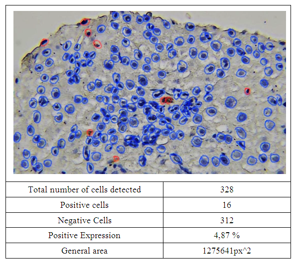 | Figure 1. Bcl-2 marker (4.87%) was expressed at a low level in the liver tissue of a non-white breed rat in an experiment with brain damage. Dab is painted by the chromogenic method. Image magnified 400 times. QuPath-0.4.0.ink. was scanned in the program and the level of expression was determined. Expressed cells are in red |
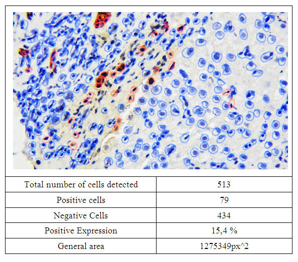 | Figure 2. In the experiment, the Bcl-2 marker (15.4%) was expressed at a low level in the liver tissue of a white breed rat with brain damage. Dab is painted by the chromogenic method. Image magnified 400 times. QuPath-0.4.0.ink. was scanned in the program and the level of expression was determined. Expressed cells are in red |
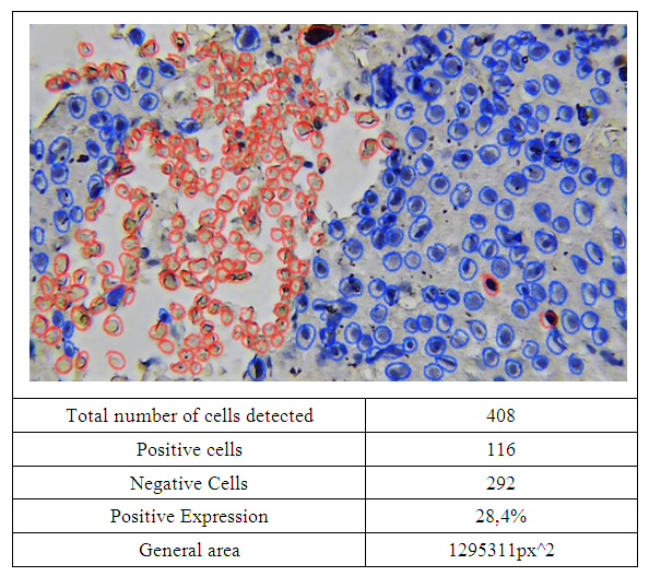 | Figure 3. Bcl-2 marker (28.4%) was moderately expressed in the liver tissue of a white breed rat with brain injury in the experiment. Dab is painted by the chromogenic method. Image magnified 400 times. QuPath-0.4.0.ink. was scanned in the program and the level of expression was determined. Expressed cells are in red |
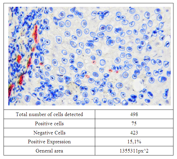 | Figure 4. Bcl-2 marker (15.1 %) was expressed at a low level in the liver tissue of a non-white breed rat in an experiment with brain damage. Dab is painted by the chromogenic method. Image magnified 400 times. QuPath-0.4.0.ink. was scanned in the program and the level of expression was determined. Expressed cells are in red |
6. Conclusions
- The expression level of immunohistochemical marker Bcl-2 was low in 80% of the materials and 28.4% in the 20% of the materials. %, that is, it was found to be moderately expressed. Therefore, it has been proven that the level of expression of the Bcl-2 marker has a low level of apoptosis (as a result of aging or the influence of various damaging factors) in liver tissue cells during brain injury, that is, the programmed death of cells. Long-term weightlessness can cause liver damage characterized by hepatocytic degeneration, portal fibrosis, glycogen depletion, and mitochondrial and endoplasmic reticulum swelling and loss of membrane integrity.